- Table View
- List View
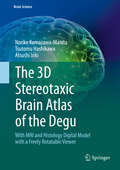
The 3D Stereotaxic Brain Atlas of the Degu: With Mri And Histology Digital Model With A Freely Rotatable Viewer (Brain Science)
by Atsushi Iriki Tsutomu Hashikawa Noriko Kumazawa-ManitaThis book is the first digital atlas of the degu brain with microscopic features simultaneously in Nissl sections and magnetic resonance imaging (MRI). As an experimental animal model, the degu contributes to a variety of medical research fields in diabetes, hyperglycemia, pancreatic function, and adaptation to high altitude, among others. Recently the degu has gained increasing importance in the field of neuroscience, particularly in studies evaluating the relationship between sociality and cognitive brain functions, and in studies pertaining to the evolutional aspects of the acquisition of tool-use abilities. Furthermore, aging-related brain dysfunction in humans can be studied using this animal model in addition to mammals with much longer lifespans. This brain atlas is constructed to provide histological and volume-rendered information simultaneously, fitting with any spatial coordination in brain positioning. It can be a useful guide to degus as well as to other rodents for studies of brain structures conducted using MRI or other contemporary examination methods with volume-rendering functions.
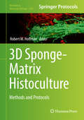
3D Sponge-Matrix Histoculture: Methods And Protocols (Methods In Molecular Biology #1760)
by Robert M. HoffmanThis volume describes numerous applications of sponge-matrix histoculture to study cancer biology and the treatment of cancer, stem cells, organoids, growth and repair of nerves, lymphoid tissues that produce antibodies, and HIV infection. The chapters in this book cover topics including, in vivo-like growth patterns of multiple types of tumors in Gelfoam® histoculture; development of the histoculture drug response assay (HDRA) for cancer patients; hair-shaft growth in Gelfoam® histoculture of skin as well as isolated hair follicles in Gelfoam® histoculture; the use of Gelfoam® histoculture to discover novel treatment of HIV infection; and imaging DNA repair after UV irradiation damage of cancer cells in Gelfoam® histoculture. The volume explains what is true 3D cell and tissue culture and corrects widespread misconceptions of 3D culture in the literature. Written in the highly successful Methods in Molecular Biology series format, chapters include introductions to their respective topics, lists of the necessary materials and reagents, step-by-step, readily reproducible laboratory protocols, and tips on troubleshooting and avoiding known pitfalls. <p><p> Cutting–edge and authoritative, 3D Sponge-Matrix Histoculture: Methods and Protocols is a valuable resource that contributes to new cell and tissue culture research, and other diseases based on the true 3D culture.
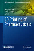
3D Printing of Pharmaceuticals (AAPS Advances in the Pharmaceutical Sciences Series #31)
by Abdul W. Basit Simon Gaisford3D printing is forecast to revolutionise the pharmaceutical sector, changing the face of medicine development, manufacture and use. Potential applications range from pre-clinical drug development and dosage form design through to the fabrication of functionalised implants and regenerative medicine. Within clinical pharmacy practice, printing technologies may finally lead to the concept of personalised medicines becoming a reality. This volume aims to be the definitive resource for anyone thinking of developing or using 3D printing technologies in the pharmaceutical sector, with a strong focus on the translation of printing technologies to a clinical setting. This text brings together leading experts to provide extensive information on an array of 3D printing techniques, reviewing the current printing technologies in the pharmaceutical manufacturing supply chain, in particular, highlighting the state-of-the-art applications in medicine and discussing modern medicine manufacture from a regulatory perspective. This book is a highly valuable resource for a range of demographics, including academic researchers and the pharmaceutical industry, providing a comprehensive inventory detailing the current and future applications of 3D printing in pharmaceuticals. Professor Abdul Basit is a Professor of Pharmaceutics at the UCL School of Pharmacy, University College London. Abdul's research sits at the interface between pharmaceutical science and gastroenterology, forging links between basic science and clinical outcomes. His research has been translated into the design of new technologies and improved disease treatments, many of which have been commercialised. Abdul is also a serial entrepreneur and has filed multiple patents, is the recepient of several research awards and has founded 3 companies (Kuecept, Intract Pharma, FabRx). He further serves as a consultant to many pharmaceutical organisations and is on the advisory boards of scientific journals, healthcare and charitable bodies.Professor Simon Gaisford holds a Chair in Pharmaceutics and is Head of the Department of Pharmaceutics at the UCL School of Pharmacy, University College London. He has published 110 papers, 8 book chapters, 4 authored books and is the recipient of multiple research awards. His research is focused on novel technologies for manufacturing medicines, particularly using ink-jet printing and 3D printing, translating his expertise by co-founding FabRx. Simon is also an expert in the physicochemical characterisation of compounds and formulations with thermal methods and calorimetry.
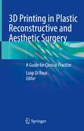
3D Printing in Plastic Reconstructive and Aesthetic Surgery: A Guide for Clinical Practice
by Luigi Di RosaThis handy volume illustrates the basics of clinical three-dimensional (3D) printing, addressing the practical aspects of establishing a simple and effective 3D printing service in a medical facility. No longer confined to makers and workshops, this very recent technology has been fast developing and rapid prototyping has proven its potential in the clinical field as well, leading to new approaches. The declared aim of this work is enabling medical professionals to create bespoke anatomical models from a series of CT or MRI images, and assisting them in choosing the best suited 3D printers and materials for each specific clinical need. The text includes original, full-color step-by-step photos for better guidance, and a complete review of related publications in literature. Single chapters devoted to specific areas of 3D printing application, such as rhinoplasty, ear reconstruction, oculoplasty, maxillofacial surgery, as well as for surgical simulations. Contents are completed by a review of the legal aspects and the safety and quality considerations, as well as a thorough examination of the variety of 3D printers, compatible materials as filaments and resins, and including the available online resources. Plastic, Ophthalmologic and Maxillofacial surgeons, and professionals dealing with surgical reconstruction, will find this guide to be a valuable companion for the understanding of 3D printing in clinical practice.
3D Printing in Oral & Maxillofacial Surgery
by Lobat Tayebi Reza Masaeli Kavosh ZandsalimiThis book is a comprehensive guide to 3D printing and 3D bioprinting methods and their application in oral and maxillofacial surgeries. Among the 3D printing methods considered are fused deposition modeling, selective laser sintering, photopolymer jetting, powder binder printing, and stereolithography, while the coverage of 3D bioprinting encompasses inkjet, microextrusion, and laser techniques. In each case, the relevance of the technique to oral and maxillofacial surgery is explained. In addition, the available inks and bioinks for 3D printing are reviewed. The roles of soft and hard tissue printing in oral and maxillofacial tissue engineering and the use of 3D printing in multi- and interfacial tissue engineering are then examined in depth. The particular value of 3D printing in the treatment of critically sized defects is discussed separately. Finally, up-to-date information is provided on guided tissue/bone regeneration using 3D printing. The book will be of interest to both oral and maxillofacial surgeons and biomedical engineers.
3D Printing in Oral Health Science: Applications and Future Directions
by Prabhat Kumar Chaudhari Dinesh Bhatia Jitendra SharanThis book on 3D printing in oral health science aims to equip the reader with a sound understanding of contemporary clinical applications in all fields of dentistry and their future directions. In the last few years, the development of 3D printing for medical and dental applications has increased tremendously. Advancements in 3D printing create the possibility of customized products, savings on small-scale productions, ease of sharing and processing of patient image data, and educational up-gradation. Looking at the dental specialties, it is evident that 3D printing has applications in all aspects of oral health science including prosthodontics, oral surgery, periodontics, endodontics, and orthodontics. This book will cover all major fields in dentistry and will help the practitioner in the process of decision-making and apply concepts in clinical or laboratory practice. It is based on current scientific evidence to provide readers with an up-to-date contemporary understanding of the subject, both from the clinical and the technological side. The book is a valuable asset for all who specialize in 3D printing and for those interested in learning more about this field.
3D Printing in Medicine and Its Role in the COVID-19 Pandemic: Personal Protective Equipment (PPE) and other Novel Medical and Non-Medical Devices
by Frank J. RybickiThis book describes how “makers” with no medical experience became and remain clinically important because they utilized 3D printing to produce supplies for healthcare, including medical and non-medical devices, and to improve the quality of life for patients with COVID-19 and those who care for them. It shows how 3D printing became vital during the pandemic due to its broad availability and the inherently digital nature of the work that enables thriving digital exchanges and work in isolation. Subsequent chapters highlight some of the “maker” communities' efforts that made a difference in their part of North America. Each contribution describes the unique experiences, challenges, and successes.While this book is written and edited mostly from a medical perspective, additional input from medical engineers, administrators, attorneys, and public safety officials enables a broad perspective to highlight some of the ingenuity from the North American 3D printing community who responded to the initial case volumes of COVID-19.
3D Printing in Medicine: A Practical Guide for Medical Professionals
by Frank J. Rybicki Gerald T. GrantThis book describes the fundamentals of three-dimensional (3D) printing, addresses the practical aspects of establishing a 3D printing service in a medical facility, and explains the enormous potential value of rendering images as 3D printed models capable of providing tactile feedback and tangible information on both anatomic and pathologic states. Individual chapters also focus on selected areas of applications for 3D printing, including musculoskeletal, craniomaxillofacial, cardiovascular, and neurosurgery applications. Challenges and opportunities related to training, materials and equipment, and guidelines are addressed, and the overall costs of a 3D printing lab and the balancing of these costs against clinical benefits are discussed. Radiologists, surgeons, and other physicians will find this book to be a rich source of information on the practicalities and expanding medical applications of 3D printing.
3D Printing in Bone Surgery
by Carmine Zoccali Pietro Ruggieri Francesco BenazzoFilling a gap in the literature, this is the first book to comprehensively discuss 3D printing applied to bone surgery. It provides both the scientific basics and practical applications, with a special focus on 3D-printed, custom-made titanium prostheses (3DPCMP) used for bone reconstruction following tumor resection. Initially applied to pelvic and scapular prostheses – because of their of highly complex anatomy – this technology is increasingly being adopted in other fields of orthopedics, such as limb surgery, traumatology and degenerative diseases. Throughout the book, experts from various fields share their knowledge, describing 3D printing applied to the reconstruction of different bone segments, reviewing each application and comparing it with traditional reconstruction. They also present real-world case studies from their clinical practice. Uniquely responding to the growing interest surrounding 3D printing for bone reconstruction, this book is invaluable for orthopedic, neuro- , head and neck as well as maxillofacial surgeons wishing to gain insights into this new and promising field.
3D Printing in Biomedical Engineering (Materials Horizons: From Nature to Nanomaterials)
by Sunpreet Singh Chander Prakash Rupinder SinghThis book gives a comprehensive overview of the rapidly evolving field of three-dimensional (3D) printing, and its increasing applications in the biomedical domain. 3D printing has distinct advantages like improved quality, cost-effectiveness, and higher efficiency compared to traditional manufacturing processes. Besides these advantages, current challenges and opportunities regarding choice of material, design, and efficiency are addressed in the book. Individual chapters also focus on select areas of applications such as surgical guides, tissue regeneration, artificial scaffolds and implants, and drug delivery and release. This book will be a valuable source of information for researchers and professionals interested in the expanding biomedical applications of 3D printing.
3D Printing at Hospitals and Medical Centers: A Practical Guide for Medical Professionals
by Frank J. Rybicki Jonathan M. Morris Gerald T. GrantThis new edition describes the fundamentals of three-dimensional (3D) printing as applied to medicine and extends the scope of the first edition of 3D Printing in Medicine to include modern 3D printing within Health Care Facilities, also called at the medical “Point-Of-Care” (POC). This edition addresses the practical considerations for, and scope of hospital 3D printing facilities, image segmentation and post-processing for Computer Aided Design (CAD) and 3D printing. The book provides details regarding technologies and materials for medical applications of 3D printing, as well as practical tips of value for physicians, engineers, and technologists. Individual, comprehensive chapters span all major organ systems that are 3D printed, including cardiovascular, musculoskeletal, craniomaxillofacial, spinal, neurological, thoracic, and abdominal. The fabrication of maxillofacial prosthetics, the planning of head and neck reconstructions, and 3D printed medical devices used in cranial reconstruction are also addressed. The second edition also includes guidelines and regulatory considerations, costs and reimbursement for medical 3D printing, quality assurance, and additional applications of CAD such as virtual reality. There is a new Forward written by Ron Kikinis, PhD and a new Afterword written by Michael W. Vannier, MD. This book offers radiologists, surgeons, and other physicians a rich source of information on the practicalities and expanding medical applications of 3D printing. It will also serve engineers, physicist, technologists, and hospital administrators who undertake 3D printing. The second edition is designed as a textbook and is expected to serve in this capacity to fill educational needs in both the medical and engineering sectors.
3D Printing and Bioprinting for Pharmaceutical and Medical Applications (Emerging Materials and Technologies)
by Jose Luis Pedraz Muñoz Laura Saenz del Burgo Martínez Gustavo Puras Ochoa Jon Zarate SesmaThe increasing availability and decreasing costs of 3D printing and bioprinting technologies are expanding opportunities to meet medical needs. 3D Printing and Bioprinting for Pharmaceutical and Medical Applications discusses emerging approaches related to these game-changer technologies in such areas as drug development, medical devices, and bioreactors. Key Features: Offers an overview of applications, the market, and regulatory analysis Analyzes market research of 3D printing and bioprinting technologies Reviews 3D printing of novel pharmaceutical dosage forms for personalized therapies and for medical devices, as well as the benefits of 3D printing for training purposes Covers 3D bioprinting technology, including the design of polymers and decellularized matrices for bio-inks development, elaboration of 3D models for drug evaluation, and 3D bioprinting for musculoskeletal, cardiovascular, central nervous system, ocular, and skin applications Provides risk-benefit analysis of each application Highlights bioreactors, regulatory aspects, frontiers, and challenges This book serves as an ideal reference for students, researchers, and professionals in materials science, bioengineering, the medical industry, and healthcare.
3D Printing and Biofabrication (Tissue Engineering and Regeneration)
by Aleksandr Ovsianikov James Yoo Vladimir MironovProvides an in-depth introduction to 3D printing and biofabrication.<P><P> Covers inkjet, extrusion and laser-based processing of cell-containing materials.<P> Includes mathematical models used in tissue engineering.<P>This volume provides an in-depth introduction to 3D printing and biofabrication and covers the recent advances in additive manufacturing for tissue engineering. The book is divided into two parts, the first part on 3D printing discusses conventional approaches in additive manufacturing aimed at fabrication of structures, which are seeded with cells in a subsequent step. The second part on biofabrication presents processes which integrate living cells into the fabrication process.
3D Printing and Bio-Based Materials in Global Health: An Interventional Approach to the Global Burden of Surgical Disease in Low-and Middle-Income Countries (SpringerBriefs in Materials #First Edition)
by Sujata K. Bhatia Krish W. RamaduraiThis book examines the potential to deploy low-cost, three-dimensional printers known as RepRaps in developing countries to fabricate surgical instruments and medical supplies to combat the “global surgical burden of disease.” <P><P>Approximately two billion people in developing countries around the world lack access to essential surgical services, resulting in the avoidable deaths of millions of individuals each year. A fundamental barrier that inhibits access to surgical care in these locations is the lack of basic surgical instruments and supplies in healthcare facilities. RepRap printers are highly versatile 3D printers assembled from basic, domestically sourced materials that can fabricate low-cost surgical instruments on-site, ultimately enhancing the interventional capacity of healthcare facilities to treat patients. <P><P> Rather than focusing on one specific field of interest, this book takes an integrative approach that incorporates topics and methods from multiple disciplines ranging from global health and development economics to materials science and applied engineering. <P> These topics include the feasibility of using bio-based plastics to fabricate surgical instruments via 3D printing sustainably, the application of "frugal innovation and engineering” in resource-poor settings, and analyses related to the social returns on investment, barriers to entry, and current and future medical device supply-chain paradigms. In taking a multi-disciplinary approach, the reader can gain a holistic understanding of the multiple facets related to implementing medical device innovations in developing countries.
3D Printing: Emerging Technologies and Functionality of Polymeric Excipients in Drug Product Development (AAPS Advances in the Pharmaceutical Sciences Series #44)
by Michael A. Repka Nigel LangleyThis inclusive text describes 3D Printing for pharmaceutical applications, including emerging 3D technologies. The book focuses on the functionality of the materials/biomaterials used for the preparation of dosage forms and devices, fundamentals for preparing these systems and novel applications using these additive manufacturing techniques. Also, the text includes clinical relevance and regulatory considerations for the future of personalized medicine.Authored by experts with a broad range of experience, extensive insight into the science of 3D printing technology used to produce these systems is provided. Highlighting viewpoints from the academic, polymer excipient, equipment, product development and regulatory communities, this comprehensive text compiles input from industry thought leaders to illustrate strategies and technologies for applying techniques of additive manufacturing for drug product and device development while also providing insight into the path forward for the technology in years to come.
3D Physical and Virtual Models in Fetal Medicine: Applications and Procedures
by Heron Werner Gabriele Tonni Jorge LopesTechnological innovations accompanying advances in medicine have given rise to the possibility of obtaining better-defined fetal images that assist in medical diagnosis and contribute toward genetic counseling offered to parents during the prenatal care. 3D printing is an emerging technique with a variety of medical applications such as surgical planning, biomedical research and medical education.Clinical Relevance: 3D physical and virtual models from ultrasound and magnetic resonance imaging have been used for educational, multidisciplinary discussion and plan therapeutic approaches. The authors describe techniques that can be applied at different stages of pregnancy and constitute an innovative contribution to research on fetal abnormalities. We will show that physical models in fetal medicine can help in the tactile and interactive study of complex abnormalities in multiple disciplines. They may also be useful for prospective parents because a 3D physical model with the characteristics of the fetus should allow a more direct emotional connection to their unborn child.
3D Multiscale Physiological Human
by Nadia Magnenat-Thalmann Osman Ratib Hon Fai Choi3D Multiscale Physiological Human aims to promote scientific exchange by bringing together overviews and examples of recent scientific and technological advancements across a wide range of research disciplines. As a result, the variety in methodologies and knowledge paradigms are contrasted, revealing potential gaps and opportunities for integration. Chapters have been contributed by selected authors in the relevant domains of tissue engineering, medical image acquisition and processing, visualization, modeling, computer aided diagnosis and knowledge management. The multi-scale and multi-disciplinary research aspects of articulations in humans are highlighted, with a particular emphasis on medical diagnosis and treatment of musculoskeletal diseases and related disorders. The need for multi-scale modalities and multi-disciplinary research is an emerging paradigm in the search for a better biological and medical understanding of the human musculoskeletal system. This is particularly motivated by the increasing socio-economic burden of disability and musculoskeletal diseases, especially in the increasing population of elderly people. Human movement is generated through a complex web of interactions between embedded physiological systems on different spatiotemporal scales, ranging from the molecular to the organ level. Much research is dedicated to the understanding of each of these systems, using methods and modalities tailored for each scale. Nevertheless, combining knowledge from different perspectives opens new venues of scientific thinking and stimulates innovation. Integration of this mosaic of multifaceted data across multiple scales and modalities requires further exploration of methods in simulations and visualization to obtain a comprehensive synthesis. However, this integrative approach cannot be achieved without a broad appreciation for the multiple research disciplines involved.
3D Imaging Technologies in Atherosclerosis
by Jasjit S. Suri Rikin Trivedi Luca SabaAtherosclerosis represents the leading cause of mortality and morbidity in the world. Two of the most common, severe, diseases that may occur, acute myocardial infarction and stroke, have their pathogenesis in the atherosclerosis that may affect the coronary arteries as well as the carotid/intra-cranial vessels. Therefore, in the past there was an extensive research in identifying pre-clinical atherosclerotic diseases in order to plan the correct therapeutical approach before the pathological events occur. In the last 20 years imaging techniques and in particular Computed Tomography and Magnetic Resonance had a tremendous improvement in their potential. In the field of the Computed Tomography the introduction of the multi-detector-row technology and more recently the use of dual energy and multi-spectral imaging provides an exquisite level of anatomic detail. The MR thanks to the use of strength magnetic field and extremely advanced sequences can image human vessels very quickly while offering an outstanding contrast resolution.
3D Imaging in Medicine, Second Edition
by Jayaram K. Udupa Gabor T. HermanThis book provides a quick and systematic presentation of the principles of biomedical visualization and three-dimensional (3D) imaging. Topics discussed include basic principles and algorithms, surgical planning, neurosurgery, orthopedics, prosthesis design, brain imaging, cardio-pulmonary structure analysis and the assessment of clinical efficacy. Students, scientists, researchers, and radiologists will find 3D Imaging in Medicine a valuable source of information for a variety of actual and potential clinical applications for 3-D imaging.
3D Imaging in Endodontics: A New Era in Diagnosis and Treatment
by Mohamed I. Fayad Bradford R. JohnsonThis book, now in an extensively revised second edition, is designed to provide the reader with a full understanding of the role of cone beam computed tomography (CBCT) in helping to solve many of the most challenging problems in endodontics. It will shorten the learning curve in application of this exciting imaging technology in a variety of contexts: difficult diagnostic cases, treatment planning, evaluation of internal tooth anatomy prior to root canal therapy, nonsurgical and surgical treatments, early detection and treatment of resorptive defects, and outcomes assessment. The ability to obtain an accurate 3D representation of a tooth and the surrounding structures by means of noninvasive CBCT imaging is changing the approach to clinical decision making in endodontics. Clinicians long accustomed to working in very small, three-dimensional spaces are no longer constrained by the limitations of two-dimensional imaging. The challenges of mastering the new technology can, however, be daunting. The detailed guidance contained in this book will help endodontists to take full advantage of the important benefits offered by CBCT.
3D Imaging in Endodontics: A New Era in Diagnosis and Treatment
by Mohamed Fayad Bradford R. JohnsonThis book is designed to provide the reader with a full understanding of the role of cone beam computed tomography (CBCT) in helping to solve many of the most challenging problems in endodontics. It will shorten the learning curve in application of this exciting imaging technique in a variety of contexts: difficult diagnostic cases, treatment planning, evaluation of internal tooth anatomy prior to root canal therapy, nonsurgical and surgical treatments, early detection and treatment of resorptive defects, and outcomes assessment. The ability to obtain an accurate 3D representation of a tooth and the surrounding structures by means of noninvasive CBCT imaging is changing the approach to clinical decision making in endodontics. Clinicians long accustomed to working in very small, three-dimensional spaces are no longer constrained by the limitations of two-dimensional imaging. The challenges of mastering the new technology can, however, be daunting. The detailed guidance contained in this book will help endodontists to take full advantage of the important benefits offered by CBCT.
3D Image Reconstruction for CT and PET: A Practical Guide with Python (Focus Series in Medical Physics and Biomedical Engineering)
by Daniele Panetta Niccolo CamarlinghiThis is a practical guide to tomographic image reconstruction with projection data, with strong focus on Computed Tomography (CT) and Positron Emission Tomography (PET). Classic methods such as FBP, ART, SIRT, MLEM and OSEM are presented with modern and compact notation, with the main goal of guiding the reader from the comprehension of the mathematical background through a fast-route to real practice and computer implementation of the algorithms. Accompanied by example data sets, real ready-to-run Python toolsets and scripts and an overview the latest research in the field, this guide will be invaluable for graduate students and early-career researchers and scientists in medical physics and biomedical engineering who are beginners in the field of image reconstruction. A top-down guide from theory to practical implementation of PET and CT reconstruction methods, without sacrificing the rigor of mathematical background Accompanied by Python source code snippets, suggested exercises, and supplementary ready-to-run examples for readers to download from the CRC Press website Ideal for those willing to move their first steps on the real practice of image reconstruction, with modern scientific programming language and toolsets Daniele Panetta is a researcher at the Institute of Clinical Physiology of the Italian National Research Council (CNR-IFC) in Pisa. He earned his MSc degree in Physics in 2004 and specialisation diploma in Health Physics in 2008, both at the University of Pisa. From 2005 to 2007, he worked at the Department of Physics "E. Fermi" of the University of Pisa in the field of tomographic image reconstruction for small animal imaging micro-CT instrumentation. His current research at CNR-IFC has as its goal the identification of novel PET/CT imaging biomarkers for cardiovascular and metabolic diseases. In the field micro-CT imaging, his interests cover applications of three-dimensional morphometry of biosamples and scaffolds for regenerative medicine. He acts as reviewer for scientific journals in the field of Medical Imaging: Physics in Medicine and Biology, Medical Physics, Physica Medica, and others. Since 2012, he is adjunct professor in Medical Physics at the University of Pisa. Niccolò Camarlinghi is a researcher at the University of Pisa. He obtained his MSc in Physics in 2007 and his PhD in Applied Physics in 2012. He has been working in the field of Medical Physics since 2008 and his main research fields are medical image analysis and image reconstruction. He is involved in the development of clinical, pre-clinical PET and hadron therapy monitoring scanners. At the time of writing this book he was a lecturer at University of Pisa, teaching courses of life-sciences and medical physics laboratory. He regularly acts as a referee for the following journals: Medical Physics, Physics in Medicine and Biology, Transactions on Medical Imaging, Computers in Biology and Medicine, Physica Medica, EURASIP Journal on Image and Video Processing, Journal of Biomedical and Health Informatics.
3D Histology Evaluation of Dermatologic Surgery
by Patrick Adam Helmut BreuningerThere hasn't been a book concerning "Microscopically Controlled Surgery" published and it is vital to publish a book that details all the different terms and methodology used in microscopically controlled surgery. The goal is to create a practical, concise and simple explanation of 3D-histology with workflows and detailed illustrative material for dermatologists. It is therefore designed to be a goal-oriented manual rather than an exhaustive reference work. It will provide the essential information for all working with patients undergoing this group of treatments.
3D Echocardiography of Structural Heart Disease: An Imaging Atlas
by Hakimeh Sadeghian Zahra Savand-RoomiThis atlas presents outstanding three-dimensional (3D) echocardiographic images of structural heart diseases, including congenital and valvular diseases and cardiac masses and tumors. The aim is to enable the reader to derive maximum diagnostic and treatment benefit from the modality through optimal image acquisition and interpretation. To this end a wide range of instructive individual cases are depicted, with sequential arrangement of all images and views of diagnostic value, including 3D zoom, full-volume, and live 3D images. For each case, a key lesson is highlighted and attention is drawn to aspects of relevance to diagnosis and treatment. In addition, readers will have online access to echocardiographic video clips for each patient. The closing part of the book examines the role of 3D echocardiography in structural heart disease interventions. The superb quality of the illustrations and the range of cases considered ensure that this atlas will be an excellent visual learning tool and an ideal aid for cardiology residents and fellows in day-to-day clinical practice.
3D Echocardiography
by Takahiro ShiotaSince the publication of the second edition of this volume, 3D echocardiography has penetrated the clinical arena and become an indispensable tool for patient care. The previous edition, which was highly commended at the British Medical Book Awards, has been updated with recent publications and improved images. This third edition has added important new topics such as 3D Printing, Surgical and Transcatheter Management, Artificial Valves, and Infective Endocarditis. The book begins by describing the principles of 3D echocardiography, then proceeds to discuss its application to the imaging of • Left and Right Ventricle, Stress Echocardiography • Left Atrium, Hypertrophic Cardiomyopathy • Mitral Regurgitation with Surgical and Nonsurgical Procedures • Mitral Stenosis and Percutaneous Mitral Valvuloplasty • Aortic Stenosis with TAVI / TAVR • Aortic and Tricuspid Regurgitation • Adult Congenital Heart Disease, Aorta • Speckle Tracking, Cardiac Masses, Atrial Fibrillation KEY FEATURES In-depth clinical experiences of the use of 3D/2D echo by world experts Latest findings to demonstrate clinical values of 3D over 2D echo One-click view of 263 innovative videos and 352 high-resolution 3D/2D color images in a supplemental eBook.