- Table View
- List View
Atlas of Comparative Diagnostic and Experimental Hematology
by Alfred Jarecki Clifford SmithA vital resource on blood and bone marrow cell morphology in laboratory animal medicine. This fully revised new edition is an essential reference for clinical pathologists in diagnostic laboratories, and medical or veterinary research. The atlas contains over 400 color images of cells from the peripheral blood and bone marrow from a variety of animals encountered in laboratory animal medicine, in health and disease. Key features: New chapter on flow cytometry and its application in terms of routine analyses as a means of identifying abnormalities in cell marker expression, which is of particular relevance for pre-clinical safety assessment Covers the most recent developments in laboratory animal hematology, including parameters measured by the latest generation of analyzers Coverage of a wide range of laboratory animal species, as well as those used in clinical veterinary trials Photomicrographs present normal and abnormal blood cells from a variety of hematological conditions along with descriptive text
Atlas of Crabs of the Persian Gulf
by Reza NaderlooThis illustrated atlas describes 256 extant brachyuran crab species in the Persian Gulf and the Gulf of Oman. Identification keys are provided for 37 brachyuran families, 142 genera and 256 species on the basis of their main synapomorphies. Brief but precise descriptions highlighting the main characteristics are also provided for every family. The atlas displays features high-quality color photos, offering a hands-on guide and equipping readers to readily diagnose crab species in the region. Importantly, a line drawing of the first male gonopod, as well as its main diagnostic characteristics, are provided for all species. Further, every species is supplemented with synonymies that encompass the original descriptions, overall revision of the given taxa, monographs and all records from the northwestern Indian Ocean including the Persian Gulf and the Gulf of Oman. For each species, the book provides detailed local and global distribution maps, together with important ecological data including habitat preference. Further, it includes a general introduction to the brachyuran crabs with schematic drawings of their external morphology, as well as a comprehensive introduction to the Persian Gulf and the Gulf of Oman as marine ecoregions (geography, hydrology, biology, and environmental condition). The book offers an indispensable guide for all professionals, researchers, and students interested in brachyuran crabs around the globe and particularly in the Persian Gulf and the Gulf of Oman.
Atlas of Diagnostic Pathology in Nonhuman Primates
by Andrew D. Miller Ivanela Kondova-Perseng Keith G. MansfieldThe Atlas of Diagnostic Pathology in Nonhuman Primates offers the first extensively illustrated collection of classic lesions in nonhuman primate diseases and pathological conditions, compiled by an international team of expert contributors. Organized by infectious and noninfectious conditions, the atlas comprehensively covers viral, bacterial, fungal, and parasitic diseases, as well as nutritional, toxic, and metabolic causes, and genetic, age-related, neoplastic, and noninfectious inflammatory conditions. Since nonhuman primates are an indispensable resource for efficacy and safety evaluation of novel therapeutic strategies targeting clinically important human diseases, research with monkeys is critical to understand how to prevent and treat emerging infectious diseases such as Zika virus disease, Ebola, Middle East Respiratory Syndrome (MERS), COVID-19/SARS-CoV-2/coronavirus, pandemic flu, and many more. This book is intended to serve veterinary practitioners in university facilities, zoos, biotechnological and pharmaceutical companies, as well as clinicians, researchers, and students engaged in nonhuman primate research.
Atlas of Ear Diseases of the Dog and Cat
by Sue Paterson Karen M. TobiasBringing together a wealth of images of normal and diseased dog and cat ears, this is an indispensible diagnostic tool for the small animal veterinary practitioner seeing ear cases on a regular basis. This fully illustrated atlas covers the anatomy of the canine and feline ear, diagnostic techniques, a range of commonly seen diseases, and ear surgery.Atlas of Ear Diseases of the Dog and Cat is one of the most complete picture references for this rapidly expanding branch of small animal medicine and surgery. It is an invaluable aid for general practitioners, as well as those specialising in dermatology, and serves as an effective revision aid for veterinary students and those studying for further qualifications in veterinary dermatology.Includes over 400 high quality colour clinical images and clear line drawingsImages are accompanied by clear explanatory text throughoutEnables veterinarians to match cases seen in practice with photos supplied to aid diagnosisWritten by highly qualified specialist veterinary dermatologist and veterinary surgeon
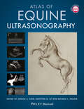
Atlas of Equine Ultrasonography
by Michele L. Frazer Kristina G. Lu Jessica A. KiddThe only visual guide to equine ultrasonography based on digital ultrasound technology. Atlas of Equine Ultrasonography provides comprehensive coverage of both musculoskeletal and non-musculoskeletal areas of the horse. Ideal for practitioners in first opinion or referral practices, each chapter features normal images for anatomical reference followed by abnormal images covering a broad range of recognised pathologies. The book is divided into musculoskeletal, reproductive and internal medicine sections and includes positioning diagrams demonstrating how to capture optimal images. With contributions from experts around the world, this book is the go-to reference for equine clinical ultrasonography.Key features include:Pictorially based with a wealth of digital ultrasound images covering both musculoskeletal and non-musculoskeletal areas and their associated pathologies.Each chapter begins with a discussion of normal anatomy and demonstrates how to obtain and interpret the images presented.A video library of over 50 ultrasound examinations is available at www.wiley.com/go/kidd/equine-ultrasonography for streaming or download and viewing on-the-go.
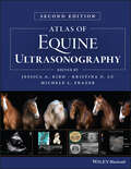
Atlas of Equine Ultrasonography
by Michele L. Frazer Kristina G. Lu Jessica A. KiddATLAS OF EQUINE ULTRASONOGRAPHY A THOROUGH EXPANSION TO THE FIRST ATLAS OF ULTRASONOGRAPHY IN THE HORSE, WITH NEW AND SIGNIFICANTLY IMPROVED IMAGES Ultrasonography is a vital diagnostic tool that can be applied in numerous functions in a veterinary practice. In conjunction with relevant clinical information—patient history and physical examination findings, for example—it can act as an important aid in the veterinarian’s decision-making process. Many vets in equine practice rely upon ultrasonography as a mainstay of equine diagnostic imaging on a wide range of structures and body systems. Ultrasonography is a useful procedure that is non-invasive and acts in complement to radiography to successfully diagnose the animal’s condition. This book’s aim is to encourage the clinician to rely further on the use of ultrasonography in their practice. The second edition of Atlas of Equine Ultrasonography provides an updated and expanded revision of the first atlas of ultrasonography in the horse. The first edition of this important resource was the first pictorially-based book to cover ultrasonography in the horse, and remains the only book currently available on the subject. The current version offers 450 additional images with greater clarity and precision in the images throughout and demonstrates how to obtain images in each body region while offering clinical ultrasonograms that show pathology. Atlas of Equine Ultrasonography readers will also find: High-quality clinical ultrasonograms for important musculoskeletal, reproductive, and medical conditions in the horse More than 1,500 images, with accompanying concise text describing the images A companion website that provides video clips showing dynamic ultrasound exams Atlas of Equine Ultrasonography is an invaluable reference to any veterinarian evaluating ultrasonograms in equine patients. As a result, this book will be of particular interest to equine specialists, veterinary radiologists, equine practitioners, and veterinary students.
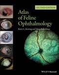
Atlas of Feline Ophthalmology
by Kerry L. Ketring Mary Belle GlazeSuccessful management of eye disease relies on the veterinarian’s ability to identify ocular features and distinguish pathologic changes. Atlas of Feline Ophthalmology, Second Edition is an invaluable diagnostic reference, providing high-quality color photographs for comparison with a presenting complaint. Presenting 394 photographs illustrating both normal and pathologic ocular conditions, this Second Edition offers a current, complete reference on ocular diseases, adding conditions recognized since publication of the first edition, a broader geographic scope, and many new images with improved quality. Carefully designed for easy reference, the contents are divided into sections corresponding to specific anatomical structures of the eye. A useful appendix new to this edition groups figures by etiology, making it easy to find every image associated with a specific agent or disease. Atlas of Feline Ophthalmology, Second Edition is a useful tool aiding general practitioners in diagnosing eye disease in cats.
Atlas of Invertebrate Viruses: Atlas Of Invertebrate Viruses (1991) (CRC Press Revivals)
by Jean R. Adams Jean R. BonamiThe Purpose of this book is to provide a helpful reference for invertebrate pathologist, virologists, and electron microscopists on invertebrate viruses. Investigators from around the world have shared their expertise in order introduce scientists to the exciting advances in invertebrate virology.
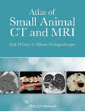
Atlas of Small Animal CT and MRI
by Allison Zwingenberger Erik WisnerAtlas of Small Animal CT & MRI is a highly illustrated diagnostic imaging guide to common clinical disorders of dogs and cats. Contains over 3,000 high quality CT, MRI and related diagnostic images Offers a unique approach emphasizing comparative imaging and pathologic correlation Focuses on important imaging features relevant to imaging diagnosis of disease in dogs and cats Written by internationally renowned experts in the field
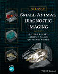
Atlas of Small Animal Diagnostic Imaging
by Clifford R. Berry Nathan C. Nelson Matthew D. WinterATLAS OF Small Animal Diagnostic Imaging Comprehensive and up-to-date resource on the interpretation of diagnostic images in small animals using survey radiographs and other modalities Atlas of Small Animal Diagnostic Imaging provides a comprehensive, multimodality atlas of small animal diagnostic imaging, with high-quality images depicting radiography, scintigraphy, ultrasonography, computed tomography, and magnetic resonance imaging. Taking a traditional body systems approach, the book offers an image-intensive resource to survey radiographs with some other imaging modalities being used to emphasize interpretation of survey radiographs. The Atlas offers clinically relevant information for small animal practitioners and students. Each body structure is thoroughly covered and well-illustrated, with discussion of the strengths and weaknesses of each modality in various scenarios. Edited by three experienced radiographers, The Atlas of Small Animal Diagnostic Imaging contains information on: Basics of diagnostic imaging, physics of diagnostic imaging, CT and MRI physics, US physics, and nuclear medicine physics Musculoskeletal normal anatomic variants, developmental orthopedic disease, joint disease, fracture and fracture healing, aggressive bone disease, and head and spine imaging Thorax anatomy, variants, and interpretation paradigm, extrathoracic structures, pleural space, pulmonary parenchyma, and mediastinum Abdomen anatomy, variants, and interpretation paradigm, extra-abdominal and body wall, peritoneal and retroperitoneal, liver and biliary, and spleen With its expansive coverage of the subject and hundreds of high-quality images to aid in efficient and seamless reader comprehension, Atlas of Small Animal Diagnostic Imaging is an invaluable and must-have resource for small animal practitioners, veterinary students, veterinary radiologists, and specialists in a number of areas.
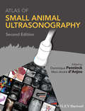
Atlas of Small Animal Ultrasonography
by Dominique Penninck Marc-André D'AnjouAtlas of Small Animal Ultrasonography provides a highly visual guide to the use of diagnostic ultrasound in small animal practice. This up-to-date atlas of the most commonly performed ultrasound examinations will be a valuable reference to well-established users of ultrasound as well as those just being introduced to the technology.Each chapter provides the reader with valuable information on the use of ultrasound diagnostics in small animal veterinary medicine. Taking a systems-based approach, Atlas of Small Animal Ultrasonography is divided into chapters on ultrasound procedures and techniques for examination of all major body parts and systems. Each chapter provides numerous illustrations and ultrasound images to aid the reader in diagnostic interpretation.Atlas of Small Animal Ultrasonography is an in-depth reference that provides thorough coverage of ultrasound techniques and interpretation. This volume will serve as an indispensable guide to the use of this important clinical modality.
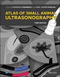
Atlas of Small Animal Ultrasonography
by Dominique Penninck Marc‐André D’AnjouComprehensive reference covering ultrasound techniques and findings in small animal practice with more than 2500 high-quality sonograms and illustrations Atlas of Small Animal Ultrasonography, Third Edition is a comprehensive reference for ultrasound techniques and findings in small animal practice. Offering more than 2500 high-quality sonograms and illustrations of normal structures and disorders, the book takes a systems-based approach to ultrasound examinations in small animals. With complete coverage of small animal ultrasonography, this reference guide is an essential resource for veterinary sonographers of all skill levels. In addition to updates reflecting current diagnostic imaging practice, the Third Edition adds two new chapters, on Point of Care Ultrasonography (POCUS) and on vascular diseases of the abdomen. Also, pertinent ultrasound-assisted interventional procedures were added in several chapters. The Third Edition of Atlas of Small Animal Ultrasonography features: More than 2500 figures of normal and abnormal ultrasound features of the thorax, abdomen, neck, eye/orbit and musculoskeletal systemComplementary imaging modalities when clinically pertinent to the clinical situation Additional surgical or histopathological specimens to best highlight the main features and complete case presentationsAccess to a companion website offering more than 150 annotated video loops of real-time ultrasound evaluations, illustrating the appearance of normal structures and common disorders Atlas of Small Animal Ultrasonography, Third Edition remains an essential teaching and reference tool for novice and advanced veterinary sonographers alike.
Atlas of Small Animal Wound Management and Reconstructive Surgery
by Michael M. PavleticA one-stop reference for the surgical treatment of wounds in small animal patients Wound management and reconstructive surgery are among the most challenging and innovative subspecialties of veterinary surgery for the management of traumatic injuries and neoplastic conditions commonly encountered in small animals. Atlas of Small Animal Wound Management and Reconstructive Surgery, Fourth Edition presents detailed procedures for surgical reconstruction and essential information on the principles of wound healing and wound management for dogs and cats. Coverage encompasses the pathophysiology and management of the wide variety of wounds encountered in small animal practice and the most current reconstructive techniques for closing the most challenging defects. This updated edition is presented with additional full color images and now includes color in each illustration, to enhance the reader’s understanding of each subject and successful execution of the surgical techniques covered. It imparts new and updated information on a wide variety of topics, including skin and muscle flap techniques, skin fold disorders, facial and nasal reconstructive surgery, foot pad surgery, urogenital reconstructive surgery, and includes a new chapter on reconstructive surgery of the pinna. Provides a key reference for general practitioners, surgeons, surgical residents, veterinary students, and small animal technicians in a single source Discusses current and new wound management and reconstructive surgical techniques for all body regions, including the face, ear, mouth, eye, nose, trunk, external genitalia, and foot Contains 35brand-new plate illustrations, updated and enhanced color plate illustrations and drawings, and additional clinical photographs throughout Features a new chapter dedicated to the surgical management of pinnal injuries and disorders, including new reconstructive surgical options developed by the author Includes information boxes to emphasize important points and the author’s personal observations, drawing on more than forty years of surgical experience Atlas of Small Animal Wound Management and Reconstructive Surgery, Fourth Edition is a valuable one-stop reference for veterinary surgeons, residents, and small animal practitioners.
Atlas of Small Animal Wound Management and Reconstructive Surgery
by Michael M. PavleticFully updated new edition of the state-of-the-art, image-driven reference on surgical reconstruction and wound management in dogs and cats Taking a visual approach to the topic with detailed line drawings and high-quality clinical photographs to demonstrate the techniques described, Atlas of Small Animal Wound Management and Reconstructive Surgery provides detailed step-by-step procedures for surgical reconstruction and key information on wound management in dogs and cats. The Atlas covers all body regions, including the face, ears, mouth, eyelids, nose, trunk, external genitalia, and feet. The Fifth Edition has been thoroughly revised to include new techniques, updated information, and references. Over 30 new technique plates have been added and the atlas now includes over 200 colored plates and illustrations. There is a dramatic increase in photographic case series supporting the illustrations. Written by an accomplished veterinary surgeon based on the author’s years of research and clinical experience, Atlas of Small Animal Wound Management and Reconstructive Surgery includes detailed information on: Anatomy of the skin, principles of wound healing and wound management, topical wound care products, management of common complications, and bandages, dressings, external splints, and protective devicesWound drainage techniques, negative pressure wound therapy, and management of specific wounds including bites, thermal, frostbite, gunshot/arrow wounds, pressure ulcers, hygromas, snake/spider bites, and porcupine quillsTension relieving techniques, skin stretching techniques, tissue expanders, local and distant skin flaps, axial pattern skin flaps, skin grafting techniques, and surgical dermatomeFacial, nasal, eyelid, and oral reconstructive surgery, including cleft lip and cleft palate repair, muscle flaps and myocutaneous flaps, pinnal reconstruction, and aural hematoma managementFoot pad reconstruction, reconstructive surgery of the prepuce and vulva, techniques for fecal incontinence and atresia ani, use of the omentum, and a supplemental reconstructive surgery case series The Fifth Edition of Atlas of Small Animal Wound Management and Reconstructive Surgery is a practical, complete, and state-of-the art reference for veterinary surgeons, surgical oncologists, residents, interns, and small animal practitioners to help them effectively manage wounds and reconstruct the variety of soft tissue defects encountered in dogs and cats.
Atlas of Surgical Approaches to Soft Tissue and Oncologic Diseases in the Dog and Cat
by Marije RisseladaThis book offers practical guidance to making approaches for surgery to treat soft tissue and oncologic conditions in canine and feline patients. Every approach is outlined with step-by-step descriptions on how to handle the incision and covers indications and patient positioning. Detailed, high-quality medical illustrations are also included for each, and topics are logically laid out with images on the left and text on the right. Atlas of Surgical Approaches to Soft Tissue and Oncologic Diseases in the Dog and Cat starts with a chapter on oromaxillofacial approaches, followed by chapters covering the cervical area and ear, forelimb, hindlimb, thorax, and abdomen. The book finishes with complete coverage of the approaches to the perineal area and pelvic canal and digits and tail, making it an excellent guide for surgeons to plan and execute their approach to soft tissue and oncologic diseases. Describes the complete approach to surgical incisions for soft tissue and oncologic disease, with alternative positions or approaches where appropriate Provides a high-quality medical line drawing depicting each approach Offers practical guidance for surgeons to direct their approach during surgery Covers indications, patient positioning, and step-by-step summaries of each approach Follows a logical two-page layout with text on one side and illustrations on the other Atlas of Surgical Approaches to Soft Tissue and Oncologic Diseases in the Dog and Cat is an essential reference for any veterinary surgeon or clinician treating soft tissue and oncologic diseases surgically.
Atlas of Terrestrial Mammal Limbs
by Anthony Herrel Christine Böhmer Jean-Christophe Theil Anne-Claire FabreAtlas of Terrestrial Mammal Limbs is the first comprehensive and detailed anatomy book on a broad phylogenetic and ecological range of mammals. This extraordinary new work features more than 400 photographs and illustrations visualizing the limb musculature of 28 different species. Standardized views of the dissected bodies and concise text descriptions make it easy to compare the anatomy across different taxa. It provides tables of nomenclature and comparative muscle maps (schematic drawings on the origins and insertions of the muscles onto bones) in a diversity of animals. Atlas of Terrestrial Mammal Limbs is a reliable reference and an indispensable volume for all students and professional researchers in biology, paleontology, and veterinary medicine. Key Features: Provides an overview of the anatomy of the mammalian limb Includes osteological correlates of the limb muscles Illustrates anatomy in 2D Guides dissection Documents anatomical diversity in mammalian limbs Related Titles: D. L. France. Human and Nonhuman Bone Identification: A Color Atlas. (ISBN 978-1-4200-6286-1) S. N. Byers. Forensic Anthropology Laboratory Manual, 4th Edition (ISBN 978-1-1386-9073-8) S. N. Byers. Introduction to Forensic Anthropology, 5th Edition (ISBN 978-1-1381-8884-6) R. Diogo, et al. Muscles of Chordates: Development, Homologies, and Evolution (ISBN 978-1-1385-7116-7)

Atlas of Trace Fossils in Well Core: Appearance, Taxonomy and Interpretation
by Dirk KnaustThis book provides the reader with a well-balanced blend of high-quality photographs, figures and accompanied text for the identification of trace fossils both in core and in outcrop. Ichnological data become more and more crucial in sedimentological and paleoenvironmental interpretations, not only in the exploration and exploitation of hydrocarbons but also in the characterization of aquifers and in scientific drilling. After an introduction to the study of trace fossils in core and an outlining of ichnological basics, principles and concepts, the book continues with a detailed description and interpretation of 39 trace fossils (ichnogenera) and associated features (such as bioturbate texture, plant roots and their traces, borings and pseudo-trace fossils) commonly encountered in well cores and in outcrop. The trace fossils are highlighted by their expression in well core, nicely illustrated with carefully prepared eye-catching core photographs. This unique information is complemented with examples of trace fossils in outcrop, as well as relevant key figures from the literature. Each description is presented in a consistent manner, stating the ichnogenus name and author in the title, followed by sections about morphology and size, ichnotaxonomy, substrate, appearance in core, similar trace fossils, producers, ethology, depositional environment, ichnofacies, age as well as reservoir quality. The book is rounded off with an extensive list of references for further reading. The material for the book originates from the author's continuous work with trace fossils in core over the last two decades.
Atlas of Veterinary Surgical Pathology
by Joseph S. HaynesATLAS OF VETERINARY SURGICAL PATHOLOGY An indispensable next-to-the-microscope diagnostic resource for veterinary pathologists Atlas of Veterinary Surgical Pathology delivers a comprehensive exploration of the lesions and diseases most commonly encountered by veterinary practitioners in small animals and horses. The book includes coverage of diseases of the skin, eye, and musculoskeletal systems, and male and female reproductive tracts. It also offers descriptions of relevant microscopic features and color microphotographs of the included lesions. More than 500 images depict lesions. With fully detailed discussions of degenerative, inflammatory, and neoplastic lesions, the book is an authoritative guide to quickly and accurately identifying common and uncommon lesions in small animals and horses. It also offers: A thorough introduction to the techniques relevant to surgical pathology, including specimen preparation and the interpretation of biopsy and the reporting of results Comprehensive explorations of skin and adjacent soft tissue, including developmental and degenerative diseases, inflammatory diseases, and neoplasms Practical discussions of reproductive tract lesions Complete treatments of lesions of the musculoskeletal system, and eye and periocular tissues Perfect for veterinary pathologists and residents, Veterinary Surgical Pathology is a practical handbook to the lesions and diseases encountered by veterinary professionals in small animal and equine surgical pathology.

Atmospheric Animals in Watercolour: Painting with Spirit & Vitality
by Jean HainesFrom domestic cats and dogs to wild lions and giraffes, best-selling artist and author Jean Haines shows the reader how to bring a vitality of life to their animal artwork.Known for the vibrant colours and exciting, innovative painting techniques that she brings to her work, Jean's latest book invites beginners and more experienced artists alike to share a journey through painting animals from around the world.Painting animals requires a delicacy of touch. Jean starts with simple monochrome artworks and progresses on to more vivid paintings that incorporate exciting textural effects. Using lessons from nature to help bring the reader closer to the animals, Jean shows how to portray the animal's spirit and bring vitality to the reader's artwork.Suitable for all abilities, from first-time painters to experienced artists, Jean shares all the materials and unique techniques she uses, and provides a wealth of expert tips and advice inspired by the animals themselves, helping the reader to progress and find their own path.Scattered throughout the book are dozens of Jean’s wonderful paintings, showcasing a veritable Noah's Ark of different animals to inspire the reader on their artistic journey.
Atomic Frenchie: Sit. Stay. Rule. (Atomic Frenchie Ser. #1)
by Tom McWeeney Tom E Sniegoski<p>Kirby is no ordinary French Bulldog—he is twenty-two pounds of fur-covered ruthless ambition. Freakishly intelligent and determined, he has one goal: Rule the World.When Kirby, a French Bulldog with a serious Napoleon Complex, moves to a new home in the quaint New England town of Strasburg, Massachusetts, and stumbles upon a forgotten secret laboratory, he realizes that his dreams of Planetary Conquest are finally within paw's reach. <p>But, suddenly, Kirby realizes he isn’t alone. Seemingly out of nowhere, a strange group of people appear, exhibiting what Kirby can only describe as superpowers! Kirby must rise up against all who stand in his way to emerge victorious in this ultimate quest for world domination. <p>The Atomic Frenchie is the first book in a series following the adventures of Kirby, professional supervillain and future ruler of Earth, as he schemes and battles his way past evil library robots, supernatural cat ladies, superpowered mail men, and more bizarre characters to achieve his sinister dreams of ruling the world. <p>This is a fixed-format ebook, which preserves the design and layout of the original print book.</p>
Atoms of Mind: The "Ghost in the Machine" Materializes
by W. R. KlemmThis book describes the author's view of how the mind "thinks" at various levels of operation. These levels include nonconscious mind (as in spinal/brainstem reflexes and neuroendocrine controls), subconscious mind, and conscious mind. In the attempt to explain conscious mind, there is considerable critique of arguments over whether or not free will is an illusion. Finally, the author summarizes current leading theories for consciousness (Bayesian probability, chaos, and quantum mechanics) and then presents his own theory based on patterns of nerve impulses in circuits that are interlaced coherently into larger networks.
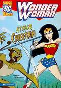
Attack of the Cheetah (Wonder Woman)
by Jane MasonWhen a cheetah exhibit opens at the National Zoo in Washington D.C., Princess Diana arrives to witness the event. At the grand opening, the rare cats suddenly escape, fleeing through a crowd of frightened zoogoers! Diana quickly transforms into her secret identity, Wonder Woman, captures the cats and saves the day. But when the cheetahs continue their odd behavior, only one thing can explain it -- the cat-like super-villain, Cheetah, is on the loose! Picture descriptions present.
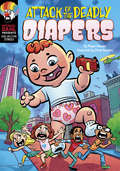
Attack of the Deadly Diapers (Michael Dahl Presents: Side-Splitting Stories)
by Megan AtwoodIn the midst of a boring summer, soon-to-be fifth grader Angie DelMar discovers something amazing about the eccentric woman who's just moved into town. She is actually a mad scientist who plans to take over the world by enlarging and controlling insects! With the help of her friends Cooper, Genevieve, and Harvey, Angie nabs the mixture and flees. But before they reach safety, the friends encounter a day care center where the mixture is accidentally released. Soon the town is overcome by gigantic toddlers! Can the friends save the town, defeat the scientist, and restore the bawling, blubbering behemoths to their cute and cuddly size?
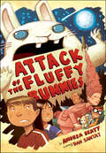
Attack of the Fluffy Bunnies (Fluffy Bunnies Ser.)
by Andrea Beaty“A lighthearted, clever send-up of zany horror conventions, this book is just the thing for kids about ready for M. T. Anderson’s Whales on Stilts.” —BooklistWhen Joules and Kevin Rockman’s parents drop them off at Camp Whatsitooya on their way to an International Spamathon, the twins expect a summer of marshmallows, campfires, and canoe trips. What they do not expect is to defend the earth from an invasion of sugar-addicted, murderous, seven-foot-tall rabbits from another galaxy. Happily, the Rockman twins, veteran watchers of the Late, Late, Late Creepy Show for Insomniacs, are unusually well-prepared for dealing with monstrous beings from outer space. If only their fellow campers were so lucky.Andrea Beaty, New York Times–bestselling author of several very funny picture books and a mystery novel, here reaches new heights of hilarity and verbal dexterity in a novel sure to become a camp—ba-dum-dum—classic.“Beaty’s tale of high silliness is sure to please, and it’s dotted with Santat’s mini-comics and spot illustrations, which move the story along.” —Kirkus Reviews“Beaty’s storytelling is lighthearted and fast-paced . . . her unconventional and entertaining narrative make it a wholly fun read.” —Publishers Weekly