- Table View
- List View
Human Exposure to Pollutants via Dermal Absorption and Inhalation
by Ian Colbeck Mihalis LazaridisThe human body is exposed to pollution on a daily basis via dermal exposure and inhalation. This book reviews the information necessary to address the steps in exposure assessment relevant to air pollution. The aim is to identify available information including data sources and models, and show that an integrated multi-route exposure model can be built, validated and used as part of an air quality management process. Many epidemiological studies have focused on inhalation exposure. Whilst this is appropriate for many substances, failure to consider the importance of exposure and uptake of material deposited on the skin may lead to an over/underestimation of the risk. Hence dermal exposure is also considered. Drinking water contamination by disinfection by-products is also discussed. Written by leading experts in the field, this book provides a comprehensive review of ambient particulate matter and will be of interest to students, researchers and policymakers involved in air quality management, environmental health and related disciplines, as well as environmental consultants and ventilation engineers.
Human Eye Imaging and Modeling
by Jasjit S. Suri U. Rajendra Acharya E.Y.K. Ng Jen Hong TanAdvanced image processing and mathematical modeling techniques are increasingly being used for the early diagnosis of eye diseases. A comprehensive review of the field, Human Eye Imaging and Modeling details the latest advances and analytical techniques in ocular imaging and modeling. The first part of the book looks at imaging of the fundus as wel
Human Factors and Ergonomics of Prehospital Emergency Care
by Joseph R. Keebler Elizabeth H. Lazzara Paul MisasiThis book provides an introduction to the field of human factors for individuals who are involved in the delivery and/or improvement of prehospital emergency care and describes opportunities to advance the practical application of human factors research in this critical domain. Relevant theories of human performance, including systems engineering principles, teamwork, training, and decision making are reviewed in light of the needs of current day prehospital emergency care. The primary focus is to expand awareness human factors and outlay the potential for novel and more effective solutions to the issues facing prehospital care and its practitioners.
Human Factors and Patient Safety: A primer for doctors and medical students
by Ibrahim ImamThis book fulfils the need of doctors, medical students, and all healthcare personnel for information that addresses fundamental patient safety concepts that are not usually covered in conventional medical curricula.There are three valuable features. Firstly, the content encompasses the main areas of human factors and patient safety in short and easily accessible language supplemented by anecdotes from safety-critical industries such as aviation and nuclear power. Secondly, each chapter highlights the problems of human error and provides solutions that help to reduce the risks to patients. Finally, the coverage highlights the important role the public should play in protecting their own safety when in contact with healthcare systems.
Human Factors in Air Transport: Understanding Behavior and Performance in Aviation
by Erik Seedhouse Anthony Brickhouse Kimberly Szathmary E. David WilliamsThis textbook provides students and the broader aviation community with a complete, accessible guide to the subject of human factors in aviation. It covers the history of the field before breaking down the physical and psychological factors, organizational levels, technology, training, and other pivotal components of a pilot and crew's routine work in the field.The information is organized into easy-to-digest chapters with summaries and exercises based on key concepts covered, and it is supported by more than 100 full-color illustrations and photographs. All knowledge of human factors required in aviation university studies is conveyed in a concise and casual manner, through the use of helpful margin notes and anecdotes that appear throughout the text.
Human Factors in Surgery: Enhancing Safety and Flow in Patient Care
by Bruce L. Gewertz Tara N. Cohen Eric J. LeyThis book delivers a comprehensive review of human factors principles as they relate to surgical care inside and outside of the operating theatre. It provides multi-dimensional human-centered insights from the viewpoint of academic surgeons and experts in human factors engineering to improve workflow, treatment time, and outcomes. To guide the reader, the book begins broadly with Human Factors Principles for Surgery then narrows to a discussion of surgical specialties and scenarios. Each chapter follows the following structure: (1) An overview of the topic at hand to provide a reference for readers; (2) a case study or story to illustrate the topic; (3) a discussion of the topic including human factors insights; (4) lessons learned, or personal “pearls” related to improving the specific system described. Written by experts in the field, Human Factors in Surgery: Enhancing Safety and Flow in Patient Care describes elements of the surgical system and highlights the lessons learned from systems engineering. It serves as a valuable resource for surgeons at any level in their training that wish to improve their practice.
Human Factors in the Health Care Setting
by Mike Davis Advanced Life Support Group Barabara Phillips Peter-Marc Fortune Jacky HansonHuman factors relates to the interaction of humans and technical systems. Human factors engineering analyzes tasks, considering the components in relation to a number of factors focusing particularly on human interactions and the interface between people working within systems. This book will help instructors teach the topic of human factors.
Human Factors und Patientensicherheit in der Akutmedizin
by Gesine Hofinger Michael St.Pierre„Human Factors“ spielen in der Akutmedizin eine große Rolle. Mindestens 30.000 Todesfälle pro Jahr sind auf Fehlhandlungen im Krankenhaus zurückzuführen. In etwa 80% der Fälle ist menschliches Versagen schuld. Das Werk beschreibt die wesentlichen Grundlagen zum Thema u.a. Fehler und Fehlerursachen, Psychologie menschlichen Handelns, Einfluss von Stress und Müdigkeit, Entscheidungsfindung und Handlungsstrategien, Kommunikation, Teamarbeit, Führung. Beleuchtet werden das Individuum, das Team, die Organisation, das Gesundheitssystem.Ursprünglich unter dem Namen "Notfallmanagement" publiziert, erscheint die 3. Auflage komplett aktualisiert und erweitert unter dem neuen Titel "Human Factors und Patientensicherheit in der Akutmedizin". Das Werk richtet sich an alle, die in der Akutmedizin tätig sind, insbesondere Ärzte, Pflegekräfte und Rettungsdienstpersonal.
Human Factors und Patientensicherheit in der Akutmedizin: Patientensicherheit Und Human Factors In Der Akutmedizin
by Gesine Hofinger Michael St.PierreEine sichere Versorgung in Notfallsituationen hängt entscheidend davon ab, dass die Behandelnden richtig entscheiden und adäquat handeln. Doch menschliches Entscheiden und Handeln wird von zahlreichen Faktoren beeinflusst, die eine sichere Versorgung gefährden. Welche „Human Factors“ einen maßgeblichen Einfluss haben, beschreibt das vorliegende Werk praxisnah und anhand zahlreicher Fallbeispiele. Die 4. Auflage erscheint komplett aktualisiert und erweitert. Das Werk richtet sich an alle, die in der Akutmedizin tätig sind, insbesondere Ärzte, Pflegekräfte und Rettungsdienstpersonal.
Human Fertility
by Zev Rosenwaks Paul M. WassarmanHuman Fertility: Methods and Protocols is intended for all practitioners of reproductive medicine and ART, as well as for embryologists and reproductive, developmental, cell and molecular biologists and others in the biomedical sciences. The volume presents straight-forward manner best practice approaches for overcoming a host of fertility challenges. Written in the highly successful Methods in Molecular Biology series format, chapters include introductions to their respective topics, lists of the necessary materials and reagents, step-by-step, readily reproducible laboratory protocols and tips on troubleshooting and avoiding known pitfalls. Authoritative and cutting-edge, Human Fertility: Methods and Protocols aids scientists in continuing to study assisted reproductive technologies.
Human Fetal Growth and Development
by Niranjan Bhattacharya Phillip G. StubblefieldThis unique book delves into the mysteries of human fetal growth and maturation. Growing knowledge in genetics indicates that factors that impact on/influence fetal growth and maturation may have a role in determining a person's health and disease in later years. Placental, maternal, environmental, nutrient as well as fetal genome factors each play a role in producing a healthy, unhealthy or abnormal baby. A study of fetal growth and maturation is therefore basic to the understanding of why fetal growth problems occur, what implications these can have for adult disease, and how clinical intervention can help to reverse growth problems. The present study will be comprehensive and will be a major contribution to the fields of gynecology, genetics, obstetrics, biochemistry, molecular biology and clinical medicine. It will include cutting edge research in the field as well as explorations on clinical interventions in fetal growth, which will not only add to existing knowledge but also prompt future research. The two Editors are distinguished in their fields and both have extensive clinical and research experience. They felt that they could use their expertise to create a book that will help students, practitioners, researchers and others to understand the subject of gestation, growth and maturation and its implications from a multi-dimensional point of view, which will help them develop their own expertise in a cutting-edge and developing field. They have brought toget her medical scientists, clinical practitioners, embryologists, endocrinologists, immunologists, gynecologists, obstetricians, reproductive and molecular biologists, geneticists and many others to create a state-of-the-art book on a subject with increasing demand for further knowledge. It aims to integrates different disciplines to give a holistic view of human fetal growth maturation.
Human Fetal Tissue Transplantation
by Phillip Stubblefield Niranjan BhattacharyaMany diseases earlier considered to be incurable are now being treated with modern innovations involving fetal tissue transplants and stem cells derived from fetal tissues. Fetal tissues are the richest source of fetal stem cells as well as other varying states of differentiated cells and support or stromal cells. The activity of such stem cells is at their peak provided they are given the correct niche. Stem cells, as we know, are immortal cells with the capacity to regenerate into any kind of differentiated cell as per niche-guidance. As such, fetal tissues have the potential capacity to mend, regenerate and repair damaged cells or tissues in adults, when directly transplanted to the site of injury, or even when transplanted in some other site, because it may have a homing capacity to migrate to the site of the specific injured organ. This is a new area of translational research and needs to be highlighted because of its immense potential. This book will bring together the new work of prominent medical scientists and clinicians who are conducting pioneering research in human fetal tissue transplantation. This will include direct transplant of healthy fetal tissue into mature patients as well as in hosts with genetic diseases. Transplant techniques, donor-host interaction, cell and tissue storage, ethical and legal issues, are some of the many matters which the book will deal with.
Human Flourishing: A Multidisciplinary Perspective on Neuroscience, Health, Organizations and Arts
by Marc Grau Grau Yasin Rofcanin Mireia Las HerasThis open access book presents a novel multidisciplinary perspective on the importance of human flourishing. The study of the good life or Eudaimonia has been a central concern at least since Aristotelian times. This responds to the common experience that we all seek happiness. Today, we are immersed in a new paradoxical boom, where the pursuit of happiness seems to permeate everything (books, media, organizations, talks), but at the same time, it is nowhere, or at least very difficult to achieve. In fact, it is not easy to even find a consensus regarding the meaning of the word happiness. Seligman (2011), one of the fathers of the positive psychology, confirmed that his original view the meaning he referred to was close to that of Aristotle. But, he recently confessed that he now detests the word happiness, since it is overused and has become almost meaningless. The aim of this open access book is to shed new light on human flourishing through the lenses of neurosciences and health, organizations, and arts. The novelty of this book is to offer a multi-disciplinary perspective on the importance of human flourishing in our lives. The book will examine further how different initiatives, policies and practices create opportunities for generating human flourishing.
Human Frontiers, Environments and Disease
by Tony McmichaelCharting the relentless trajectory of humankind across time and geography, Tony McMichael highlights the changing survival patterns of our ancient ancestors, who roamed the African savannahs several million years ago, to today's populous, industrialized, and globalized world. McMichael explores the changes in human biology, culture, and surrounding environments that have influenced patterns of health and disease over the course of humankind's history, arguing that the health of populations is primarily a product of the interaction of human societies with the wider environment, its various ecosystems, and other life-support processes. Tony McMichael is professor of epidemiology, London School of Hygiene and Tropical Medicine. He has held positions in Australia, USA, and UK, and has taught widely in Asia, Africa, and Europe. He has advised WHO, UNEP, the World Bank and Intergovernmental Panel on Climate Change on public health issues. His previous book, Planetary Overload (Cambridge University Press, 1993) was a widely acclaimed and influential account of global environmental change and the health of the human species.
Human Frontiers: The Future of Big Ideas in an Age of Small Thinking
by Michael Bhaskar'A fascinating book . . . Bhaskar is a reassuringly positive and often witty guide'Observer'A fascinating, must-read book covering a vast array of topics from the arts to the sciences, technology to policy. This is a brilliant and thought-provoking response to one of the most critical questions of our age: how we will come up with the next generation of innovation and truly fresh ideas?'Mustafa Suleyman, cofounder of DeepMind and Google VP'Have "big ideas" and big social and economic changes disappeared from the scene? Michael Bhaskar's Human Frontiers is the best look at these all-important questions.'Tyler Cowen, author of The Great Stagnation and The Complacent Class'Michael Bhaskar explores the disturbing possibility that a complacent, cautious civilization has lost ambition and is slowly sinking into technological stagnation rather than accelerating into a magical future. He is calling for bold, adventurous innovators to go big again. A fascinating book'Matt Ridley, author of How Innovation WorksWhere next for humanity? Is our future one of endless improvement in all areas of life, from technology and travel to medicine, movies and music? Or are our best years behind us? It's easy to assume that the story of modern society is one of consistent, radical progress, but this is no longer true: more academics are researching than ever before but their work leads to fewer breakthroughs; innovation is incremental, limited to the digital sphere; the much-vaunted cure for cancer remains elusive; space travel has stalled since the heady era of the moonshot; politics is stuck in a rut, and the creative industries seem trapped in an ongoing cycle of rehashing genres and classics. The most ambitious ideas now struggle. Our great-great-great grandparents saw a series of transformative ideas revolutionise almost everything in just a few decades. Today, in contrast, short termism, risk aversion, and fractious decision making leaves the landscape timid and unimaginative.In Human Frontiers, Michael Bhaskar draws a vividly entertaining and expansive portrait of humanity's relationship with big ideas. He argues that stasis at the frontier is the result of having already pushed so far, taken easy wins and started to hit limits. But new thinking is still possible. By adopting bold global approaches, deploying cutting edge technology like AI and embracing a culture of change, we can push through and expand afresh.Perfect for anyone who has wondered why we haven't gone further, this book shows in fascinating detail how the 21st century could stall - or be the most revolutionary time in human history.
Human Fungal Diseases: Diagnostics, Pathogenesis, Drug Resistance and Therapeutics
by Saif HameedThis reference book reviews the latest advancements in the fungal infections and their impact on human health. It presents epidemiology, diagnosis, pathogenesis, risk factors, virulence mechanisms, treatment, and strategies for the disease management and prevention of fungal infections. The book further reviews host-pathogen interactions, biofilm formation, and quorum sensing. It also covers the clinical manifestations, diagnostic approaches, and management strategies of opportunistic fungal infections, emerging fungal infections, and allergic fungal infections. It presents the latest advancements in diagnostic methods and therapeutic strategies, covering both conventional techniques and state-of-the-art approaches.Further, the book elucidates antifungal stewardship, nanotechnology, and omics technologies, providing insights into cutting-edge strategies for prevention, control, and management of multidrug-resistant fungi. This book is useful for researchers, students, and health professionals working in the fields of mycology, infectious diseases, immunology, dermatology, and pulmonology.
Human Fungal Pathogen Identification
by Thomas LionThis detailed volume presents timely and authoritative content offering a comprehensive overview of the current state of the art in fungal diagnostics. Moreover, it addresses on-going developments expected to provide a basis for targeted treatment strategies resulting in improved outcome of invasive mycoses. The knowledge of host-related predisposing factors and stratified treatment options facilitating timely onset of adequate antifungal therapy are critical for successful clinical management and outcome of invasive fungal disease (IFD), requiring not only rapid diagnosis of a fungal infection and identification of the causative species, but also assessment of pathogen/host factors related to pathogenicity, susceptibility, and response to treatment. Written for the highly successful Methods in Molecular Biology series, chapters include introductions to their respective topics, lists of the necessary materials and reagents, step-by-step, readily reproducible protocols, and tips on troubleshooting and avoiding known pitfalls. Authoritative and practical, Human Fungal Pathogen Identification: Methods and Protocols serves as an ideal reference for researchers investigating the ever-growing worldwide healthcare problems involving fungal infections.
Human Fungal Pathogens
by Oliver KurzaiWhereas plant and insect infections are commonly caused by fungi, only a small minority of the vast diversity of fungal species is pathogenic to humans. Despite this, fungal infections cause considerable morbidity and mortality worldwide. This volume is dedicated to the biology, clinical presentation and management of invasive fungal infections. Major pathogenic fungi are introduced by world-leading experts and the basic principles of fungal virulence are reviewed in the light of new results and experimental technologies that offer unprecedented insights into invasive infections caused by Aspergillus, Candida, Cryptococcus, Pneumocystis and Mucorales. In parallel, the clinical presentation of invasive fungal infections and current approaches to their diagnosis and treatment are summarized to provide an overview of human pathogenic fungi, linking pathogen biology to the clinical presentation of disease.
Human Gametes and Preimplantation Embryos
by David K. Gardner Emre Seli Denny Sakkas Dagan WellsIn recent years, the advancing science and increasing availability of assisted reproduction have given new hope to infertile couples. However, the use of IVF and ART has also led to marked increases in the number of multiple-infant live births. This poses a public health concern, as these neonates have a higher rate of pre-term delivery, compromising their survival chances and increasing their risk of lifelong disability. By optimizing the selection of gametes and embryos with high probabilities of implantation, it is possible to reduce the number of embryos transferred and, by extension, the number of high-risk multiple gestations, while maintaining or increasing pregnancy rates. Human Gametes and Preimplantation Embryos: Assessment and Diagnosis provides a broad yet concise overview of established and developing methodologies for assessment of gamete and embryo viability in assisted reproduction. This book elucidates the best practices for precisely selecting viable specimens based on morphology and cleavage rate and covers the spectrum of emerging adjunctive technologies for predicting reproductive potential. The authors present their extensive knowledge of "omics" approaches (genomics, transcriptomics, proteomics, and metabolomics), with unbiased delineation of the associated advantages and potential pitfalls. This valuable clinical resource is well suited to infertility specialists, Ob/Gyn physicians, IVF laboratory technicians, and researchers in the fields of embryology and reproductive medicine.
Human Genes and Neoliberal Governance: A Foucauldian Critique
by Antoinette RouvroyOriginal and interdisciplinary, this is the first book to explore the relationship between a neoliberal mode of governance and the so-called genetic revolution. Looking at the knowledge-power relations in the post-genomic era and addressing the pressing issues of genetic privacy and discrimination in the context of neoliberal governance, this book demonstrates and explains the mechanisms of mutual production between biotechnology and cultural, political, economic and legal frameworks. In the first part Antoinette Rouvroy explores the social, political and economic conditions and consequences of this new ‘perceptual regime’. In the second she pursues her analysis through a consideration of the impact of ‘geneticization’ on political support of the welfare state and on the operation of private health and life insurances. Genetics and neoliberalism, she argues, are complicit in fostering the belief that social and economic patterns have a fixed nature beyond the reach of democratic deliberation, whilst the characteristics of individuals are unusually plastic, and within the scope of individual choice and responsibility. This book will be of interest to all students of law, sociology and politics.
Human Genetics and Genomics
by Bruce R. Korf Mira B. IronsThis fourth edition of the best-selling textbook, Human Genetics and Genomics, clearly explains the key principles needed by medical and health sciences students, from the basis of molecular genetics, to clinical applications used in the treatment of both rare and common conditions.A newly expanded Part 1, Basic Principles of Human Genetics, focuses on introducing the reader to key concepts such as Mendelian principles, DNA replication and gene expression. Part 2, Genetics and Genomics in Medical Practice, uses case scenarios to help you engage with current genetic practice.Now featuring full-color diagrams, Human Genetics and Genomics has been rigorously updated to reflect today's genetics teaching, and includes updated discussion of genetic risk assessment, "single gene" disorders and therapeutics. Key learning features include:Clinical snapshots to help relate science to practice'Hot topics' boxes that focus on the latest developments in testing, assessment and treatment'Ethical issues' boxes to prompt further thought and discussion on the implications of genetic developments'Sources of information' boxes to assist with the practicalities of clinical research and information provisionSelf-assessment review questions in each chapterAccompanied by the Wiley E-Text digital edition (included in the price of the book), Human Genetics and Genomics is also fully supported by a suite of online resources at www.korfgenetics.com, including:Factsheets on 100 genetic disorders, ideal for study and exam preparationInteractive Multiple Choice Questions (MCQs) with feedback on all answersLinks to online resources for further studyFigures from the book available as PowerPoint slides, ideal for teaching purposesThe perfect companion to the genetics component of both problem-based learning and integrated medical courses, Human Genetics and Genomics presents the ideal balance between the bio-molecular basis of genetics and clinical cases, and provides an invaluable overview for anyone wishing to engage with this fast-moving discipline.
Human Genetics: Concepts and Applications
by Ricki LewisHuman Genetics: Concepts and Applications embraces the broadening of human genetics from an academic and medical discipline to an informational science that can be highly personal, yet have a societal impact. By coming to know genetic backgrounds, people can control their environments in healthier ways. Genetic knowledge is, therefore, both informative and empowering.
Human Genome Structure, Function and Clinical Considerations
by Luciana Amaral HaddadThis book provides a detailed evidence-based overview of the latest developments in how the structure of the human genome is relevant to the health professional. It features comprehensive reviews of genome science including human chromosomal and mitochondrial DNA structure, protein-coding and noncoding genes, and the diverse classes of repeat elements of the human genome. These concepts are then built upon to provide context as to how they functionally relate to differences in phenotypic traits that can be observed in human populations. Guidance is also provided on how this information can be applied by the medical practitioner in day-to-day clinical practice. Human Genome Structure, Function and Clinical Considerations collates the latest developments in genome science and current methods for genome analysis that are relevant for the clinician, researcher and scientist who utilises precision medicine techniques and is an essential resource for any such practitioner.
Human Growth and Nutrition in Latin American and Caribbean Countries
by Sudip Datta BanikThis book analyzes biological and sociocultural factors that influence nutritional status, physical growth, development and maturation of children and adolescents in Latin American and Caribbean (LAC) countries in the perspective of human ecology. Chapters in this book bring together both theoretical and empirical studies that take into account human biological and environmental conditions to understand how ethnic diversity, culturally determined lifestyle and dietary habits influence biological variation of human growth and nutrition in nine LAC countries: Argentina, Brazil, Chile, Cuba, Dominican Republic, El Salvador, Guatemala, Mexico, and Peru. The book is divided into three sections. Chapters in the first section analyze nutritional and epidemiological aspects of child growth in the region. Articles in the second section focus on methods to evaluate human growth, development, and maturation. Finally, the third section brings together a series of studies representing different LAC countries, analyzing biocultural impacts on child growth and nutrition. By bringing together studies about the relationship between human biology, cultural diversity, nutrition and health in a region with huge environmental challenges, this volume addresses many of the challenges to achieve the United Nation’s Sustainable Development Goals 2 (Zero Hunger) and 3 (Good Health and Well-Being). Chapters in this volume present and discuss data on the effects of malnutrition on children's and adolescent's health and development, such as chronic undernutrition or stunting (growth deficit) and excess weight (overweight and obesity) as the risk factors for child morbidity and mortality m due to non-communicable diseases. Human Growth and Nutrition in Latin American and Caribbean Countries will be a valuable resource for both students and researchers in different disciplines dedicated to the interdisciplinary research on the intersection between human biology, cultural diversity, nutrition and health. It will also be a useful source of information for both health professionals and policy makers developing and implementing interventions and public policies to achieve UN’s SDGs 2 and 3, particularly in the LAC regions.
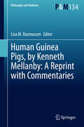
Human Guinea Pigs, by Kenneth Mellanby: A Reprint with Commentaries (Philosophy and Medicine #134)
by Lisa M. RasmussenThis book reprints Human Guinea Pigs, by Kenneth Mellanby, a seminal work in the history of medical ethics and human subject research that has been nearly unavailable for over 40 years. Detailing the use of World War II conscientious objectors who volunteered for experimentation on scabies transmission, Mellanby’s book offers insight into one approach to human subject experimentation before the development of ethical oversight regulations. His work was initially published prior to the articulation of the Nuremberg Code, which makes his subsequent position as a reporter for the British Medical Journal at the Nuremberg Trials very interesting, particularly given his sometimes controversial opinions on Nazi medical experimentation. This book reprints the second edition together with commentary essays that situate Mellanby’s ethical approach in historical context and relative to contemporary approaches. This volume is of particular interest to scholars of the history of human subject research.

