- Table View
- List View
Human Parasites: Diagnosis, Treatment, Prevention
by Heinz MehlhornThe new edition of this textbook provides an up-to-date overview of the most important parasites in humans and their potential vectors. Climate change and globalization steadily favor the opportunities for parasites to thrive. These challenges call for the latest information on pathogen transmission routes and timely preventive measures.For each parasite, this book offers a concise summary in eleven sections:1. Naming2. Geographic distribution and epidemiology3. Morphology, biology and life cycle4. Disease symptoms5. Diagnosis6. Infection pathways7. Prophylaxis8. Incubation period9. Prepatency10. Patency11. Therapeutic optionsNumerous tables, diagrams and over 200 colorful illustrations highlight the main aspects of parasitic infestations and present suitable control measures. Moreover, 60 questions help to test readers’ theoretical knowledge of the field. Readers can additionally download the free Springer Nature Flashcards App and benefit from the digital study questions.In short, this work is highly recommended for anyone looking to delve into the field of human parasitology. It is intended for students of biology and human medicine, medical doctors, pharmacists and laboratory staff alike. Furthermore, persons who plan to visit or live longer in endemic regions will find essential information on necessary preventive and control measurements.
Human Parasites: From Organisms To Molecular Biology (G - Reference,information And Interdisciplinary Subjects Ser.)
by Dunne Fong Marion M. ChanWhy does the World Health Organization (WHO) put emphasis on neglected tropical diseases (NTDs)? What are the NTDs? Are NTDs found in the United States? Is there any relationship between coronavirus disease 2019 (COVID-19) and NTDs? These are some of the questions being addressed in the book.The aim of this textbook is to introduce a modern synthesis on human parasites of medical importance. Species of parasitic protozoa and helminths are presented in detail, from history and discovery to aspects of genomes and molecular biology, together with life cycle, therapy, drug resistance, and case studies of parasitic diseases useful to the clinicians.
Human Pathobiochemistry: From Clinical Studies to Molecular Mechanisms
by Hirokazu Tsukahara Toshitaka Oohashi Francesco Ramirez Chad L. Barber Fumio OtsukaThis textbook uses a case-study approach to present the core principles of biochemistry and molecular biology in the context of human disease to students who will be involved in patient care. The 29 clinical cases have been carefully selected to cover key scientific concepts and some common, and other not so common, diseases. While the principal focus is on topics relating to metabolic disease, further subjects such as connective tissue disorders, neurological disorders, auto-inflammatory disorders, infective diseases, and cancer are also addressed. Each chapter provides a specific patient report that includes the natural history, pertinent clinical laboratory data, physical findings, subsequent diagnosis, and therapy. This is followed by a comprehensive discussion of the normal biochemical processes and reactions pertaining to the case, along with the pathophysiological mechanisms of the disease. Graphical diagrams are provided in each chapter for ease of comprehension.
Human Pathophysiology: Introductory Concepts And Clinical Perspectives
by Theresa Capriotti Joan Parker FrizzellBridge the gap between the science of disease and clinical patient care! Interpret clinical manifestations. Determine the correct inventions. Understand treatment options. . Connect drug actions and therapeutic goals. This easy-to-understand text introduces the pathophysiologic processes of the common conditions that you'll encounter in clinical settings. Chapters organized by organ systems help you understand the relationships between the pathologic event and the patient assessment data, laboratory findings, and diagnostic testing results. You'll develop the foundational knowledge you need to effectively select the appropriate nursing interventions to deliver the best patient care.
Human Pharmaceuticals in the Environment
by Bryan W. Brooks Duane B. HuggettHuman interaction with the environment remains one of the most pervasive facets of modern society. In a world characterized by rapid population growth, unprecedented global trade and digital communications, energy security, natural resource scarcities, climatic changes and environmental quality, emerging diseases and public health, biodiversity and habitat modifications are routinely touted by the popular press as they canvas global political agendas and scholarly endeavors.
Human Pharmacology
by Paul R. GardPharmacology, in its own right, is a massive subject area and has been the focus of several major textbooks. Human Pharmacology is a readable, introductory text covering all of the main aspects of pharmacology in a way that enthuses the reader to study the subject further. Each chapter includes line drawings and figures to illustrate concepts and m
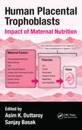
Human Placental Trophoblasts: Impact of Maternal Nutrition
by Asim K. Duttaroy Sanjay BasakHuman Placental Trophoblasts: Impact of Maternal Nutrition explores the vital roles of trophoblasts play in fetal growth and pregnancy, giving you new insight into the modulation of placental trophoblast functions by nutrients. It also reviews the role of fatty acids, folic acids, and specific vitamins in this aspect. The book highlights the critic
Human Population Genetic Research in Developing Countries: The Issue of Group Protection (Biomedical Law and Ethics Library)
by Yue WangHuman population genetic research (HPGR) seeks to identify the diversity and variation of the human genome and how human group and individual genetic diversity has developed. This book asks whether developing countries are well prepared for the ethical and legal conduct of human population genetic research, with specific regard to vulnerable target group protection. The book highlights particular issues raised by genetic research on populations as a whole, such as the potential harm specific groups may suffer in genetic research, and the capacity for current frameworks of Western developed countries to provide adequate protections for these target populations. Using The People’s Republic of China as a key example, Yue Wang argues that since the target groups of HPGR are almost always from isolated and rural areas of developing countries, the ethical and legal frameworks for human subject protection need to be reconsidered in order to eliminate, or at least reduce, the vulnerability of those groups. While most discussion in this field focuses on the impact of genetic research on individuals, this book breaks new ground in exploring how the interests of target groups are also seriously implicated in genetic work. In evaluating current regulations concerning prevention of harm to vulnerable groups, the book also puts forward an alternative model for group protection in the context of human population genetic research in developing countries. The book will be of great interest to students and academics of medical law, ethics, and the implications of genetic research.
Human Preimplantation Embryo Selection (Reproductive Medicine and Assisted Reproductive Techniques Series)
by Kay Elder Jacques CohenThe most profound dilemma in assisted reproduction to date is the inability to recognize potentially viable embryos before their replacement into the reproductive tract. Application of increasingly advanced new technology has allowed the field of embryo evaluation to evolve rapidly and dramatically over the past five years.Human Preimplantation Emb
Human Psychology As Seen Through The Dream (International Library Of Psychology Ser.)
by Turner, JuliaFirst published in 1999. Routledge is an imprint of Taylor & Francis, an informa company.
Human Resource Development in the Public Sector: The Case of Health and Social Care (Routledge Studies in Human Resource Development)
by Jim Stewart Sally SambrookAcross Europe and the world, countries are attempting to develop their health and social policies and practices to address the global challenge of increasing demand and pressurized supply, created by ageing populations, emerging technologies and finite resources (financial and human). This text provides examples of attempts to develop HRD practices in health and social care contexts within France, Ireland, The Netherlands, Romania, Russia, the UK and the USA. Thus, the book is European and international in both scope and appeal.
Human Resource Management
by Robert L. Mathis John H. Jackson Sean R. Valentine Patricia A. MeglichPrepare for HR and career success with the book that has set the standard for excellence in human resource management. Valentine/Meglich/Mathis/Jackson's HUMAN RESOURCE MANAGEMENT, 16E offers today's most current look at HRM and its impact on the success of organizations today. A leading resource in preparing for professional HR certification, this edition ensures you are familiar with all major topics for professional examinations from the Society for Human Resource Management and Human Resource Certification Institute. You examine the latest HR research as well as HR theory in contemporary practice. This edition highlights emerging trends driving change in HRM today, including technology, globalization, competencies and HR metrics. Accompanying MindTap digital resources offer a personalized, online learning platform with a tailored presentation created by your instructor. MindTap's Learning Path Navigator guides you in completing reading assignments, annotating readings, finishing homework and checking your understanding with quizzes and assessments.
Human Resource Management in Health Care
by L. Fleming Fallon Charles R. McConnellThis practical, hands-on book introduces human resources to those who are preparing to work in any area of health care or health service. Written for practitioners and students in all disciplines related to health, it covers important topics such as recruitment, training, termination, legal issues, labor unions, and more. Each chapter is introduced by a case study related to the material that follows. The case study is resolved at the conclusion of each chapter along with expert commentary and practical suggestions that can be used in the real world. Chapters also feature learning objectives, discussion points, and questions to ponder. Many examples and a number of sample forms and documents are included. <P><P> The Second Edition has been completely re-organized to reflect a better chapter flow and organization. It also offers: • All data updated throughout • New section on health care legislation • New section in each chapter, “Customer Service Box”, that emphasizes the importance of customer service in the context of the material presented in the chapter. • Completely revised instructor ancillary material.
Human Resources In Healthcare: Managing for Success
by Myron D. Fottler Association of University Programs in Health Administration Staff Bruce FriedHuman Resources in Healthcare: Managing for Success, Fourth Edition, presents the techniques and practices behind effective management of people—the healthcare profession’s most important asset. It provides the concepts and practical tools necessary for meeting the unique challenges in today’s healthcare environment. <p><p> This edition has been thoroughly revised and includes the following new content: <p><p> An expanded chapter on employment law and employee relations A new chapter on credentialing of healthcare providers A thorough update on staff recruitment, selection, and retention practices An expanded section on performance management, including workplace bullying A new chapter on workforce planning in a rapidly changing healthcare system A new chapter on nurse staffing in healthcare organizations New problem-based learning cases to engage students and expand learning comprehension Updated short cases, discussion questions, and exercises throughout
Human Resources Management for Health Care Organizations
by Donald N. Lombardi Joan E. PynesThis book is a comprehensive guide to the essential areas of health care human resources management, and is an immediately useful practical handbook for practitioners as well as a textbook for use health care management programs. Written by the authors of Handbook for the New Health Care Manager and Human Resources Management for Public and Nonprofit Organizations, the book covers the context of human resources management in the unique health care business arena from a strategic perspective includes SHRM and human resources planning, organizational culture and assessment, and the legal environment of human resources management. Managing volunteers and job analysis performance appraisal instruments, training and development programs, and recruitment, targeted selection and hiring techniques are covered. Compensation policies and practices, employer-provided benefits management, implementation of training and organizational development programs, as well as labor-management relations for health care organizations and healthcare human resource information technology are covered, with practical examples and proven strategies amply provided in each chapter.
Human Respiratory Syncytial Virus: Methods and Protocols (Methods in Molecular Biology #2948)
by Bo Liang Rachel FearnsThis volume presents a comprehensive collection of state-of-the-art techniques used to investigate respiratory syncytial virus (RSV) at multiple levels, from atomic-resolution structural studies to in vivo infection models. It serves as an essential resource for virologists studying viral genome replication, transcription, and protein interactions, as well as immunologists exploring immune responses, pathogenesis, and vaccine development. Structural biologists will find valuable insights into the use of cryo-electron microscopy, X-ray crystallography, and other biophysical approaches to characterize viral components, while translational researchers and pharmaceutical scientists will benefit from protocols designed to facilitate the development of novel antivirals and vaccine candidates. Clinicians and epidemiologists studying RSV transmission, disease outcomes, and therapeutic interventions will also find this volume highly relevant to their work. As part of the renowned Methods in Molecular Biology series, this book follows a structured format, ensuring accessibility and reproducibility. Each chapter provides a concise introduction to the method&’s background, relevance, and applications, followed by a detailed, step-by-step protocol accompanied by a comprehensive list of reagents, materials, and specialized equipment. Expert troubleshooting tips and insights are included to help optimize experimental success and avoid common pitfalls.Bringing together contributions from leading researchers in the field, this volume offers both practical guidance and inspiration for future discoveries. Whether you are an experienced scientist or a newcomer to RSV research, Human Respiratory Syncytial Virus: Methods and Protocols provides the essential tools and knowledge to advance our understanding of RSV biology and accelerate the development of novel therapeutic and preventive strategies.
Human Respiratory Viral Infections
by Sunit SingUsing a multidisciplinary approach, Human Respiratory Viral Infections is set at the level between the definitive reference work and an essential clinical manual. Exploring recent advances in human respiratory viral research, the text builds on the basic sciences of epidemiology, virology, molecular biology, and immunology to cover clinical diagnosis, mechanism of pathogenesis, manifestations of disease, impact, treatment, and management strategies. <P><P> <li>Provides a comprehensive review of all the major human respiratory viral infections <li>Supplies a detailed description of the anatomy of the respiratory system <li>Discusses the salient features of specific viruses <li>Examines the latest diagnostic tools <li>Covers risk factors associated with incidence of respiratory viral infections in children and adults in different occupational and environmental settings <P><P>Presenting the latest knowledge in human respiratory viral infections, this text will be immensely valuable for pulmonologists, biomedical scientists, research scholars, virologists, vaccinologists, immunologists, clinicians, pharmacologists, and public health specialists.
Human Retrotransposons in Health and Disease
by Gael CristofariThis unique book explores the role of retrotransposons in human health and disease. The ability of retrotransposons to affect the structure of human genes is recognized since the late 80's. However, the advances of deep-sequencing technologies have shed new light on the extent of retrotransposon-mediated genome variations. These progresses have also led to the discovery that retrotransposon activity is not restricted to the germline - resulting in inheritable genetic variations - but can also mobilize in somatic tissues, such as embryonic stem cells, neuronal progenitor cells, or in many cancers. This book covers topics related to the effects of retrotransposon insertions, and their consequences on germline and somatic genome dynamics, but also discuss the role and impact of retrotransposons sequences in a broader context, including a number of novel topics that emerged recently (long non-coding RNA, neuronal disorders, exaptation) with unexpected connections between retrotransposons, stem cell maintenance, placentation, circadian cycles or aging.
Human Retroviruses
by Elisa Vicenzi Guido Poli"Human Retroviruses: Methods and Protocols" collects key experimental protocols that have provided the basis of the major discoveries of the field. Split into five sections, this detailed volume covers mapping of the HIV life cycle, isolation, co-receptor use, and cell tropism of HIV-1, in vivo quantification of HIV-1, biological aspects of HIV-1, as well as HTLVs. Some articles explore assay and function of accessory genes, largely involving the interface between retroviral and host factors, the extracellular role of Tat and Tax, resembling the function of cytokines, and the biotechnological exploitation of HIV as lentiviral vector to carry foreign genes with therapeutic value. Written in the highly successful "Methods in Molecular Biology" series format, chapters include introductions to their respective topics, lists of the necessary materials and reagents, step-by-step, readily reproducible laboratory protocols, and tips on troubleshooting and avoiding known pitfalls. Comprehensive and authoritative, "Human Retroviruses: Methods and Protocols" provides state-of-art methodological protocols from world leaders in human retrovirology, essential for any lab working this vital field. "
Human Rights Behind Bars: Tracing Vulnerability in Prison Populations Across Continents from a Multidisciplinary Perspective (Ius Gentium: Comparative Perspectives on Law and Justice #103)
by Yves Haeck Clara Burbano HerreraThis book brings together leading authorities from the fields of international human rights law, criminology, legal medicine, and political science with international human rights judges and UN experts to analyze the current situation of detainees in Europe, the Americas and Africa.This comprehensive volume offers a platform for reflecting on the complexity of the prison problem from a multidisciplinary perspective. The authors address detention-related issues with the aim of generating new ideas that contribute to both academic discussion and critical analysis. Academic dialogue across the globe provides insights into various national and international carceral systems and how they deal with human rights behind bars. At the same time, the critical comparison helps to identify basic needs and practices that can work in multiple settings. The contributors are respected experts and leading scholars in their fields, and each has pursued prison and human rights research over the last decades. However, this is the first time that they have come together in a multidisciplinary academic project. This book aims to stimulate diverse actors to imagine alternative ways of engaging with persons deprived of their liberty, in academia and in practice.
Human Rights and Drug Control: A New Perspective
by Melissa L. BoneThis book uses a human rights perspective – developed philosophically, politically and legally – to change the way in which we think about drug control issues. The prohibitionist approach towards tackling the ‘drugs problem’ is not working. The laws and mentality that see drugs as the problem and tries to fight them, makes the ‘drugs problem’ worse. While the law is the best-placed mechanism to regulate our actions in relation to particular drugs, this book argues against the stranglehold of the criminal law, and instead presents a human rights perspective to change the way we think about drug control issues. Part I develops a conceptual framework for human rights in the context of drug control – philosophically, politically and legally – and applies this to the domestic (UK) and international drug control system. Part II focuses on case law to illustrate both the potential and the limitations of successfully applying this unique perspective in practice. The conclusion points towards a bottom-up process for drug policy which is capable of reconfiguring the mentality of prohibition. This book will be of interest to students and scholars of human rights, criminal law, criminology, politics and socio-legal studies.
Human Rights in Psychiatry: Prospects and Dilemmas of Abolishing Coercion in Mental Health Care
by Dirk RichterThe book describes the ethical lines of conflict, shows why coercion can no longer be justified and analyzes the consequences and dilemmas of a possible abolition of coercive measures in psychiatric care. The use of coercion in mental health care is one of the most controversial topics in psychiatric nursing and psychiatry. The conflict line centers around the UN-Convention in the Rights of People with Disabilities (CRPD). Advocates of the CRPD are pushing for the complete abolition of coercion while opponents see central medical and legal aspects of care for people with mental health problems at risk. Clinicians in conventional psychiatry, including many mental health nurses, primarily justify these measures because of the assumed benefits of coercion-associated care and with the argument that many people affected are unable to make appropriate decisions for their own health in a crisis situation. This argument also applies to human rights, for example by basing coercive measures in the event of suicidality on the right to life. Three central topics are developed in the book. First, it is shown that psychiatric coercion can no longer be justified because the current practice of psychiatric care does not meet the ethico-legal requirements for the use of coercion. Second, a human rights-based approach of psychiatric care is outlined, which is fundamentally based on the will and preferences of people with mental health problems. Third, the consequences and dilemmas are indicated, e.g., the issue of how to deal with suicidality or dementia without the use of coercion. This book is aimed to receive a specific attention from the psychiatric nursing community.
Human Ring Chromosomes: A Practical Guide for Clinicians and Families
by Thomas Liehr Peining LiThis book presents chromosome-wise clinical cases following an evidence-based protocol, in addition to providing the scientific background on the mechanisms of human ring chromosome (RC) formation. Presence of RCs in a genome can lead to several rare genetic diseases. This book, edited by the leading experts Prof. Peining Li and Prof. Thomas Liehr, is the first comprehensive book on this topic.Over the past 60 years, banding cytogenetics, fluorescence in situ hybridization, chromosome microarray analysis, and whole genome sequencing have been used to diagnose cases with a RC. Ring syndrome of sever growth retardation and variable intellectual disability has been considered a common clinical feature for all RCs. Clinical heterogeneity of chromosome-specific deletion and duplication syndromes, gene-related organ and tissue defects, cancer predisposition to different types of tumors, and reproduction failure has been reported in the literature. However, the cases of RCs reportedin the literature account for less than 1% of its real occurrence. Current diagnostic practice lacks laboratory standards for analyzing cellular behavior and genomic imbalances of RCs to evaluate its compound effects on patients. The under-representation of clinical cases and the lack of comprehensive diagnostic analysis make challenging to establish accurate clinico-cytogenomic correlations. Given recent advances in genomic technology and organized efforts from peer experts, standardized cytogenomic diagnosis and evidence-based clinical management could be envisioned for all patients with RCs.Furthermore, supernumerary small ring chromosomes and the patient’s perspective are addressed—the latter by including family stories of RC-carrier relatives. Acquired RCs in various cancers are also discussed, as well as the potential role of RCs in research applications like iPSC cellular modeling and genomic editing.This book is a valuable reference for clinical geneticists, personnel in cytogenetics and molecular genetics laboratories, genetic counselors, and researchers in related fields.
Human Risk Assessment: The Role Of Animal Selection And Extrapolation
by M. V. Roloff; A. G. E. Wilson; W. E. Ribelin; W. P. Ridley; F. A. RueckerThis book explores the factors which are critical in the selection of an appropriate animal species for toxicology studies and the subsequent extrapolation of the data to humans. It provides some future directions for risk assessment activities at the Environmental Protection Agency.
Human Saliva, Volume I
by Jorma O. TenovuoThis book covers current knowledge of the origins of various salivary compounds, their normal values, age dependency, relation to flow rate, diseases or hormonal status, and assay problems regarding human saliva. It discusses practical aspects of salivary analyses.