- Table View
- List View
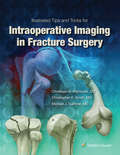
Illustrated Tips and Tricks for Intraoperative Imaging in Fracture Surgery
by Michael J. GardnerAn optimal view on the fluoroscope screen simplifies the procedure for the surgeon and improves the outcome for the patient. Illustrated Tips and Tricks for Intraoperative Imaging in Fracture Surgery is a unique resource that expertly covers the use of intra-operative fluoroscopy in fracture surgery, bridging the gap between strategic patient positioning and maximizing the efficiency of fluoroscopy. Using an easy-to-follow, reader-friendly format, it provides a real-world understanding of the intra-operative C-arm’s capabilities and limitations, considering factors such as the fracture type, the operating table, positioning adjuncts, patient body habitus, and the functional range of the fluoroscope.
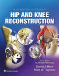
Illustrated Tips and Tricks in Hip and Knee Reconstructive and Replacement Surgery
by Mark W. Pagnano Daniel J. BerryPart of the popular Tips and Tricks series, Illustrated Tips and Tricks in Hip and Knee Reconstruction provides succinct and practical advice acquired from years of professional practice in hip and knee surgery. Led by Drs. Daniel Berry and Marc Pagnano of the Mayo Clinic, this visually stunning reference focuses exclusively on detailed descriptions of technical tips and tricks for all aspects of hip and knee reconstruction. This unique approach is highly useful to orthopaedic surgery fellows and residents – anyone who would benefit from exposure to the wisdom that experienced attending surgeons pass on to those who are training in this complex field.

Illustrated Tips and Tricks in Sports Medicine Surgery
by Frederick AzarPublisher's Note: Products purchased from 3rd Party sellers are not guaranteed by the Publisher for quality, authenticity, or access to any online entitlements included with the product. This new quick-reference is the latest volume in the Illustrated Tips and Tricks series. You’ll find succinct, precise information from a wide range of experts and prestigious institutions on tackling technical problems in sports medicine surgery. Drawings, operative photos, and videos are used liberally throughout the book to illustrate surgical techniques and provide a handy visual complement to the text.
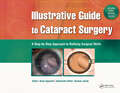
Illustrative Guide to Cataract Surgery: A Step-by-Step Approach to Refining Surgical Skills
by Amar AgarwalIllustrative Guide to Cataract Surgery by Editor, Dr. Amar Agarwal and Associate Editor, Soosan Jacob is a unique book that includes matching step-by-step clinical photographs, medical illustrations, and videos to explain the most common techniques and steps in cataract surgery. With more than 650 illustrations, ophthalmologists and residents will visually learn the most essential procedures in cataract surgery, step-by-step.The content, comprised from leading ophthalmic surgeons, is structured into systematically divided sections such as phaco surgery, microincision cataract surgery, challenging cases, and complications. It allows for quick reference, without having to search through voluminous books. Each image is supplemented with concise, informative text that helps further explain the techniques.In addition, video instruction is offered through a companion website, with each book purchase. Using the same approach as the book, the website presents videos that match each technique or step in cataract surgery.Just a few of the procedures explained:• Vertical chopping• Biaxial microincisional cataract surgery• Posterior polar cataract• Iris hooks in small pupil phaco• Subluxated cataracts• Torn rhexis• Glued IOL implantation• Intraocular lens implantationWith a combination of matching photos and illustrations alongside brief text and companion website, Illustrative Guide to Cataract Surgery stands apart from traditional books.
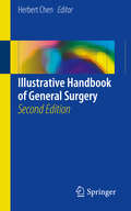
Illustrative Handbook of General Surgery
by Herbert ChenWith the large number of surgical atlases in the market focusing on a variety of surgical procedures, it is almost impossible to find an area that is not comprehensively illustrated. However, almost all of these atlases are very large and serve to be 'library reference' books, which are commonly displayed in the office and utilized in that manner. There is no atlas which is easily transportable, yet one would imagine that every medical student and resident would potentially want a copy of such a title to carry on them at all times. The book aims to cover all facets of general surgery and each chapter has five to ten photographs illustrating the basic anatomical context, as well as the various techniques specific to the operation. Each chapter author provides a brief narrative describing what is going on during the operation, and also outlines common pitfalls and personal suggestions.
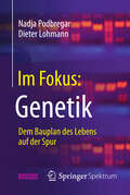
Im Fokus: Dem Bauplan des Lebens auf der Spur
by Nadja Podbregar Dieter LohmannWelche Macht haben unsere Gene? Sind sie die schicksalhafte Blaupause, die bestimmt, wie intelligent, wie alt oder wie schön wir sind? Lange Zeit war dies die gängige Lehrmeinung. Doch das Dogma ihrer monolithischen Allmacht ist heute längst gefallen. Immer häufiger stoßen Forscher auf Hinweise, wie unser Leben den Genen "ins Handwerk" pfuscht und wie eng die Wechselwirkungen zwischen Erbgut, Stoffwechsel und Umwelt manchmal sind. Die moderne Biotechnologie eröffnet neue Wege der Forschung, wirft aber auch ethische und gesellschaftliche Fragen auf. Dieses Buch erklärt unter anderem, warum bei den Genen die Verpackung manchmal wichtiger ist als der Inhalt, was Viren in unserem Erbgut verloren haben und weshalb es "das Methusalem-Gen" nicht gibt.
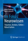
Im Fokus: Neurowissen
by Nadja Podbregar Dieter LohmannKönnen wir im Schlaf lernen? Wie weit reicht der Einfluss der Hormone? Und wodurch wird bestimmt, ob wir hochbegabt sind? Im Zentrum dieser Fragen steht unser Gehirn. In den letzten Jahren sind Neurowissenschaftler immer tiefer in die Struktur und Physiologie des Gehirns vorgedrungen. Sie entdeckten neue, überraschende Zusammenhänge und Wechselwirkungen, die auch unser Bild von uns selbst ständig verändern. Dieses Buch nimmt Leser mit auf eine Erkundungsreise zu faszinierenden und rätselhaften Phänomenen unseres Denkens, Fühlens und Bewusstseins.
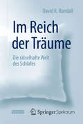
Im Reich der Träume: Die rätselhafte Welt des Schlafes
by David K. RandallDer New York Times-Bestseller über die erstaunlichen Erkenntnisse der SchlafforschungJede Nacht widmen wir ihm etliche Stunden: Dennoch gibt der Schlaf uns allen – Schläfern wie Wissenschaftlern gleichermaßen – verblüffend viele Rätsel auf. Obwohl wir nahezu ein Drittel unseres Lebens schlafend verbringen, wissen wir nicht wirklich, inwiefern dies bedeutsam für uns ist. Warum müssen wir überhaupt schlafen? Warum bedroht Schlafmangel unsere Gesundheit, warum ist Schlafentzug Folter? Was geschieht mit uns, wenn wir träumen? Lernen wir im Schlaf? Und was hat es mit dem Schlafwandeln auf sich?Kaum etwas beeinflusst unser Leben so sehr wie die Qualität unseres Schlafs. Schlafstörungen scheinen in Zusammenhang mit Diabetes, Bluthochdruck, Schlaganfällen und Demenz zu stehen. Und in unseren sozialen Beziehungen birgt der Schlafplatz eine Menge Zündstoff: Sollten Eltern ihr Bett mit ihrem Säugling teilen? Was tun, wenn der andere schnarcht? Ist eine Ehe zum Scheitern verurteilt, wenn die Partner in getrennten Betten schlafen?Der Wissenschaftsjournalist David Randall gewährt uns vielfältige Einblicke in die Forschungsarbeiten, die jene nächtlichen Stunden zu erhellen versuchen. Auf einer Entdeckungsreise, die von Kriegsschauplätzen bis ins Kinderzimmer führt, offenbart Im Reich der Träume, dass Schlafen nicht im Entferntesten so banal ist, wie es uns erscheinen mag, wenn wir abends das Licht ausmachen.
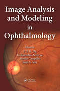
Image Analysis and Modeling in Ophthalmology
by Jasjit S. Suri U. Rajendra Acharya Aurélio Campilho E. Y. K. NgDigital fundus images can effectively diagnose glaucoma and diabetes retinopathy, while infrared imaging can show changes in the vascular tissues. Likening the eye to the conventional camera, Image Analysis and Modeling in Ophthalmology explores the application of advanced image processing in ocular imaging. This book considers how images can be us
Image Analysis and Recognition: 16th International Conference, ICIAR 2019, Waterloo, ON, Canada, August 27–29, 2019, Proceedings, Part II (Lecture Notes in Computer Science #11663)
by Aurélio Campilho Fakhri Karray Alfred YuThis two-volume set LNCS 11662 and 11663 constitutes the refereed proceedings of the 16th International Conference on Image Analysis and Recognition, ICIAR 2019, held in Waterloo, ON, Canada, in August 2019. The 58 full papers presented together with 24 short and 2 poster papers were carefully reviewed and selected from 142 submissions. The papers are organized in the following topical sections: Image Processing; Image Analysis; Signal Processing Techniques for Ultrasound Tissue Characterization and Imaging in Complex Biological Media; Advances in Deep Learning; Deep Learning on the Edge; Recognition; Applications; Medical Imaging and Analysis Using Deep Learning and Machine Intelligence; Image Analysis and Recognition for Automotive Industry; Adaptive Methods for Ultrasound Beamforming and Motion Estimation.
Image Analysis for Moving Organ, Breast, and Thoracic Images: Third International Workshop, RAMBO 2018, Fourth International Workshop, BIA 2018, and First International Workshop, TIA 2018, Held in Conjunction with MICCAI 2018, Granada, Spain, September 16 and 20, 2018, Proceedings (Lecture Notes in Computer Science #11040)
by Andrew P. Bradley Jens Petersen Kensaku Mori Gustavo Carneiro Kanwal Bhatia Bernhard Kainz Lena Maier-Hein Tom Vercauteren Danail Stoyanov Zeike Taylor Anne Martel Gabriel Maicas Reinhard R. Beichel Ozan Oktay Jacinto Nascimento Hang Min Matthew S. Brown Colin Jacobs Bianca Lassen-Schmidt Raúl San José Estépar Alexander Schmidt-Richberg Catarina VeigaThis book constitutes the refereed joint proceedings of the Third International Workshop on Reconstruction and Analysis of Moving Body Organs, RAMBO 2018, the Fourth International Workshop on Breast Image Analysis, BIA 2018, and the First International Workshop on Thoracic Image Analysis, TIA 2018, held in conjunction with the 21st International Conference on Medical Imaging and Computer-Assisted Intervention, MICCAI 2018, in Granada, Spain, in September 2018. The 5 full papers (out of 10 submissions) presented at RAMBO, the 9 full papers (out of 18 submissions) presented at BIA, and the 20 full papers (out of 21 submissions) presented at TIA were carefully reviewed and selected. The RAMBO papers cover aspects of medical imaging where motion plays a role in the image formation or analysis. The BIA papers deal with topics such as computer-aided detection and diagnosis of breast cancer, quantitative analysis of breast imaging modalities, and large scale breast image screening and analysis. The TIA papers cover aspects of image analysis research for lung and cardiac diseases including segmentation, registration, quantification, modeling of the image acquisition process, visualization, validation, statistical modeling, biophysical lung modeling (computational anatomy), deep learning and novel applications.
Image Analysis for Ophthalmological Diagnosis
by Robert KoprowskiThis monograph focuses on the use of analysis and processing methods for images from the Corvis® ST tonometer. The presented analysis is associated with the quantitative, repeatable and fully automatic evaluation of the response of the eye, eyeball and cornea to an air-puff. All the described algorithms were practically implemented in MATLAB®. The monograph also describes and provides the full source code designed to perform the discussed calculations. As a result, this monograph is intended for scientists, graduate students and students of computer science and bioengineering as well as doctors wishing to expand their knowledge of modern diagnostic methods assisted by various image analysis and processing methods.
Image Analysis in Stroke Diagnosis and Interventions: 4th International Workshop, SWITCH 2024, and 6th International Challenge, ISLES 2024, Held in Conjunction with MICCAI 2024, Marrakesh, Morocco, October 10, 2024, Proceedings (Lecture Notes in Computer Science #15408)
by Theo van Walsum Roland Wiest Markus D. Schirmer Ruisheng Su Ezequiel de la Rosa Leonhard Rist Ewout Heylen Frank Te Nijenhuis Danny Ruijters Richard McKinley Susanne WegenerThis book constitutes the refereed proceedings of the 4th International MICCAI Stroke Workshop on Imaging and Treatment Challenges, SWITCH 2024, as well as the Ischemic Stroke Lesion Segmentation Challenge, ISLES 2024, held in conjunction with MICCAI 2024, in Marrakesh, Morocco, on October 10, 2024. The 12 revised full papers presented in this volume were selected form 16 submissions. The papers describe research advancements in image analysis for the diagnosis and intervention of ischemic and haemorrhagic stroke and present the latest developments in segmentation, disease prognosis, stroke diagnosis and treatment, and other clinically relevant applications.
Image Analysis: Methods and Applications, Second Edition
by Donat-P. HaderAutomatic image analysis has become an important tool in many fields of biology, medicine, and other sciences. Since the first edition of Image Analysis: Methods and Applications, the development of both software and hardware technology has undergone quantum leaps. For example, specific mathematical filters have been developed for quality enhanceme
Image Based Computing for Food and Health Analytics: IBCFHA
by Rajeev Tiwari Deepika Koundal Shuchi UpadhyayIncrease in consumer awareness of nutritional habits has placed automatic food analysis in the spotlight in recent years. However, food-logging is cumbersome and requires sufficient knowledge of the food item consumed. Additionally, keeping track of every meal can become a tedious task. Accurately documenting dietary caloric intake is crucial to manage weight loss, but also presents challenges because most of the current methods for dietary assessment must rely on memory to recall foods eaten. Food understanding from digital media has become a challenge with important applications in many different domains. Substantial research has demonstrated that digital imaging accurately estimates dietary intake in many environments and it has many advantages over other methods. However, how to derive the food information effectively and efficiently remains a challenging and open research problem. The provided recommendations could be based on calorie counting, healthy food and specific nutritional composition. In addition, if we also consider a system able to log the food consumed by every individual along time, it could provide health-related recommendations in the long-term. Computer Vision specialists have developed new methods for automatic food intake monitoring and food logging. Fourth Industrial Revolution [4.0 IR] technologies such as deep learning and computer vision robotics are key for sustainable food understanding. The need for AI based technologies that allow tracking of physical activities and nutrition habits are rapidly increasing and automatic analysis of food images plays an important role. Computer vision and image processing offers truly impressive advances to various applications like food analytics and healthcare analytics and can aid patients in keeping track of their calorie count easily by automating the calorie counting process. It can inform the user about the number of calories, proteins, carbohydrates, and other nutrients provided by each meal. The information is provided in real-time and thus proves to be an efficient method of nutrition tracking and can be shared with the dietician over the internet, reducing healthcare costs. This is possible by a system made up of, IoT sensors, Cloud-Fog based servers and mobile applications. These systems can generate data or images which can be analyzed using machine learning algorithms. Image Based Computing for Food and Health Analytics covers the current status of food image analysis and presents computer vision and image processing based solutions to enhance and improve the accuracy of current measurements of dietary intake. Many solutions are presented to improve the accuracy of assessment by analyzing health images, data and food industry based images captured by mobile devices. Key technique innovations based on Artificial Intelligence and deep learning-based food image recognition algorithms are also discussed. This book examines the usage of 4.0 industrial revolution technologies such as computer vision and artificial intelligence in the field of healthcare and food industry, providing a comprehensive understanding of computer vision and intelligence methodologies which tackles the main challenges of food and health processing. Additionally, the text focuses on the employing sustainable 4 IR technologies through which consumers can attain the necessary diet and nutrients and can actively monitor their health. In focusing specifically on the food industry and healthcare analytics, it serves as a single source for multidisciplinary information involving AI and vision techniques in the food and health sector. Current advances such as Industry 4.0 and Fog-Cloud based solutions are covered in full, offering readers a fully rounded view of these rapidly advancing health and food analysis systems.
Image Fusion in Preclinical Applications
by Claudia Kuntner-Hannes York HaemischThis book provides an accessible and comprehensive overview of the state of the art in multimodal, multiparametric preclinical imaging, covering all the modalities used in preclinical research. The role of different combinations of PET, CT, MR, optical, and optoacoustic imaging methods is examined and explained for a range of applications, from research in oncology, neurology, and cardiology to drug development. Examples of animal studies are highlighted in which multimodal imaging has been pivotal in delivering otherwise unobtainable information. Hardware and software image registration methods and animal-specific factors are also discussed. The readily understandable text is enhanced by numerous informative illustrations that help the reader to appreciate the similarities to, but also the differences from, clinical applications. Image Fusion in Preclinical Applications will be of interest to all who wish to learn more about the use of multimodal/multiparametric imaging as a tool for in vivo investigations in preclinical medical and pharmaceutical research.
Image Guided Dermatologic Treatments
by Robert L. BardThis book showcases the latest digital skin imaging, optical/laser systems and advanced immunologic therapies including topics ranging from the basic dermatologic sciences to advanced microscopic and laser optics. The addition of radiologic breakthroughs serves as comprehensive source for the dermatologic community, helping them access sonographic, CT, MRI and nuclear medicine procedures refined for dermatologic and subcutaneous pathologies. In addition, it assists radiologists determine the appropriate imaging technologies for specific clinical dermal disorders. A detailed and up-to-date overview of image-guided treatments is provided. The initial chapters on benign and inflammatory diseases are precursors to advanced chapters on hidradenitis suppurativa and pigmented lesion analysis. A dedicated chapter on melanoma skin cancer and malignant melanoma is followed by updated concepts of melanoma treatment, including genetic markers and PET/CT to monitor therapeutic success. Further chapters address such topics as dermal trauma from foreign bodies and burns, scar imaging, fillers complications and podiatric imaging. Chapters on optical coherence tomography and reflectance confocal microscopy complete the coverage. All chapters were written by dermatologists trained in ultrasound diagnosis, interventional radiologists, dermatopathologists and specialists in advanced optical and microscopic dermatologic analysis, providing a reference guide to noninvasive diagnosis techniques and image guided minimally invasive treatment options. As such, Image Guided Dermatologic Treatments will be an invaluable asset for clinicians in medical and allied fields where dermatologic diagnosis using the least invasive option is required.
Image Guided Interventions of the Spine: Principles and Clinical Applications
by Scott H. Faro Majid Khan Sergiy V. KushchayevThis book is a comprehensive review of image guided interventions of the spine. Beginning with a chapter dedicated to the history of image guided spinal interventions, authors set the stage for the role these procedures have and will play in the field. Chapters cover the key procedures, techniques, and considerations to maximize effectiveness and patient care. Some major topics covered include: imaging osseo-ligamentous spine anatomy, percutaneous vertebroplasty, image guided tumor ablation, and vascular spine intervention. Additional features include high-quality illustrations with concise descriptions and clinical cases discussions. This is an ideal guide for interventional neuroradiologists, radiologists, pain management physicians, neurosurgeons, orthopedic spine surgeons, and related residents, fellows, and students wanting in depth information on image guided interventions of the spine.
Image Guided Prostate Cancer Treatments
by Dan Sperling Jurgen J. Fütterer Robert L. BardImage-Guided Prostate Cancer Treatments is a comprehensive reference and practical guide on the technology and application of ultrasound and MRI in the male pelvis, with special attention to the prostate. The book is organized into three main sections, the first of which is devoted to general aspects of imaging and image-guided treatments. The second section provides a systematic overview of the application of ultrasound and MRI to the diagnosis and treatment of diseases of the lower urinary tract. Performance of the ultrasound and MRI studies is explained, and the normal and abnormal pathological anatomy is reviewed. Correlation with the ultrasound in the same plane is provided to assist in understanding the MRI sequences. Biopsy and interventional procedures, ultrasound-MRI fusion techniques, and image-guided therapies, including focused ultrasound, photodynamic therapy, microwave and laser ablation, are all fully covered. The third section focuses on securing treatment effectiveness and the use of follow-up imaging to ensure therapeutic success and detect tumor recurrence at an early stage, which is vital given that prompt focal treatment of recurrence is very successful. Here, particular attention is paid to the role of Doppler ultrasound and DCE-MRI technologies. This book, containing a wealth of high-quality illustrations based on high-end equipment, will acquaint beginners with the basics of prostate ultrasound and MRI, while more advanced practitioners will learn new skills, means of avoiding pitfalls, and ways of effectively relating the imaging and image-guided treatments to the clinical situation. The information provided will permit a tailored approach in dealing with specific pathologic issues.
Image Principles, Neck, and the Brain
by Luca SabaMagnetic resonance imaging (MRI) is a technique used in biomedical imaging and radiology to visualize internal structures of the body. Because MRI provides excellent contrast between different soft tissues, the technique is especially useful for diagnostic imaging of the brain, muscles, and heart.In the past 20 years, MRI technology has improved si
Image Processing and Acquisition using Python (Chapman & Hall/CRC The Python Series)
by Ravishankar Chityala Sridevi PudipeddiImage Processing and Acquisition using Python provides readers with a sound foundation in both image acquisition and image processing—one of the first books to integrate these topics together. By improving readers’ knowledge of image acquisition techniques and corresponding image processing, the book will help them perform experiments more effectively and cost efficiently as well as analyze and measure more accurately. Long recognized as one of the easiest languages for non-programmers to learn, Python is used in a variety of practical examples. A refresher for more experienced readers, the first part of the book presents an introduction to Python, Python modules, reading and writing images using Python, and an introduction to images. The second part discusses the basics of image processing, including pre/post processing using filters, segmentation, morphological operations, and measurements. The second part describes image acquisition using various modalities, such as x-ray, CT, MRI, light microscopy, and electron microscopy. These modalities encompass most of the common image acquisition methods currently used by researchers in academia and industry. Features Covers both the physical methods of obtaining images and the analytical processing methods required to understand the science behind the images. Contains many examples, detailed derivations, and working Python examples of the techniques. Offers practical tips on image acquisition and processing. Includes numerous exercises to test the reader’s skills in Python programming and image processing, with solutions to selected problems, example programs, and images available on the book’s web page. New to this edition Machine learning has become an indispensable part of image processing and computer vision, so in this new edition two new chapters are included: one on neural networks and the other on convolutional neural networks. A new chapter on affine transform and many new algorithms. Updated Python code aligned to the latest version of modules.
Image Processing and Acquisition using Python (Chapman And Hall/crc Mathematical And Computational Imaging Sciences Ser.)
by Ravishankar Chityala Sridevi PudipeddiImage Processing and Acquisition using Python provides readers with a sound foundation in both image acquisition and image processing-one of the first books to integrate these topics together. By improving readers' knowledge of image acquisition techniques and corresponding image processing, the book will help them perform experiments more effectiv
Image Processing in Diabetic Related Causes
by Amit Kumar Fahimuddin ShaikThis book is a collection of all the experimental results and analysis carried out on medical images of diabetic related causes. The experimental investigations have been carried out on images starting from very basic image processing techniques such as image enhancement to sophisticated image segmentation methods. This book is intended to create an awareness on diabetes and its related causes and image processing methods used to detect and forecast in a very simple way. This book is useful to researchers, Engineers, Medical Doctors and Bioinformatics researchers.
Image Processing in Radiation Therapy (Imaging in Medical Diagnosis and Therapy)
by Kristy K. BrockImages from CT, MRI, PET, and other medical instrumentation have become central to the radiotherapy process in the past two decades, thus requiring medical physicists, clinicians, dosimetrists, radiation therapists, and trainees to integrate and segment these images efficiently and accurately in a clinical environment. Image Processing in Radiation
Image Processing with MATLAB: Applications in Medicine and Biology
by Omer Demirkaya Musa H. Asyali Prasanna K. SahooImage Processing with MATLAB: Applications in Medicine and Biology explains complex, theory-laden topics in image processing through examples and MATLAB algorithms. It describes classical as well emerging areas in image processing and analysis. Providing many unique MATLAB codes and functions throughout, the book covers the theory of probability an