- Table View
- List View
Hypoxia and Cancer
by Giovanni MelilloThe imbalance between rapidly proliferating tumor cells and inadequate and inefficient tumor vasculature leads to a decrease in oxygen levels (hypoxia and/or anoxia) in tumor tissues. Intra-tumor hypoxia profoundly affects the biological behavior of cancer cells, which become resistant to conventional therapies and acquire a more invasive and metastatic phenotype. Hypoxia is a hallmark of the malignant phenotype and a key feature of the tumor microenvironment. Hypoxia Inducible Factor 1 (HIF-1) is a master regulator of the transcriptional response to oxygen deprivation. HIF triggers the expression of genes whose products induce angiogenesis, decrease oxygen consumption, switch metabolism to glycolysis, maintain a stem cell phenotype and select for more invasive and metastatic cells. Therapeutic approaches targeting HIF, directly or downstream mediators of its transcriptional activity, are being developed. Intra-tumor hypoxia is a topic has been gaining scientific interest over the last few years for its wide involvement in many physiological and pathological processes. This volume will cover the latest research and translational aspects associated with intra-tumor hypoxia, along with opportunities for drug development offered by this unique feature of the tumor microenvironment. The ongoing efforts to translate our understanding of the biology underlying intra-tumor hypoxia in viable therapeutic options face many challenges, but this book will provide an opportunity for an in-depth analysis of the fundamental mechanisms implicated in the adaption to low oxygen levels and will scrutinize the potential for opportunities that are being pursued in both research and the drug development industry.
Hypoxia and Cancer Metastasis (Advances in Experimental Medicine and Biology #1136)
by Daniele M. GilkesThe present book is an attempt to provide a detailed review of studies that clarify our current understanding of the role of hypoxia in the progression of primary cancer to metastatic disease. It will enable researchers to discover the critical cellular changes that occur under hypoxic conditions and play a role in metastatic dissemination, from the activation of hypoxia-inducible factors, HIF-1 and HIF-2, to the transcriptional profile changes that occur in cancer cells and promote cancer cell survival under detrimental conditions. Readers will discover the methods and challenges involved in imaging and quantifying the degree of hypoxia in a primary tumor. We will provide an understanding of the hypoxia-induced phenotypes that influence heterogeneity, alter the secretome and tumor microenvironment, modify cellular metabolism, and promote immune suppression and resistance to chemotherapy. Finally, we will uncover the therapeutic strategies that are being devised to target the hypoxic microenvironment in the hopes of preventing metastasis and improving the efficacy of standard-of-care cancer treatments. This work is an up to date source of information on the challenges and complexity of the hypoxic tumor microenvironment. Basic and translational scientists, post-doctoral fellows, graduate students, and those interested in how tumors metastasize will find this book a reference that details how hypoxia influences metastatic disease.
Hypoxia and Tumor Microenvironment: Implications for Cancer Therapy
by Jagat Rakesh Kanwar Sukhes MukherjeeThis book provides a comprehensive overview of the hypoxic tumor microenvironment and its impact on various aspects of cancer biology, including DNA damage response, genome instability, tumor immunity, non-coding RNA regulation, metabolic regulation of CAR-T cell function, and angiogenesis. It explores the regulation and therapeutic potential of hypoxia-inducible factors, as well as strategies to improve cancer immunotherapy by targeting the hypoxic tumor microenvironment. This book is a valuable resource for students, researchers, and Medical science professionals interested in understanding the complex interplay between hypoxia and cancer biology.
Hypoxia in Cancer: Significance and Impact on Cancer Therapy
by Jagat Rakesh Kanwar Sukhes MukherjeeThis book reviews the central role of hypoxia in cancer initiation and progression. It discusses the mechanisms of hypoxia in chemoresistance, radioresistance, angiogenesis, vasculogenesis, metastasis, metabolic, and genomic instability. It also explores the potential of hypoxia in the diagnosis and treatment of cancer. The book provides an overview of hypoxia imaging, its biological relevance, and mechanism of action. It helps in understanding the molecular mechanisms of the regulation of senescence by hypoxia. It explores the contribution of hypoxia to immune resistance and immune suppression/tolerance and determines the hypoxia-responsive long non-coding RNAs in regulating hypoxic gene expression at chromatin, transcriptional, and post-transcriptional levels. Further, it presents the functional link between hypoxia and miRNA expressions and hypoxia-regulated miRNAs in cancer cell survival in a low oxygen environment. Lastly, it discusses the applications of tumor-on-a-chip technology for the understanding of hypoxia-tumor microenvironment. This book is a valuable source for oncologists and scientists working to understand the role of hypoxia in cancer and therapeutic approaches.
Hypoxia: Methods and Protocols (Methods in Molecular Biology #2755)
by Daniele M. GilkesThis volume explores the latest ways to detect hypoxia in the context of cancer, including techniques to study gene expression changes, protein responses, and cellular adaptations to low-oxygen conditions. This book also covers new protocols to characterize hypoxia in tumors with high spatial resolution and novel therapeutic approaches that consider the complex microenvironments of solid tumors. Written in the highly successful Methods in Molecular Biology series format, chapters include introductions to their respective topics, lists of the necessary materials and reagents, step-by-step, readily reproducible laboratory protocols, and tips on troubleshooting and avoiding known pitfalls.Comprehensive and cutting-edge, Hypoxia: Methods and Protocols is a valuable tool to help cancer researchers and other scientists detect hypoxia in cancer and other disease pathologies.
Hypoxic Respiratory Failure in the Newborn: From Origins to Clinical Management
by Shyamala DakshinamurtiWe have all been hypoxic. Fetal tolerance for intrauterine hypoxia arises from evolutionarily conserved physiological mechanisms, the antecedents of which can be learned from diving mammals or species at high altitudes. Understanding fetal hypoxia leads to understanding the huge physiological shifts of neonatal transition and the dangers of perinatal hypoxia. This comprehensive volume of topical review articles by expert authors addresses the origins of hypoxia tolerance, the impact of oxygen on circulatory transition at birth, and the biochemistry of hypoxia in the pulmonary circuit, as well as the classification, diagnosis, and clinical management of hypoxic respiratory failure and persistent pulmonary hypertension in the term neonate. The goal of Hypoxic Respiratory Failure in the Newborn is to connect our understanding of hypoxia from animals in extreme environments, with how the human fetus handles its hypoxic environment; and why the human newborn suddenly cannot. The book will educate health care professionals on how to care for newborns with hypoxic respiratory failure, including the use of up-to-date diagnostic tools and therapies. It also highlights areas of controversy and ongoing research in hypoxic respiratory failure and pulmonary hypertension of the newborn, including challenging case studies. Key Features Explores evolutionary context and comparative physiology of hypoxia tolerance in the fetus and neonate, from basic research to clinical scenarios Provides guidance to trainees, physicians, and allied health professionals engaged in NICU care; pediatricians, cardiologists, pulmonologists, anesthesiologists, neonatologists, and physiologists to effectively manage infants in hypoxic respiratory failure Includes case scenarios emphasizing current diagnostic and therapeutic controversies and algorithmic approaches to decipher difficult clinical cases

Hypsodonty in Mammals
by Richard H. MaddenThe evolution of high-crowned teeth, hypsodonty, is a defining characteristic of many terrestrial herbivores. To date, the most prominent focus in the study of the teeth of grazing herbivores has been co-evolution with grasses and grasslands. This book develops the idea further and looks at the myriad ways that soil can enter the diet. Madden then expands this analysis to examine the earth surface processes that mobilize sediment in the environment. The text delivers a global perspective on tooth wear and soil erosion, with examples from the islands of New Zealand to the South American Andes, highlighting how similar geological processes worldwide result in convergent evolution. The final chapter includes a review of elodonty in the fossil record and its environmental consequences. Offering new insights into geomorphology and adaptive and evolutionary morphology, this text will be of value to any researcher interested in the evolution of tooth size and shape.
Hysterectomy
by Ibrahim Alkatout Liselotte MettlerThis book initiates the descriptions of the practical performance of different hysterectomies with conventional and robotically assisted laparoscopy, laparotomy and vaginal surgery. Laparoscopic hysterectomy has been out as an additional technique for hysterectomies for the last couple of decades. As the necessary light, augmentation and advanced skill has only been introduced into this already 200 year old surgical procedure within the last few decades by laparoscopy, the editors aim to look at the laparoscopic procedures followed by the traditional techniques of hysterectomy with laparotomy and vaginal surgery.
Hysterectomy: Exploring Your Options (A Johns Hopkins Press Health Book)
by Edward E. Wallach Esther Eisenberg Isabel Green Stacey A. ScheibThis patient-oriented guide helps women of all ages understand their options and make informed decisions about their health care.Hysterectomy is the second most common major surgical procedure performed on women in the United States. For some women, the decision to have a hysterectomy is an easy one; for others, it is a difficult choice associated with concerns about risks, discomfort, and female identity. Yet many disorders of the uterus—fibroid tumors, uterine and cervical cancer, pelvic inflammatory disease, endometriosis, adenomyosis, and uterine prolapse—may require surgical treatment. In this thoroughly updated edition of Hysterectomy: Exploring Your Options, gynecologists Edward E. Wallach, Esther Eisenberg, Isabel Green, and Stacey A. Scheib describe and explain every aspect of the procedure, including:• Symptoms of gynecological disorders that may require uterine fibroid removal or hysterectomy• The full range of diagnostic and therapeutic imaging techniques, including MRI-focused ultrasound• Thorough explanations of specific alternative measures that may be used to avoid the need for hysterectomy• The various techniques for hysterectomy, including single-incision surgery and robotic hysterectomy• How to prepare for surgery and what to expect while in the hospital• Details on the surgery and postoperative recovery, including information about pain medications, when to resume daily activities, how sexual function may be affected, future reproductive possibilities, and the benefits and risks of hormone replacement therapyIncluded in this compassionate, comprehensive guide to treatment and recovery for women having—or deciding whether to have—a hysterectomy are stories of women whose own experiences with hysterectomy offer useful advice for anyone considering the procedure.
Hysterectomy: Exploring Your Options, The Information You Need for the Decisions You Face (A Johns Hopkins Press Health Book)
by MD Edward E. Wallach MD Esther Eisenberg MD Isabel Green MD Scheib A Stacey&“An excellent reference not only for patients but also for nurses, medical assistants, and clerical staff who work in a busy gynecologist&’s office.&” —Wanda Ronner, MD, Pennsylvania Hospital of the University of Pennsylvania Health System Hysterectomy is the second most common major surgical procedure performed on women in the United States. For some women, the decision to have a hysterectomy is an easy one; for others, it is a difficult choice associated with concerns about risks, discomfort, and female identity. Yet many disorders of the uterus—fibroid tumors, uterine and cervical cancer, pelvic inflammatory disease, endometriosis, adenomyosis, and uterine prolapse—may require surgical treatment. In this thoroughly updated edition of Hysterectomy: Exploring Your Options, gynecologists Edward E. Wallach, Esther Eisenberg, Isabel Green, and Stacey A. Scheib describe and explain every aspect of the procedure, including: Symptoms of gynecological disorders that may require uterine fibroid removal or hysterectomyThe full range of diagnostic and therapeutic imaging techniques, including MRI-focused ultrasoundThorough explanations of specific alternative measures that may be used to avoid the need for hysterectomyThe various techniques for hysterectomy, including single-incision surgery and robotic hysterectomyHow to prepare for surgery and what to expect while in the hospitalDetails on the surgery and postoperative recovery, including information about pain medications, when to resume daily activities, how sexual function may be affected, future reproductive possibilities, and the benefits and risks of hormone replacement therapyIncluded in this compassionate, comprehensive guide to treatment and recovery for women having—or deciding whether to have—a hysterectomy are stories of women whose own experiences with hysterectomy offer useful advice for anyone considering the procedure. &“A valuable reference.&” —The New York Times
Hysteresis Phenomena in Biology
by Hamid Reza NooriThe occurrence of hysteresis phenomena has been traditionally associated with mechanical and magnetic properties of materials. However, recent studies on the dynamics of biological processes suggest switch-like behavior that could be described by mathematical models of hysteresis. This book presents the milestones and perspectives of biological hysteresis and provides a comprehensive and application-oriented introduction to this subject. The target audience primarily comprises researchers but the book may also be beneficial for graduate students.
Hysteria Complicated by Ecstasy: The Case of Nanette Leroux
by Jan GoldsteinA unique account of a peasant girl's mental illness in nineteenth-century FranceHysteria Complicated by Ecstasy offers a rare window into the inner life of a person ordinarily inaccessible to historians: a semiliterate peasant girl who lived almost two centuries ago, in the aftermath of the French Revolution. Eighteen-year-old Nanette Leroux fell ill in 1822 with a variety of incapacitating nervous symptoms. Living near the spa at Aix-les-Bains, she became the charity patient of its medical director, Antoine Despine, who treated her with hydrotherapy and animal magnetism, as hypnosis was then called. Jan Goldstein translates, and provides a substantial introduction to, the previously unpublished manuscript recounting Nanette's strange illness—a manuscript coauthored by Despine and Alexandre Bertrand, the Paris physician who memorably diagnosed Nanette as suffering from "hysteria complicated by ecstasy." While hysteria would become a fashionable disease among urban women by the end of the nineteenth century, the case of Nanette Leroux differs sharply from this pattern in its early date and rural setting.Filled with intimate details about Nanette's behavior and extensive quotations of her utterances, the case is noteworthy for the sexual references that contemporaries did not recognize as such; for its focus on the difference between biological and social time; and for Nanette's fascination with the commodities available in the region's nascent marketplace. Goldstein's introduction brilliantly situates the text in its multiple contexts, examines it from the standpoint of early nineteenth-century medicine, and uses the insights of Foucault and Freud to craft a twenty-first-century interpretation.A compelling, multilayered account of one young woman's mental afflictions, Hysteria Complicated by Ecstasy is an extraordinary addition to the cultural and social history of psychiatry and medicine.
Hysteroscopy
by Andrea Tinelli Luis Alonso Pacheco Sergio HaimovichThis book offers a cutting-edge guide to hysteroscopy and provides readers with the latest and most essential information on procedure techniques, clinical advances and international developments in practice and treatment of endometrial pathology. Providing comprehensive coverage, it explains in detail every aspect of hysteroscopy, from diagnostics to hysteroscopic surgery. As such, it addresses the bases of hysteroscopy; pre-, intra- and post-hysteroscopy medications; intracavitary pathologies; fertility issues; and surgical implications and complications. At the same time, it also explores challenging and controversial topics, such as hysteroscopy and ART, submucous myomas, and uterine malformations. All topics are discussed by prominent experts in the field, and clearly organized and illustrated to help readers gain the most from each chapter. Accordingly, the book offers a valuable resource for all gynecologists working at hysteroscopy units, reproductive units, gynecological and oncological units, as well as a quick reference guide for all other physicians interested in the topic.
Hysteroscopy Simplified by Masters
by Sunita Tandulwadkar Bhaskar PalThe book covers all aspects of hysteroscopy, adenomyomas, diagnosis, management, fact and fiction, and related technological advances. It includes detailed descriptions of the history and evolution, instrumentation, and energy sources used in hysteroscopy. Further chapters cover the process of setting up hi-tech hysteroscopy units, and the maintenance and sterilization of all instruments used during surgery. The book also examines the role of hysteroscopy in infertility and recurrent pregnancy loss, uterine malformations and endometrial polyps in detail. All chapters were written by respected international experts, and are richly illustrated with colour hysteroscopic images. Given its scope, the book offers a valuable resource for all gynaecologists and graduate students.
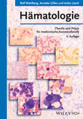
Hämatologie: Theorie und Praxis für medizinische Assistenzberufe
by Anita Läsch Rolf Mahlberg Annette GillesSeit dem Erscheinen der ersten Auflage 1994 ist das Lehrbuch „Hämatologie” nicht mehr aus dem Ausbildungsangebot für Medizinisch-Technische Assistenten wegzudenken. Die dritte Auflage integriert nicht nur den neuesten Stand der MTA-Ausbildung, sondern präsentiert sich mit mehr didaktischen Merkmalen und einem übersichtlicheren Layout. Darüber hinaus wurden die folgenden Themen erweitert bzw. eingeführt: • Hämatologische Cytogenetik • Hämatologische Molekulargenetik • Antikörper-Diff erenzierung • Nachweis von Kälte-Agglutininen • Säure-Elution • Interne und externe Qualitätskontrolle in der Blutgruppenserologie.
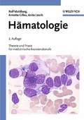
Hämatologie: Theorie und Praxis für medizinische Assistenzberufe
by Anita Läsch Rolf Mahlberg Annette GillesSeit dem Erscheinen der ersten Auflage 1994 ist das Lehrbuch "Hämatologie" nicht mehr zu stoppen. Die vorliegende neue, stark überarbeitete und aktualisierte Auflage integriert den neuesten Stand der MTA-Ausbildung. Das Buch orientiert sich thematisch am Lehrinhaltskatalog des Deutschen Verbands Technischer Assistentinnen und Assistenten in der Medizin (dvta). Didaktisch ausgeklügelt ist der Band eine unerlässliche Hilfe in der Ausbildung. Drei Jahre dauert die Ausbildung zur/zum MTA. Es sind drei lernintensive Jahre mit einem anspruchsvollen Mix aus Theorie und Praxis. "Hämatologie" ist das bewährte Lehrbuch, das nun schon einige Generationen von MTA-Auszubildenden begleitet hat. Das Buch gliedert sich in einen theoretischen und einen praktischen Teil. Ersterer behandelt die Grundlagen der Hämatologie und ihrer Krankheitsbilder. Der praktische Teil vermittelt die in der hämatologischen Diagnostik und Therapie verwendeten Techniken. Es gibt zahlreiche Handlungsanleitungen für die tägliche Arbeit. Daneben wird kompetent in Physiologie, Pathophysiologie und Labordiagnostik eingeführt. Der übersichtliche Aufbau sowie etliche erläuternde Tabellen und Abbildungen erleichtern das Verständnis. Und die überzeugende Stoffvermittlung macht vor allem eines deutlich: Die Autoren bringen jahrelange Erfahrung aus der MTA-Ausbildung mit. Dabei trägt die neue Auflage den umfangreichen Fortschritten Rechnung, die insbesondere in der molekularbiologischen Diagnostik erreicht wurden.
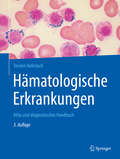
Hämatologische Erkrankungen: Atlas und diagnostisches Handbuch
by Torsten HaferlachErfahrene Experten beschreiben in dem Band die wichtigsten hämatologischen Krankheitsbilder und ihre Diagnose unter Berücksichtigung der aktuellen WHO-Klassifikation. Welche morphologischen Merkmale sind z. B. bei Knochenmarkinsuffizienz im Blutausstrich sichtbar, welche Antigene werden bei Leukämie gebildet, gibt es genetische Veränderungen auf chromosomaler oder molekularer Ebene? Antworten auf diese Fragen können rasch nachgeschlagen werden. Der Band enthält viele morphologische Abbildungen sowie eine Liste von Laboren für Spezialuntersuchungen.
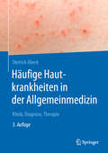
Häufige Hautkrankheiten in der Allgemeinmedizin: Klinik, Diagnose, Therapie
by Dietrich AbeckWeil Hauterkrankungen immer öfter auftreten, wird auch für Allgemeinärzte dermatologisches Wissen immer wichtiger. Der Autor behandelt in dem Band die Klinik, Pathogenese, Diagnose und Therapie der häufigsten dermatologischen Probleme. Zahlreiche Abbildungen unterstützen Ärzte bei der schnellen Zuordnung der Symptome. Der Band enthält Empfehlungen zum Ablauf der Therapie, zu Wirkstoffen und zur Hautpflege sowie Hinweise zu weiterführender Literatur. Die 2., aktualisierte Auflage wurde um drei Kapitel zu weiteren häufigen Hautkrankheiten ergänzt.
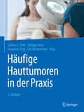
Häufige Hauttumoren in der Praxis
by Rüdiger Hein Johannes Ring Tilo Biedermann Sabine G. PlötzIn der täglichen Praxis sehen Ärzte viele verschiedene Hauttumoren oder tumorartige Hautveränderungen. Die exakte, frühzeitige Diagnose, die Einleitung einer passgenauen Therapie oder auch die Beruhigung eines Patienten mit einem harmlosen Befund setzen eine genaue Kenntnis der häufigsten Hauttumoren voraus. Der Band bietet eine prägnante Darstellung des klinischen Bildes mit hervorragenden Farbfotos, übersichtliche Tabellen zur schnellen Orientierung und eine Hinführung zur passenden Therapie. Mit Hinweisen zur Durchführung des Hautkrebsscreenings.
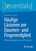
Häufige Läsionen am Daumen- und Fingerendglied: Differentialdiagnosen und Therapien (essentials)
by Christian k. SpiesIn diesem essential werden die häufigsten Erkrankungen und Verletzungen an den Fingerendgliedern und am Daumen vogestellt. Die Darstellungen der feinen anatomischen Strukturen der Fingerendglieder, des Daumens und des Nagelplattenorgans ermöglichen ein tieferes Verstehen dieser Erkrankungen/Verletzungen und deren Therapien. Vorgestellt werden die häufigsten Differentialdiagnosen in dieser Region. Die verschiedenen konservativen und operativen Therapiemöglichkeiten, die zielgenau und präzise erläutert werden, runden das Thema ab.
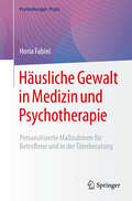
Häusliche Gewalt in Medizin und Psychotherapie: Personalisierte Maßnahmen für Betroffene und in der Täterberatung (Psychotherapie: Praxis)
by Horia FabiniDieses Fachbuch für Fachleute klärt auf: Häusliche Gewalt ist keine psychische Störung, sondern ein Bedrohungsszenario, welches mit erheblichen Risiken für die Betroffenen einhergeht. Psychotherapie ist sowohl mit Blick auf die Betroffenen als auch auf die Täter nur im Ausnahmefall die Methode der ersten Wahl und eine unsachgemäße Schwerpunktsetzung, kann vorhandene Gefährdungen verschärfen. In Fällen von häuslicher Gewalt müssen deswegen stets Sicherheitsaspekte im Vordergrund stehen – und zwar unabhängig davon, ob es um die Betreuung von Opfern oder um Täterberatung geht. Aus dem Inhalt: Gefährdungs- und Gefährlichkeitseinschätzung, Risikomanagement, Krisenintervention und Beratung zu Schutzmaßnahmen in unterschiedlichen Bedrohungslagen, professionelle Unterstützung Betroffener, Beratung von Tätern, Zusammenarbeit mit Behörden und Hilfseinrichtungen, professionelle Kooperation und Vernetzung. Über den Autor: Horia Fabini ist Psychologischer Psychotherapeut, Gruppentherapeut (BAG), Psychotraumatologe (DeGPT), Fachpsychologe Notfallpsychologie, Kriminalpsychologe sowie Supervisor und Lehrtherapeut (DVT). Er ist darüber hinaus tätig als Präventionsmanager Extremismus/Radikalisierung, Gutachter für die Schwerpunkte Legal- und Gefährlichkeitsprognose und wissenschaftlicher Leiter des Curriculums Notfallpsychologie an der Bodelschwingh-Akademie in Berlin sowie Dozent in weiteren Bildungseinrichtungen.
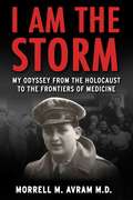
I Am the Storm: My Odyssey from the Holocaust to the Frontiers of Medicine
by Morrell Michael AvramMorrell Avram, born in Bucharest, could have easily become one of the 200,000 Romanian Jews killed by the German Nazis or their Romanian allies. I AM THE STORM is the riveting true story of how he survived—and later triumphed as a pioneering doctor—through a combination of grit and persistence. At age 11, Avram was separated from his mother and baby sister because the US Embassy would only allow them to immigrate on the condition that they leave Morrell and his father behind. What the family hoped would be a brief separation became six terrifying years. Amid the horrors of the war, Morrell had to fend mostly for himself, shuttling from relative to relative, hiding place to hiding place. Among his close calls: He longed to buy a ticket on the Struma, a ship taking Jewish refugees from Romania to Palestine, that was torpedoed and sank along with many of his friends. He walked into his bar mitzvah ceremony with dozens of Nazi soldiers stationed outside the synagogue. He was strafed and nearly killed by an American warplane. Upon finally escaping Romania and reuniting with his mother and sister, Avram faced a host of new challenges in New York. After getting through high school with minimal English, he was thrilled to get into college but found it impossible to juggle classes while working to help support his family. By age 21, it looked as if his dream of becoming a doctor was doomed. But relief came from an unlikely source—a draft notice from the US Army, which transformed him from an anxious &“subway rat&” into a focused soldier, driven by the words of his drill sergeant: &“You are the storm! You are invincible!&” Avram&’s unlikely journey continued as a med student in Brussels and Geneva, as a young doctor in Brooklyn, and as one of the leaders of the new field of nephrology. He became a pathbreaking specialist in dialysis and kidney transplants, saving tens of thousands of patients personally and millions more through treatments he helped devise.
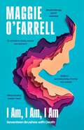
I Am, I Am, I Am: The Breathtaking Number One Bestseller
by Maggie O'FarrellAS FEATURED ON DESERT ISLAND DISCS, BIG SCOTTISH BOOK CLUB AND THE ZOE BALL BOOKCLUB, A BOOK OF THE YEAR IN THE SUNDAY TIMES, THE TIMES, GUARDIAN, IRISH TIMES, OBSERVER, RED and THE TELEGRAPH.*SHORTLISTED FOR THE PEN ACKERLEY PRIZE FOR MEMOIR AND AUTOBIOGRAPHY 2018*I AM, I AM, I AM is a memoir with a difference - the unputdownable story of an extraordinary woman's life in near-death experiences. Insightful, inspirational, gorgeously written, it is a book to be read at a sitting, a story you finish newly conscious of life's fragility, determined to make every heartbeat count.A childhood illness she was not expected to survive. A teenage yearning to escape that nearly ended in disaster. A terrifying encounter on a remote path. A mismanaged labour in an understaffed hospital. Shocking, electric, unforgettable, this is the extraordinary memoir from Costa Novel-Award winner and Sunday Timesbestselling author Maggie O'Farrell. It is a book to make you question yourself. What would you do if your life was in danger, and what would you stand to lose?
I Am, I Am, I Am: The Breathtaking Number One Bestseller
by Maggie O'FarrellAS SELECTED FOR THE ZOE BALL BOOKCLUB, A BOOK OF THE YEAR IN THE SUNDAY TIMES, THE TIMES, GUARDIAN, IRISH TIMES, OBSERVER, RED and THE TELEGRAPH.I AM, I AM, I AM is a memoir with a difference - the unputdownable story of an extraordinary woman's life in near-death experiences. Insightful, inspirational, gorgeously written, it is a book to be read at a sitting, a story you finish newly conscious of life's fragility, determined to make every heartbeat count.A childhood illness she was not expected to survive. A teenage yearning to escape that nearly ended in disaster. A terrifying encounter on a remote path. A mismanaged labour in an understaffed hospital. Shocking, electric, unforgettable, this is the extraordinary memoir from Costa Novel-Award winner and Sunday Timesbestselling author Maggie O'Farrell. It is a book to make you question yourself. What would you do if your life was in danger, and what would you stand to lose? I AM, I AM, I AM will speak to readers who loved Cheryl Strayed's WILD or Max Porter's GRIEF IS THE THING WITH FEATHERS.(P)2017 Headline Publishing Group Ltd
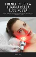
I Benefici Della Terapia Della Luce Rossa: Come perdere peso e curare l’acne, le cicatrici e l’artrite a casa
by Dane JonVuoi sbarazzarti dell'acne e delle cicatrici, perdere peso, trattare l'artrite e il dolore? Le tecniche di terapia della luce rossa vengono da tempo utilizzate per curare tantissime patologie, sono poco invasive e donano enormi benefici. La terapia della luce rossa offre un valido aiuto contro le malattie croniche e l’infiammazione ed è un ottimo sostegno al buon umore. Ma c’è molto di più! Ecco il segreto dei professionisti della salute per sentirsi più in forma che mai! Questo eBook illustra il metodo di utilizzo della terapia della luce rossa più veloce ed efficace per sentirti bene! Imparerai a migliorare il tuo benessere e la tua salute in poche settimane. E non solo: ogni singolo aspetto della tua vita migliorerà, letteralmente. Grazie a questa guida imparerai tecniche di utilizzo collaudate senza dover spendere altri soldi in libri e corsi. Nella guida scoprirai come: Curare le cicatrici. Perdere peso. Trattare l’artrite. Guarire dall’acne. Sapere tutto ciò che c’è da sapere. + MOLTO DI PIÙ Se vuoi sentirti più in forma migliorando contemporaneamente l’aspetto della tua pelle e il tuo benessere, allora questa guida fa proprio al caso tuo.