- Table View
- List View
I Can Make You Thin: The Revolutionary System Used by More Than 3 Million People
by Paul MckennaPaul McKenna, PhD, is the most popular nonfiction author in the UK and his television shows have been watched by millions of people in 42 countries around the world. He has worked with Hollywood movie stars, Olympic gold medalists, rock stars, business leaders, and royalty. Now it's your turn!
I Can't Save You: A Memoir
by Anthony Chin-QueeThe raw and gripping memoir of a Black physician who confronts his past mistakes and relationships as he learns to find his own path forward At first glance, Anthony Chin-Quee looks like a traditional success story: a smart, ambitious kid who grew up to become a board-certified otolaryngologist—an ear, nose, and throat surgeon. Yet the truth is more complicated. As a self-described &“not white, mostly Black, and questionably Asian man,&” Chin-Quee knows that he doesn&’t fit easily into any category. Growing up in a family with a background of depression, he struggled with relationships, feelings of inadequacy, and a fear of failure that made it difficult for him to forge lasting bonds with others. To repair that, he began his own unflinching examination of what it means to be both a physician and a Black man today. What saved him and his sanity was not medicine but storytelling: by sharing stories from his life and career, Chin-Quee learned how powerful the truth can be in helping to forgive yourself and others as you chart a new way forward. By turns harrowing and hilarious, honest and human, I Can&’t Save You is the fascinating true story of how looking within can change you and your life for the better.
I Cannot Control Everything Forever: A Memoir of Motherhood, Science, and Art
by Emily C. BloomAn eloquent and intimate debut memoir about navigating the gap between expectation and reality in modern motherhood.I Cannot Control Everything Forever is Emily Bloom’s journey towards and through motherhood, a path that has become, for the average woman, laden with data and medical technology. Emily faces decisions regarding genetic testing and diagnosis, technologies that offer the illusion of certainty but carry the weight of hard decisions. Her desire to know more thrusts her back into the history of science, as she traces the discoveries that impacted the modern state of pregnancy and motherhood. With the birth of their daughter, who is diagnosed with congenital deafness and later, Type 1 diabetes, Emily and her husband find their life centered around medical data, devices, and doctor’s visits, but also made richer and fuller by parenting an exceptional child. As Emily learns, technology and data do not reduce the labor of caretaking. These things often fall, as the pandemic starkly revealed, on mothers. Trying to find a way out of the loneliness and individualism of 21st century parenthood, Emily finds joy in reaching outwards, towards art and literature–such as the maternal messiness of Louise Bourgeois or Greek myths about the power of fate–as well as the collective sustenance of friends and community.With lyrical and enchanting prose, I Cannot Control Everything Forever is an inspired meditation on art, science, and motherhood.
I Ching Acupuncture - The Balance Method: Clinical Applications of the Ba Gua and I Ching
by David TwickenI Ching Acupuncture - The Balance Method is a system of acupuncture point selection based on the principles of Chinese philosophy and classic Chinese texts, including the I Ching, Nei Jing Su Wen and Ling Shu. In this unique book Dr. Twicken presents classic Chinese philosophical models that explain the relationships between philosophy, Chinese medical principles, acupuncture channels and the human body. The models are the He Tu, Luo Shu Nine Palaces, Early Heaven Ba Gua, Later Heaven Ba Gua, Twelve-Stage Growth Cycle, Stems and Branches and the Chinese calendar. These models and theories clearly show the relationships between the acupuncture channels and the human body and provide guiding theory for acupuncture strategies and point selection. I Ching Acupuncture presents six Balance Methods. This clinically effective system of acupuncture is based on minimal and distal acupuncture treatments. I Ching Acupuncture - The Balance Method is a valuable and effective acupuncture system that can complement any practice.
I Contain Multitudes: The Microbes Within Us and a Grander View of Life
by Ed YongNew York Times BestsellerNew York Times Notable Book of 2016 • NPR Great Read of 2016 • Named a Best Book of 2016 by The Economist, Smithsonian, NPR's Science Friday, MPR, Minnesota Star Tribune, Kirkus Reviews, Publishers Weekly, The Guardian, Times (London)From Pulitzer Prize winner Ed Yong, a groundbreaking, wondrously informative, and vastly entertaining examination of the most significant revolution in biology since Darwin—a “microbe’s-eye view” of the world that reveals a marvelous, radically reconceived picture of life on earth.Every animal, whether human, squid, or wasp, is home to millions of bacteria and other microbes. Pulitzer Prize-winning author Ed Yong, whose humor is as evident as his erudition, prompts us to look at ourselves and our animal companions in a new light—less as individuals and more as the interconnected, interdependent multitudes we assuredly are.The microbes in our bodies are part of our immune systems and protect us from disease. In the deep oceans, mysterious creatures without mouths or guts depend on microbes for all their energy. Bacteria provide squid with invisibility cloaks, help beetles to bring down forests, and allow worms to cause diseases that afflict millions of people.Many people think of microbes as germs to be eradicated, but those that live with us—the microbiome—build our bodies, protect our health, shape our identities, and grant us incredible abilities. In this astonishing book, Ed Yong takes us on a grand tour through our microbial partners, and introduces us to the scientists on the front lines of discovery. It will change both our view of nature and our sense of where we belong in it.
I Die, but My Memory Lives On: The World Aids Crisis And The Memory Book Project
by Henning Mankell&“A deeply moving account of Henning Mankell&’s personal responses to AIDS and its victims, both parents and children left behind far too soon.&” —Archbishop Desmond Tutu The internationally famous creator of the bestselling Kurt Wallander mysteries tells the true story of a heartrending tradition spawned by a major health crisis: the invaluable Memory Book Project, which gives those dying of AIDS an opportunity to record their lives in words and pictures for the children they leave behind. In Uganda, Mankell finds village after village populated only by children and the elderly—those left behind after AIDS swept away an entire generation. These slim, intensely personal volumes can contain words, pictures, a pressed butterfly, or even grains of sand as ways to represent the lives lost to this devastating plague. Excerpts from Ugandan memory books appear throughout I Die, but My Memory Lives On and, together with Mankell&’s narrative, they tell the stories of individual lives while sounding a powerful warning about the threat of AIDS. Featuring a foreword by Archbishop Desmond Tutu, the book includes an appendix listing AIDS organizations and resources. A portion of the book&’s proceeds will be donated to AIDS charities in Africa.
I Died?: The Personal Testimony of a Survivor
by J. F. BollesA spot of illness before Thanksgiving marks the beginning of one woman’s heart-rending, real-life journey from pain and death to survival and happiness. J. F. Bolles is a mother, a grandmother and a Christian. J. F. Bolles died, twice! She received a second chance from God to live and to love. This is her personal testimony.
I Domain Integrins
by Donald GullbergThe integrin family is composed of 24 members and approximately ten years ago (2003) we published a book devoted to the nine I domain integrin subunits. In this second edition, I am pleased that most of the original authors have been able to contribute to the updated version. I domain containing integrins include collagen receptors and leukocyte receptors. In 2003 the knockout mouse phenotypes for all of the I domain integrins had not yet been published; they are now, and are summarized and discussed in this edition. Interestingly, a recent 10 integrin mutation in dogs has indicated that collagen-binding integrins in the musculoskeletal system might have much more severe phenotypes in larger animals/humans compared to the mild integrin phenotypes observed in collagen-binding integrin deficient mice. This finding is further discussed in the book. In the cancer field, the microenvironment is taking center stage, and here collagen receptors on fibroblasts are predicted to play important roles in paracrine signaling, in regulating tissue stiffness and matrix remodeling. New technologies, new mouse models in combination with analyses of I integrins in larger animals/humans are thus predicted to increase our knowledge about this group of receptors. With this in mind we look forward to another 10 years of research with I domain integrins.
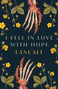
I Fell in Love with Hope
by LancaliAgainst the unforgiving landscape of a hospital, I fell in love with a mischievous, sun-eyed boy who became my only joy in that desolate place. That&’s what made it all the more soul-crushing when he committed suicide in front of me. Since then, I've sworn never to love anyone again. With three exceptions: My friends, Sony, Neo, and Coeur, a little gang of rebellious, dying kids. Sony leads the charge with the air of freedom and only one lung to breathe it. Neo, a bad-tempered and wheel-chaired writer, keeps track of our great deeds from stealing to terrorizing our nurse. Coeur is the beautiful boy, the muscle, the gentle giant with a failing heart. Before death inevitably knocks down our doors, my thieves and I have one last heist planned. A great escape that will take us far from abusive parents, crippling loss, and the realities of our diseases. So what happens when someone else walks through the door? What happens when a girl joins our party and renders me speechless with her mischievous smile? What happens when she has suns in her eyes, and as terrified as I am to lose again, I start to fall? Trigger warnings found in foreword.
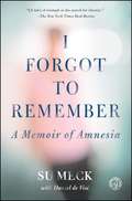
I Forgot to Remember: A Memoir of Amnesia
by Su Meck Daniel de ViséThe courageous memoir of a woman who was robbed of all her memories by a traumatic brain injury—and her more than twenty-five-year struggle to reclaim her life: “[A tale] of triumph in the search for identity” (The New York Times Book Review).In 1988, Su Meck was twenty-two and married with two children when a ceiling fan fell and struck her on the head, erasing all her memories of her life. Although her body healed rapidly, her memories never returned. After just three weeks in the hospital, her physicians released Su and she returned home to take care of her two toddlers. What would you do if you lost your past? Adrift in a world about which she understood almost nothing, Su became an adept mimic, gradually creating routines and rituals that sheltered her and her family from the near-daily threat of disaster—or so she thought. Though Su would eventually relearn to tie her shoes, cook a meal, read, and write, nearly twenty years would pass before a series of personally devastating events shattered the “normal” life she had worked so hard to build, and she realized that she would have to grow up all over again. In her own indelible voice, Su offers a unique view from the inside of a terrible injury as she “recounts her grueling climb back to normalcy…in this heart-wrenching true story” (O, The Oprah Magazine). Piercing, heartbreaking, but finally uplifting, I Forgot to Remember is the story of a woman determined to live life on her own terms.
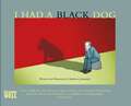
I Had a Black Dog
by Matthew Johnstone'I Had a Black Dog says with wit, insight, economy and complete understanding what other books take 300 pages to say. Brilliant and indispensable.' - Stephen Fry'Finally, a book about depression that isn't a prescriptive self-help manual. Johnston's deftly expresses how lonely and isolating depression can be for sufferers. Poignant and humorous in equal measure.' Sunday TimesThere are many different breeds of Black Dog affecting millions of people from all walks of life. The Black Dog is an equal opportunity mongrel.It was Winston Churchill who popularized the phrase Black Dog to describe the bouts of depression he experienced for much of his life.Matthew Johnstone, a sufferer himself, has written and illustrated this moving and uplifting insight into what it is like to have a Black Dog as a companion and how he learned to tame it and bring it to heel.
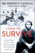
I Had to Survive: How a Plane Crash in the Andes Inspired My Calling to Save Lives
by Pablo Vierci Dr. Roberto CanessaDr. Roberto Canessa recounts his side of the famous 1972 plane crash of Uruguayan Air Force Flight 571 in the Andean Mountains and how, decades later, the harrowing journey to survive propelled him to become one of the world&’s leading pediatric cardiologists, seeing in his patients the same fierce will to live he witnessed in the Andes.As he tended to his wounded Old Christians teammates amidst the devastating carnage, rugby player Roberto Canessa, a second-year medical student at the time, realized that no one on earth was luckier: he was alive—and for that, he should be eternally grateful. As the starving group struggled beyond the limits of what seemed possible, Canessa played a key role in safeguarding his fellow survivors, eventually trekking with a companion across the hostile mountain range for help. No one could have imagined that there were survivors from the accident in such extreme conditions. Canessa's extraordinary experience on the fine line between life and death became the catalyst for the rest of his life. This uplifting tale of hope and determination, solidarity and ingenuity, gives vivid insight into the world-famous story that inspired the movie Alive! Canessa also draws a unique and fascinating parallel between his work as a doctor diagnosing very complex congenital cardiopathies in unborn and newborn infants and the difficult life-changing decisions he was forced to make in the Andes. With grace and humanity, Canessa prompts us to ask ourselves: what do you do when all the odds are stacked against you?
I Hate Myself: Overcome Self-Loathing and Realize Why You're Wrong About You
by Blaise AguirreLearn to understand the unaddressed symptom of mental health In I Hate Myself: Overcome Self-Hatred and Realize Why You're Wrong About You, internationally known Assistant Professor of Psychiatry at Harvard Medical School Dr. Blaise Aguirre tackles the pervasive and often ignored issue of self-hatred. This book provides crucial insights into identifying and overcoming this deeply disturbing feeling, explaining why common practices of "self-care" or "self-love" often fall short in cases where self-hatred has become an integral part of a person's identity. Dr. Aguirre shares compelling first-hand accounts from patients who have battled and conquered self-hatred, revealing the severe impact this feeling has on people from all walks of life and their loved ones. The book delves into the roots of self-hatred, associated mental health disorders, and offers practical strategies for overcoming these challenges. In the book, you will: Learn to identify the origins and signs of self-hatred Understand the connection between self-hatred and suicidal behavior as well as to co-occurring disorders like borderline personality disorder and depression Discover effective strategies for transforming self-loathing into self-compassion Perfect for those struggling with self-hatred and their loved ones, as well as mental health professionals, I Hate Myself offers a compassionate and practical approach to achieving self-acceptance. Start your journey towards healing today and embrace the self-worth you deserve.
I Have Cancer, Now What?: 12 Things You, Your Spouse, and Your Family Must Know in Your Battle with Cancer from Doctors to Finances, Romance to Household Needs, Getting the Word Out to Caregiver Burnout and Everything In between
by Carson Boss Cindy BossYou have cancer, and you need the help and support of your spouse and family more than ever. I Have Cancer, Now What? includes information on how to overcome the shock and fear of diagnosis, how to talk to your spouse and extended family, how to consult on what you want from your doctors, where and how your family can give you support, the transition of normal household duties and how to manage those, the real costs of cancer, both financial and emotional, what you need from your spouse and family emotionally and physically, including romance, how to manage full-time jobs, and other long-term issues that will help you get the support you need.
I Have Something to Tell You: A Memoir
by Regan HofmannFor ten years, Regan Hofmann lived a double life. To the world, she was a woman from Princeton who went to prep school, summered in the Hamptons and rode Thoroughbred horses. She had a great job, a loving family and friends and looks that made men turn their heads. From the outside, she seemed to have it all. On the inside, though, coursing through her veins and weighing heavily on her mind, was the truth: that she was HIV-positive. At first, Hofmann faced her mortality alone, shamed by a disease society considered the exclusive property of gay men, injection drug users and sex workers. Burdened by her secret, she withdrew from the world she once knew. Over time, though, Hofmann began to accept her mortality -- and HIV -- and reconsidered the way she wanted to live her life. After nearly a decade of silence, Hofmann did what she never imagined having the courage to do: she came out to the world about what she was going through. Regan Hofmann not only has the courage to fight HIV and the debilitating stigma that surrounds it, but she writes about her experience with unflinching honesty and a deep affection for the family and friends who support her. I Have Something to Tell You is a memoir of disease and survival, and an inspiring account of a life driven by a sense of purpose and a search for love in the face of the unthinkable. More than anything, it is a story that reminds us that while life can change in an instant, we each hold the power to decide how we use the time we have. With humor, vitality and an unquenchable passion, Regan shows us a life fully lived.
I Hear a Song in My Head: A Memoir in Stories of Love, Fear, Doctoring, and Flight
by M.D. Nergesh Tejani“[Tejani] shares her stories of succeeding as a doctor in Uganda during the 1960s . . . a must for those seeking a medical memoir collection.” —Midwest Book ReviewSet in Uganda of the sixties with bookends in India and New York, this doctor’s story tells of a turbulent political time when colonial Uganda graduated to self-rule. It is also the personal story of an Indian woman living in an independent African country wanting and needing assimilation but regretfully recognizing rejection. It is the story of the exhilaration of living in a country more beautiful than Eden, if sometimes a threatened Eden. But most of all it tells doctoring tales made delicate by seeing them through the heart. It was a time in medicine before evidential imperatives removed the romance. “Dr. Tejani’s unique meld of skill and compassion radiates throughout this text which will touch both physician and lay readers alike.” —Frank A. Chervenak, MD, New York Weill Cornell Medical Center “With clarity, drama, and humor, this book creates a family story, a picture of an African nation in the throes of political upheaval, and an original and illuminating view of medical needs and practices in circumstances that exist today in many parts of the world. The complex harmonies of the song in Dr. Tejani’s head will resonate for a wide variety of readers.” —Carol Sicherman, author of Rude Awakenings“Nergesh Tejani is a terrific writer . . . Her subject is often exotic, often with international themes and full of pithy observations and wisdom.” —Abraham Verghese, MD, Professor of the Theory and Practice of Medicine, Stanford University Medical Center
I Heard There Was a Secret Chord: Music as Medicine
by Daniel J. LevitinNATIONAL BESTSELLER One of Smithsonian's 10 Best Science Books of 2024 Neuroscientist and New York Times best-selling author of This Is Your Brain on Music Daniel J. Levitin reveals the deep connections between music and healing. Music is one of humanity’s oldest medicines. From the Far East to the Ottoman Empire, Europe to Africa and the pre-colonial Americas, many cultures have developed their own rich traditions for using sound and rhythm to ease suffering, promote healing, and calm the mind. In his latest work, neuroscientist and New York Times best-selling author Daniel J. Levitin (This Is Your Brain on Music) explores the curative powers of music, showing us how and why it is one of the most potent therapies today. He brings together, for the first time, the results of numerous studies on music and the brain, demonstrating how music can contribute to the treatment of a host of ailments, from neurodegenerative diseases such as Parkinson’s and Alzheimer’s, to cognitive injury, depression, and pain. Levitin is not your typical scientist—he is also an award-winning musician and composer, and through lively interviews with some of today’s most celebrated musicians, from Sting to Kent Nagano and Mari Kodama, he shares their observations as to why music might be an effective therapy, in addition to plumbing scientific case studies, music theory, and music history. The result is a work of dazzling ideas, cutting-edge research, and jubilant celebration. I Heard There Was a Secret Chord highlights the critical role music has played in human biology, illuminating the neuroscience of music and its profound benefits for those both young and old.
I Is for Inquiry: An Illustrated ABC of Inquiry-Based Instruction for Elementary Teachers and Schools
by Bruce Shore Mark Aulls Diana Tabatabai Juss Kaur MagonI Is for Inquiry takes a unique approach to helping teachers in the elementary grades create lessons and sustain inquiry in their classrooms. This colorful, illustrated alphabet book explores 26 (including X and Z) key ideas and skills in inquiry-based teaching and learning, such as collaboration, dialogue, evidence, hypothesis, and scaffolding. Each short chapter:Summarizes one inquiry element that can be built into students' experiences.Uses straightforward language and examples.Includes a classroom vignette and suggestions for using the concept.Shares selected references and related Internet-based resources.Helps teachers build self-confidence about teaching through inquiry.This book will serve as a familiar and fun resource for busy teachers at any point in their careers. Using the inquiry vocabulary and repertoire of concepts, teachers can build curriculum and share ideas with colleagues, making inquiry in the classroom as approachable as ABC!
I Know a Secret: A Rizzoli & Isles Novel (Rizzoli & Isles #12)
by Tess Gerritsen<P>Jane Rizzoli and Maura Isles—the inspiration for the smash hit TNT series—continue their bestselling crime-solving streak, as they pursue a shadowy psychopath keeping secrets and taking lives. <P> Two separate homicides, at different locations, with unrelated victims, have more in common than just being investigated by Boston PD detective Jane Rizzoli and medical examiner Maura Isles. In both cases, the bodies bear startling wounds—yet the actual cause of death is unknown. It’s a doubly challenging case for the cop and the coroner to be taking on, at a fraught time for both of them. <P>As Jane struggles to save her mother from the crumbling marriage that threatens to bury her, Maura grapples with the imminent death of her own mother—infamous serial killer Amalthea Lank. While Jane tends to her mother, there’s nothing Maura can do for Amalthea, except endure one final battle of wills with the woman whose shadow has haunted her all her life. Though succumbing to cancer, Amalthea hasn’t lost her taste for manipulating her estranged daughter—this time by dangling a cryptic clue about the two bizarre murders Maura and Jane are desperately trying to solve. <P>But whatever the dying convict knows is only a piece of the puzzle. Soon the investigation leads to a secretive young woman who survived a shocking abuse scandal, an independent horror film that may be rooted in reality, and a slew of martyred saints who died cruel and unusual deaths. And just when Rizzoli and Isles think they’ve cornered a devilish predator, the long-buried past rears its head—and threatens to engulf more innocent lives, including their own. <P><b>A New York Times Bestseller</b>
I Love a Cop, Revised Edition
by Ellen Kirschman Kevin GilmartinNothing worth doing is easy--and that includes loving a cop. Being a member of the law enforcement community is a source of pride for officers and families alike. But long hours, unpredictable shifts, and the crisis-driven nature of the profession can turn life on the home front into an emotional roller coaster ride. Dr. Ellen Kirschman, a psychologist who's worked with police officers for more than 20 years, gives you practical ways to deal with the challenges that come with the territory. Packed with stories from cops and their significant others, this book explains how to reduce spillover from on-the-job stress and cope with loneliness or worry during extended deployments. Dr. Kirschman acknowledges the tough realities of post-9/11, post-Katrina law enforcement, and she offers frank, realistic suggestions for handling serious issues like alcohol abuse and domestic violence. She also covers special topics for women and minorities on the force. Whether you read it from cover to cover or reach for it when problems arise, I Love a Cop is an indispensable tool that everyone in your family can depend on.
I Love a Cop, Revised Edition
by Ellen KirschmanNothing worth doing is easy--and that includes loving a cop. Being a member of the law enforcement community is a source of pride for officers and families alike. But long hours, unpredictable shifts, and the crisis-driven nature of the profession can turn life on the home front into an emotional roller coaster. Dr. Ellen Kirschman, a psychologist who's worked with police officers for more than 30 years, gives you practical ways to deal with the challenges that come with the territory. Packed with stories from cops and their significant others, this book explains how to reduce spillover from on-the-job stress and cope with loneliness or worry during extended deployments. Dr. Kirschman acknowledges the tough realities of 21st-century law enforcement and offers frank, realistic suggestions for handling serious issues like alcohol abuse and domestic violence. She also covers special topics for women and minorities on the force. Whether you read it from cover to cover or reach for it when problems arise, I Love a Cop is an indispensable tool that everyone in your family can depend on. Mental health professionals, see also Counseling Cops: What Clinicians Need to Know, by Ellen Kirschman, Mark Kamena, and Joel Fay.
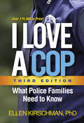
I Love a Cop, Third Edition: What Police Families Need to Know
by Ellen KirschmanPolice families are brave, resilient, and proud--and they face remarkable challenges, sometimes on a daily basis. Now thoroughly updated for today's turbulent times, this is the resource that cops and their loved ones have relied on for decades. Trusted expert Ellen Kirschman gives you practical ways to manage the stress of the job and create a healthy, supportive home environment. The third edition features the latest information, new stories from police families, two new chapters, and fully updated resources. Dr. Kirschman acknowledges the tough realities of life on the force and offers frank, realistic suggestions for handling everyday relationship dilemmas as well as serious issues like trauma, domestic violence, and alcohol abuse. Whether you read this book cover to cover or reach for it when problems arise, you will find no-nonsense guidance to help your family thrive. Mental health professionals, see also Counseling Cops: What Clinicians Need to Know, by Ellen Kirschman, Mark Kamena, and Joel Fay.
I Love a Fire Fighter
by Ellen KirschmanHow can fire fighter families manage the stress that comes with life in the service? How do you keep a grip on fears and worries during long hours of separation from your spouse? Where can you turn when times get tough? With this practical, no-nonsense, yet compassionate guide, Dr. Ellen Kirschman provides the first self-help book written to address the questions and concerns of today's fire fighter families. From the effects of shift work on your marriage, to the emotional side of physical injuries and trauma, to ways to deal with job pressures and resolve conflicts at home, read on to see what you can do to help yourself, your mate, and your children navigate the highs and lows of "the best job in the world."

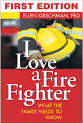
I Love a Fire Fighter, Second Edition: What the Family Needs to Know
by Ellen KirschmanHow can fire fighter families cope with the emotional toll of loving a first responder? There are ceaseless worries--about the physical dangers of the profession, the cumulative stress, and the long hours spent away from home. In this compassionate and knowledgeable guide--now fully revised and updated--psychologist Ellen Kirschman shares sage advice and practical strategies for when times get tough. From dealing with occupational hazards like trauma, marital tension, and substance use problems, to the psychological effects of fighting wildland fires, Dr. Kirschman understands the unique challenges of life on the front lines. With candor and wisdom, she shows fire fighters and their loved ones how to navigate the highs and lows of &“the best job in the world.&”
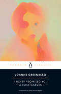
I Never Promised You a Rose Garden (Sparknotes Literature Guide Ser.)
by Joanne GreenbergThe multimillion-copy bestselling modern classic of autobiographical fiction about a young woman&’s struggle with mental health, featuring a new foreword by Esmé Weijun Wang, the New York Times bestselling author of The Collected Schizophrenias, and a new afterword by the authorAlso available from Penguin Classics: Joanne Greenberg&’s bestselling modern classic In This SignAfter making an attempt on her own life, sixteen-year-old Deborah Blau is diagnosed with schizophrenia. With the reluctant and fearful consent of her parents, she enters a psychiatric hospital many hours from her home in suburban Chicago. Here she will spend the next three years, trying, with the help of a gifted psychiatrist, to find a path back to her &“normal&” life, and to emerge from the imaginary Kingdom of Yr in which she has sought refuge.A semiautobiographical novel originally published under the pen name Hannah Green just a year after Sylvia Plath&’s The Bell Jar--a very different portrait of psychological breakdown--I Never Promised You a Rose Garden remains, more than half a century later, a timeless and ultimately hopeful book, ripe for rediscovery by a new generation eager to erase the stigma of mental illness.For more than seventy-five years, Penguin has been the leading publisher of classic literature in the English-speaking world. With more than 2,000 titles, Penguin Classics represents a global bookshelf of the best works throughout history and across genres and disciplines. Readers trust the series to provide authoritative texts enhanced by introductions and notes by distinguished scholars and contemporary authors, as well as up-to-date translations by award-winning translators.