- Table View
- List View
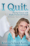
I Quit: Facing Cancer with Faith, Family & Friends
by Kristina Schnack KotlusThis candid, funny account of coping with serious illness is a rallying cry for anyone facing a difficult situation. When she found herself diagnosed with brain cancer for the second time, Kristina Kotlus chose to quit on day one. But quitting didn&’t mean giving up. It meant a whole new lease on life. Rejecting the impulse to worry or try to control things she couldn&’t, resisting all the advice to &“fight&” and be a &“warrior,&” she simply resolved to do what she could, admit she needed help (and lots of it), and put her faith in God. In this inspiring memoir, Kristina shares how she survived both diagnoses—with the support of her family, friends, and faith—in a relatable, funny way, from her original diagnosis to finding doctors to telling her kids (hint: make someone else do it). She shares openly and honestly, with just a touch of sarcasm and a heavy dose of humor and faith, and encourages readers to decide that it&’s time to stand up, wash the tears off their face, and keep going.
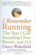
I Remember Running: The Year I Got Everything I Ever Wanted -- and ALS
by Darcy WakefieldDarcy Wakefield was a single, 33-year-old, athletic, workaholic English professor who was diagnosed with ALS. I Remember Running is Darcy's story of change and loss and challenges during her first year with ALS, as she struggles to make sense of her diagnosis and redefine herself in the face of this terminal illness. This book will move readers to see the world in a different light and proves that it is possible to live a rich, meaningful life after being diagnosed with a terminal illness.

I Served on Bataan
by Juanita RedmondThe true story of an Army nurse trapped in the Philippines during the beginning of America's entrance in WWII.
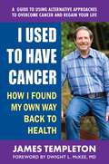
I Used to Have Cancer
by James TempletonBy all standards of success, James Templeton seemed to have it all. He was a highly successful businessman, had a beautiful wife and daughter, and, only in his early thirties, had his whole life in front of him. To avoid the same fate as his father and grandfather, who both died of heart attacks at a young age, James became an avid runner—a passion that he believed helped him stay fit and healthy. Imagine his shock when, during a routine physical, his doctor noticed a mole on his body that turned out to be a melanoma—a dangerous form of skin cancer. The mole was removed immediately and James, who was diligent in his follow-up exams, appeared to be cancer-free—but only for a short while. When the cancer reappeared and had spread, on the advice of his doctor, James followed the conventional medical protocol, which included surgery and chemotherapy. He was also involved in a clinical trial. When he learned that the treatments weren&’t working, James was obviously devastated. He had reached a new low point in his life, and as he lay in the hospital bed, he prayed fervently for help. As if by some miracle, help came to James in the form of three different visitors who would change the course of his life—and help direct him on a path back to health.I Used to Have Canceris James Templeton&’s memoir—an inspiring look back at his unique journey in overcoming stage 4 melanoma. James takes you with him on a trip crisscrossing America, during which he shares the various natural approaches he followed to battle his cancer—from diet and supplements to meditation and lifestyle adjustments. As his journey continued, you will see first-hand how James&’ definition of success changed from making money to seeing the next sunrise. And how he continues finding success by reaching out to others to share the lessons he has learned.While this book largely focuses on the various methods James used to overcome his own cancer, it is also an inspiring story of not giving up when all other avenues of conventional medicine fail. It is about taking control of your life and finding a way back from the brink of death. It is about being able to tell your friends, &“I used to have cancer.&”
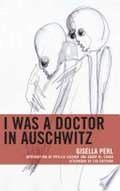
I Was A Doctor In Auschwitz (Lexington Studies in Jewish Literature Series)
by Eva Hoffman Danny M. Cohen Phyllis Lassner Gisella PerlGisella Perl’s memoir is the extraordinarily candid account of women’s extreme efforts to survive Auschwitz. With writing as powerful as that of Charlotte Delbo and Ruth Kluger, her story individualizes and therefore humanizes a victim of mass dehumanization. Perl accomplished this by representing her life before imprisonment, in Auschwitz and other camps, and in the struggle to remake her life. It is also the first memoir by a woman Holocaust survivor and establishes the model for understanding the gendered Nazi policies and practices targeting Jewish women as racially poisonous. Perl’s memoir is also significant for its inclusion of the Nazis’ Roma victims as well as in-depth representations of Nazi women guards and other personnel. Unlike many important Holocaust memoirs, Perl’s writing is both graphic in its horrific detail and eloquent in its emotional responses. One of the memoir’s major historical contributions is Perl’s account of being forced to work alongside Dr. Josef Mengele in his infamous so-called clinic and using her position to save the lives of other women prisoners. These efforts including infanticide and abortion, topics that would remain silenced for decades and, unfortunately, continue to be marginalized from all too many Holocaust accounts. After decades out of print, this new edition will ensure the crucial place of Perl’s testimony on Holocaust memory and education.
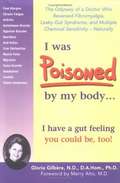
I Was Poisoned by My Body: The Odyssey of a Doctor Who Reversed Fibromyalgia, Leaky Gut Syndrome and Multiple Chemical Sensitivity, Naturally
by Gloria Gilbere Beata Golau Tama Bergstrand Merry AltoWell-documented explanation of Leaky Gut Syndrome, MCS, symptoms and remedies.
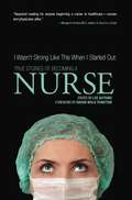
I Wasn't Strong Like This When I Started Out: True Stories of Becoming a Nurse
by Lee GutkindThis collection of true narratives reflects the dynamism and diversity of nurses, who provide the first vital line of patient care. Here, nurses remember their first "sticks," first births, and first deaths, and reflect on what gets them though long, demanding shifts, and keeps them in the profession. The stories reveal many voices from nurses at different stages of their careers: One nurse-in-training longs to be trusted with more "important" procedures, while another questions her ability to care for nursing home residents. An efficient young emergency room nurse finds his life and career irrevocably changed by a car accident. A nurse practitioner wonders whether she has violated professional boundaries in her care for a homeless man with AIDS, and a home care case manager is the sole attendee at a funeral for one of her patients. What connects these stories is the passion and strength of the writers, who struggle against burnout and bureaucracy to serve their patients with skill, empathy, and strength.
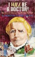
I Will Be A Doctor!
by Dorothy C. Wilson"I Will Be A Doctor" is a biography of Elizabeth Blackwell, the first woman physician in The United States. While the story may be classified as juvenile literature, I found it a pleasure to read and very enjoyable. The biography traces Elizabneth Blackwell's life from her childhood in England, to her migration to The United States, and much later back to England. Her life as a pioneer in her chosen vocation is fastinating. Because she lived primarily in the 19th century, there are many social issues which are discussed--slavery, women's rights, extreme poverty, infant death, and the huge issue of hygeine. Enjhoy!
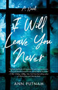
I Will Leave You Never: A Novel
by Ann PutnamIn the middle of a perilous drought in the Northwest, an arsonist begins setting fires all around. It gives Zoe Penney nightmares about her home—seated right next to tinder-dry woods—rising up in explosions of fire, as well as haunting dreams of a little boy deep in the forest.Winter brings the longed-for rains but also a cancer diagnosis for Zoe’s husband, Jay, which plunges the family into disbelief and fear. The children lean in close to their parents, can’t stop touching them. As Jay’s treatment begins, nature lets loose with strange and startling encounters, while a shadowy figure hovers about the corners of the house.First, Zoe’s fear turns to anger: How can I love you if I am to lose you? How can I live in joy when the sky is falling? But she gradually learns that it’s possible to love anything, even terrible things—if you can love them for what they are teaching you.
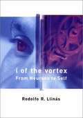
I of the Vortex
by Rodolfo R. LlinásIn I of the Vortex, Rodolfo Llinas, a founding father of modern brain science, presents an original view of the evolution and nature of mind. According to Llinas, the "mindness state" evolved to allow predictive interactions between mobile creatures and their environment. He illustrates the early evolution of mind through a primitive animal called the "sea squirt." The mobile larval form has a brainlike ganglion that receives sensory information about the surrounding environment. As an adult, the sea squirt attaches itself to a stationary object and then digests most of its own brain. This suggests that the nervous system evolved to allow active movement in animals. To move through the environment safely, a creature must anticipate the outcome of each movement on the basis of incoming sensory data. Thus the capacity to predict is most likely the ultimate brain function. One could even say that Self is the centralization of prediction.At the heart of Llinas's theory is the concept of oscillation. Many neurons possess electrical activity, manifested as oscillating variations in the minute voltages across the cell membrane. On the crests of these oscillations occur larger electrical events that are the basis for neuron-to-neuron communication. Like cicadas chirping in unison, a group of neurons oscillating in phase can resonate with a distant group of neurons. This simultaneity of neuronal activity is the neurobiological root of cognition. Although the internal state that we call the mind is guided by the senses, it is also generated by the oscillations within the brain. Thus, in a certain sense, one could say that reality is not all "out there," but is a kind of virtual reality.
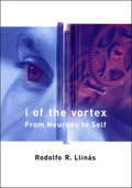
I of the Vortex: From Neurons to Self
by Rodolfo R. LlinasA highly original theory of how the mind-brain works, based on the author's study of single neuronal cells.In I of the Vortex, Rodolfo Llinas, a founding father of modern brain science, presents an original view of the evolution and nature of mind. According to Llinas, the "mindness state" evolved to allow predictive interactions between mobile creatures and their environment. He illustrates the early evolution of mind through a primitive animal called the "sea squirt." The mobile larval form has a brainlike ganglion that receives sensory information about the surrounding environment. As an adult, the sea squirt attaches itself to a stationary object and then digests most of its own brain. This suggests that the nervous system evolved to allow active movement in animals. To move through the environment safely, a creature must anticipate the outcome of each movement on the basis of incoming sensory data. Thus the capacity to predict is most likely the ultimate brain function. One could even say that Self is the centralization of prediction.At the heart of Llinas's theory is the concept of oscillation. Many neurons possess electrical activity, manifested as oscillating variations in the minute voltages across the cell membrane. On the crests of these oscillations occur larger electrical events that are the basis for neuron-to-neuron communication. Like cicadas chirping in unison, a group of neurons oscillating in phase can resonate with a distant group of neurons. This simultaneity of neuronal activity is the neurobiological root of cognition. Although the internal state that we call the mind is guided by the senses, it is also generated by the oscillations within the brain. Thus, in a certain sense, one could say that reality is not all "out there," but is a kind of virtual reality.
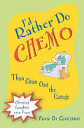
I'd Rather Do Chemo Than Clean Out the Garage: Choosing Laughter Over Tears (I'd Rather Do Chemo Than Clean Out The Garage Ser.)
by Fran Di GiacomoAn inspiring and witty memoir by a woman battling cancer—with laughter.Fran Di Giacomo made it through one case of cancer at forty—then got hit with a worse case in her fifties. Tired of the somber, weepy books she kept getting from well-meaning friends, she stumbled upon a book that made her laugh out loud—and realized that was what she&’d been missing. Laughter felt good—and that was how she wanted to feel.Inspired, she wrote this unique memoir, an unsentimental, sharply funny take on her experience—including her favorite techniques for shamelessly exploiting the chemo lifestyle. She reveals the way that indulging her sense of humor not only kept her sane during the hardest moments, but also allowed her to continue her successful career as an artist, even through thirteen hospitalizations, ten surgeries, and constant chemotherapy. Her book is terrifically entertaining—as her oncologist warns in the foreword, you should avoid reading it in the immediate postoperative period due to the risk of popping a suture. It can also help other cancer patients, or anyone dealing with hardship, to cultivate a zesty enthusiasm for life and empower themselves to keep fighting.
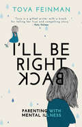
I'll Be Right Back: Parenting with Mental Illness
by Tova FeinmanAre we doomed to repeat the failings of our parents?After a whirlwind romance and marriage, Tova soon found herself pregnant. And she was determined to be the best mother she could be.But then she discovered uncomfortable secrets about her new husband. Tova was thrown out, and left to look after their daughter, Katie on her own.Wracked with memories of her unhappy childhood and suffering with postnatal depression, Tova struggled to be the mother she knew her daughter deserved.And then Katie lost her eyesight, and Tova had to fight the medical establishment and her own inner demons to secure a promising future for her daughter. Which would win, Tova's love for Katie or Tova's mental illness?
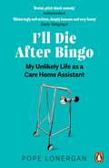
I'll Die After Bingo: My unlikely life as a care home assistant
by Pope LonerganNominated for the Chortle Comedy Book Award 2023'Blisteringly well written, deeply humane and very funny' Daily Telegraph'Enough to make you die laughing' Daily Mail'Funny and moving' Daily ExpressWhether he's initiating a coup d'état against new regulations with the residents, or forging a bond with the 98-year old who once called him a fat slut, Pope Lonergan's work is infinitely varied. This no-holds-barred account shows what life inside a care home is really like, for both residents and carers. Featuring night-time drama, incontinence pads and the uniquely dark humour of one double-amputee Alzheimer's patient, here you can learn everything you ever wanted to know (and a few things you probably really didn't) about Britain's care system.This important memoir challenges us all to think differently about the value of our elderly, and also the carers who look after them.
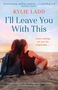
I'll Leave You With This: A must-read life-affirming story about family, loss and second chances
by Kylie Ladd'Heart-warming, uplifting, inspiring . . . a compelling read' GRAEME SIMSION'Told with warmth, compassion and humanity, I'll Leave You With This drew me in from the first page. Ladd blends depth and pace in this thought-provoking novel that is filled with hope and humour' JOANNA NELLThree years after the death of their beloved brother, all Daniel's sisters have left of him are their memories. They know he's helped others by donating his organs, but as miracles come true for the recipients, his own family are struggling with their devastating loss. When Clare suggests that they find the people Daniel's death saved, her sisters have their doubts. Will meeting them help to bring the sisters back together, or will old tensions and surfacing secrets splinter the fragile family ties forever?'Move over Jodi Picoult. Kylie Ladd creates ethical complications, then shows us both sides with uncanny insight into human nature. I inhaled this book!' FLEUR McDONALD'A page turner from the first to the last - I fell in love with all the characters and was gripped by the plot. Everyone will be reading this fantastic, heart-warming book. This is Kylie Ladd at her very best' SALLY HEPWORTH
I'll Leave You With This: A totally heartbreaking and gripping page-turner
by Kylie LaddA totally heartbreaking but uplifting story about family, loss and second chances.Three years after Daniel was killed by a senseless act of violence, all his sisters have left of him are their memories - and responsibility for Daniel's mischievous dachshund John Thomas. Daniel donated his organs, his death facilitating life-saving miracles for other families, while his own loved ones struggle to come to terms with their devastating loss, each at a crossroads of her own: It's been twelve years since film director Bridie had a hit, and while she's still invited to glitzy media events, nowadays it is as her successful actor husband's plus one. Clare and her wife Sophie have been through four rounds of IVF and while Clare remains hopeful to keep trying, it may be at the cost of her marriage . . .Conscientious Allison looks after everyone. Her younger siblings, her children and the many patients she treats in her job as Chief Obstetrician at a big teaching hospital. But who's looking out for Allison?Emma is looking for love, but isn't having much luck on dates. And there's something she can't tell anyone, whether romantic prospect or even her family, about the real reason she quietly abandoned a musical career that she loved . . . When Clare suggests that they try to make contact with the recipients of Daniel's organs to honour their brother, not everyone thinks it's a good idea. It's a heart-wrenching process, but will meeting those that their brother saved help to bring the sisters back together, or will old tensions and surfacing secrets splinter the fragile family ties forever?(P) 2022 Penguin Random House Australia
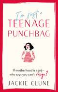
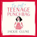
I'm Just a Teenage Punchbag: POIGNANT AND FUNNY: A NOVEL FOR A GENERATION OF WOMEN
by Jackie Clune'Obligatory reading for all parents of teenagers!' NIGELLA LAWSON'Bloody marvellous. Horribly familiar, funny, touching, sad, brutally honest...clutch this book to your stained T-shirt and never let it go.' JO BRAND'Terrific. A remarkable blend of hilarity and heartbreak with a really satisfying plot. Being childless never felt so good.' GRAHAM NORTON'Warm and witty... The competitive mothering, the hell that is other people's children, the fights and accusations of Homeland inquisition all rang deliciously true... a most entertaining read.' KATHY LETTE'Very poignant... A moving read as well as a funny one.' JANE GARVEY 'Honest, hilarious and painful' WOMAN & HOMEWarning!! This novel may lead you to make rash and life-changing decisions!**Probably don't read if you fear you may be ripe for liberation. Or if you sometimes wee when you laugh...First there was Having It All, then there was Bridget Jones' s Diary and I Don't Know How She Does It. Now there is Teenage Punchbag.I'm Just A Teenage Punchbag is a laugh-out-loud, sob-on-the bus journey through the so-called life of a middle-aged woman.Ciara is mother to three ungrateful, entitled teenagers, is married to steady Martin, a man with hairy udders, and is grieving for her mum who now lives in the wardrobe in a cardboard box from the crematorium. She finds solace in her anonymous blog, and in the daily chats she has with her mum's ashes (often the best conversations she has all day.)Despite the menopause, the invisibility of middle age and the daily self-esteem bashings, courtesy of her kids, Ciara manages to navigate the stormy waters of grief and family life - until her mask slips and she is cast out from the family bosom. She embarks on a mission to fulfil her mum's dying wishes to have her remains sprinkled from the top of the Empire State Building, finding company, distraction and - ultimately - herself in the process.If motherhood is a job - who says you can't resign?
I'm Just a Teenage Punchbag: POIGNANT AND FUNNY: A NOVEL FOR A GENERATION OF WOMEN
by Jackie CluneWarning!! This novel may lead you to make rash and life-changing decisions!**Probably don't read if you fear you may be ripe for liberation. Or if you sometimes wee when you laugh...First there was Having It All, then there was Bridget Jones' s Diary and I Don't Know How She Does It. Now there is Teenage Punchbag.I'm Just A Teenage Punchbag is a laugh-out-loud, sob-on-the bus journey through the so-called life of a middle-aged woman.Ciara is mother to three ungrateful, entitled teenagers, is married to steady Martin, a man with hairy udders, and is grieving for her mum who now lives in the wardrobe in a cardboard box from the crematorium. She finds solace in her anonymous blog, and in the daily chats she has with her mum's ashes (often the best conversations she has all day.)Despite the menopause, the invisibility of middle age and the daily self-esteem bashings, courtesy of her kids, Ciara manages to navigate the stormy waters of grief and family life - until her mask slips and she is cast out from the family bosom. She embarks on a mission to fulfil her mum's dying wishes to have her remains sprinkled from the top of the Empire State Building, finding company, distraction and - ultimately - herself in the process.If motherhood is a job - who says you can't resign?(P) 2020 Hodder & Stoughton Ltd

I'm Not in the Mood: What Every Woman Should Know About Improving Her Libido
by Judith ReichmanThe "hormone of desire," testosterone, acts on the brain to stimulate sexual interest, sensitivity to sexual stimulation, and orgasmic ability in both sexes. The amount of testosterone circulating in a woman's blood declines by about 50 percent between her twenties and fifties. The most common complaint associated with this decline is a seemingly unexplainable decrease or loss of sexual desire and enjoyment.In I'm Not in the Mood, Dr. Reichman reveals the effectiveness of small doses of testosterone in reviving sexual desire and pleasure for women. Questions answered and topics discussed include:Why and when do women make male hormones?Where do all our male hormones go?Behavior, life changes, and medical problems that affect our libidoMedications that affect our libidoWill creams, pills, lozenges, patches, or shots help?When you should see a psychiatrist, psychologist, or sex therapistHow to discuss libido issues with your doctorHow to reach your biologic sexual potential The "hormone of desire," testosterone, acts on the brain to stimulate sexual interest, sensitivity to sexual stimulation, and orgasmic ability in both sexes. The amount of testosterone circulating in a woman's blood declines by about 50 percent between her twenties and fifties. The most common complaint associated with this decline is a seemingly unexplainable decrease or loss of sexual desire and enjoyment.In I'm Not in the Mood, Dr. Reichman reveals the effectiveness of small doses of testosterone in reviving sexual desire and pleasure for women. Questions answered and topics discussed include:Why and when do women make male hormones? Where do all our male hormones go? Behavior, life changes, and medical problems that affect our libido Medications that affect our libido Will creams, pills, lozenges, patches, or shots help? When you should see a psychiatrist, psychologist, or sex therapist How to discuss libido issues with your doctor How to reach your biologic sexual potential
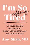
I'm So Effing Tired: A Proven Plan to Beat Burnout, Boost Your Energy, and Reclaim Your Life
by Dr. Amy ShahA guide to conquering burnout and increasing your energy from a leading medical doctor and nutrition expertEXHAUSTION DOESN&’T HAVE TO BE YOUR NEW NORMALDoes it feel like your life is too busy, your days are too short, and you're feeling overworked, overstressed, and overtired? Chances are you&’ve asked your doctor for help, only to be told that it&’s because of your age, or your workload, or, worse, that it&’s just &“normal.&”If so, you&’re not alone. Women of all ages are suffering from an epidemic of fatigue and burnout. But exhaustion doesn&’t have to be your new normal. Inspired by her personal wellness journey, integrative medical doctor Amy Shah has created this program so that you can regain your energy and reclaim your life.The key is tapping into the powerful energy trifecta: the complex relationship between your gut, your immune system, and your hormones. Drawing on the latest science and her work helping thousands of clients, Dr. Shah explains how to transform your life by changing:What You Eat: Increase your vegetable intake and sip Dr. Shah&’s hormone-balance tea to tamp down inflammation and heal your gut, without giving up your wine and chocolate!When You Eat: Changing when you eat and practicing intermittent fasting—the right way—will help you feel energized all day long.How you manage stress: Simple, stress-busting exercises and herbs like Ashwagandha and Amla berry help calm the Adrenal system and ease anxiety.In just two weeks, you&’ll feel your energy surge. In three months, you&’ll feel like a whole new person. It&’s time to regain the energy you&’ve lost, so you can get back to the life you want to live.
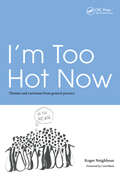
I'm Too Hot Now: Themes and Variations from General Practice
by Roger NeighbourTomorrow's general practitioners will inhabit a world of ever greater sophistication and complexity. New skills will be demanded to manage the changing expectations of patients and governments. In an age of information overload, new patterns of creative, intelligent working will need to develop. This book provides a framework, illustrated by practical examples, for such a career path to develop and be supported. It examines a number of innovative schemes which highlight varied ways forward, both for training and personal enrichment. It addresses not only the need of today's young doctors, but also the question of how to equip all general practitioners for the challenges of the future.
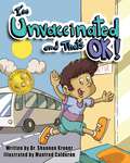
I'm Unvaccinated and That's OK!
by Dr. Shannon KronerI'm Unvaccinated and That's OK! is the story of an unvaccinated child named Nicholas Novaks, who shares the many reasons why his parents have chosen not to vaccinate him. Nicholas explains his parents&’ personal concerns about vaccine injury, the importance of finding a doctor they can trust and openly speak with, the research they did before making this decision, and what life is like for an unvaccinated child who has an older, vaccine-injured sibling. Inspired by the personal stories of vaccine-injured children, which have been shared with Dr. Shannon Kroner over many years of working with special needs families, Dr. Kroner aims to raise awareness of the importance of vaccine choice and the necessity of doing the research before making an important decision such as vaccination. Join Nicholas as he shares what it means to be an unvaccinated child in today&’s world and why one's personal choice regarding vaccination must always be respected.I&’m Unvaccinated and That&’s OK! is published through ICAN PRESS, an imprint of Skyhorse Publishing. ICAN (Informed Consent Action Network) is a nonprofit organization investigating the safety of medical procedures, pharmaceutical drugs, and vaccines while advocating for people&’s right to informed consent.
I've Seen the End of You: A Neurosurgeon's Look at Faith, Doubt, and the Things We Think We Know
by W. Lee WarrenThis gripping inspirational memoir grapples with the tension between faith and science—and between death and hope—as a seasoned neurosurgeon faces insurmountable odds and grief both in the office and at home.&“Beautiful, haunting, powerful . . .&”—Daniel G. Amen, MDDr. W. Lee Warren, a practicing brain surgeon, assumed he knew most outcomes for people with glioblastoma, head injuries, and other health-care problems. Yet even as he tried to give patients hope, his own heart would sink as he realized, I've seen the end of you.But it became far more personal when the acclaimed doctor experienced an unimaginable family tragedy. That's when he reached the end of himself.Page-turning medical stories serve as the backdrop for a raw, honest look at how we can remain on solid ground when everything goes wrong and how we can find light in the darkest hours of life. I've Seen the End of You is the rare book that offers tender empathy and tangible hope for those who are suffering. No matter what you're facing, this doesn't have to be the end. Even when nothing seems to makes sense, God can transform your circumstances and your life. And he can offer a new beginning.
I.C.U. Chest Radiology
by Harold MoskowitzA practical, highly useful guide to the principles of I.C.U. chest radiology, complete with case studies and radiographs on CDFor critically ill patients in a hospital's I.C.U., a portable chest radiograph is the most helpful, and most commonly used, x-ray examination. Cardiopulmonary complications and the malposition of lines, tubes, and catheters are often initially detected on a portable chest film. It is essential for hospital personnel to know how to approach and read these films, and yet little attention has been paid to teaching the accurate evaluation of this crucial diagnostic tool.The first book in more than a decade to specifically address this topic, I.C.U. Chest Radiology is an authoritative and concise guide to interpreting portable chest film; identifying and correcting any abnormal positions in the various devices inserted into the vascular and respiratory systems; and diagnosing abnormalities of the cardiopulmonary system. Radiology expert Dr. Harold Moskowitz outlines his approach and philosophy toward x-ray interpretation of the I.C.U. patient--one that can be used daily and in any I.C.U. setting.Divided into ten straightforward chapters, the book begins with a discussion of the physics necessary to obtain a proper film and moves on to the more clinical problems encountered each day in the I.C.U.--such as airspace disease, barotrauma, pneumonia, congestive failure, and malalignment of tubes and lines. Throughout, Moskowitz points out specific findings that can often make a difference in a patient's management. Supporting these detailed chapters is a CD featuring real-life case studies and radiographic images that simulate common problems in the I.C.U. This is a unique way for readers to prepare to handle the all-too-common scenario: the 2:00 a.m. call from an I.C.U. nurse that a patient has "crashed" and needs attention. Using knowledge gleaned from the chapters, the reader is encouraged to study the radiograph in each case, identify the various problems, determine the clinical condition that caused deterioration in the patient, and plan a course of action. Readers can test themselves with the cases and then listen as Moskowitz discusses the pertinent findings on the film.I.C.U. Chest Radiology is essential reading for those who work in or are associated with I.C.U.s--radiologists, intensivists, hospitalists, emergency room physicians, residents, medical students, physician assistants, respiratory therapists, and nurses. It will also be a valuable guide for personnel who work in step down units and emergency rooms.