- Table View
- List View
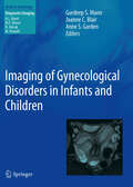
Imaging of Gynecological Disorders in Infants and Children
by Gurdeep S. Mann Anne S. Garden Joanne C. BlairThis textbook provides a comprehensive review of gynecological imaging in infancy, childhood, and adolescence. Experts from the disciplines of pediatric radiology, gynecology, surgery, and endocrinology have come together to produce a textbook that, while written primarily from the perspective of the radiologist, will be of value to all professionals involved in the management of these patients. The normal development of the female reproductive tract is described in detail through embryological development, normal childhood appearances, and puberty. Congenital abnormalities are addressed in chapters reviewing structural abnormalities of the reproductive tract and disorders of sex development. A symptoms-based approach is followed in chapters devoted to the assessment of the patient with gynecological pain and disorders of menstruation. Disorders of the breast and the imaging of patients with gynecological neoplasia are considered in dedicated chapters.
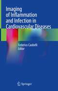
Imaging of Inflammation and Infection in Cardiovascular Diseases
by Federico CaobelliThis book addresses the most relevant imaging techniques in order to detect inflammation and infection in the most common cardiovascular diseases. The book presents data concerning molecular imaging (SPECT and PET) and cardiac magnetic resonance imaging (MRI) for each disease, with a special focus on the emerging role of hybrid PET/MR imaging. Different non-ischemic and ischemic diseases as well as cardiac infections are addressed in detail; these include: Cardiac sarcoidosis, Cardiac amyloidosis, the vulnerable plaque, Post-infarction inflammatory alterations, Pericarditis, Myocarditis, Cardiac devices infections, and Endocarditis. The book also provides a comprehensive discussion on new targets and new tracers, to date mostly investigated at a pre-clinical stage, thus constituting an excellent basis for translational imaging. Imaging of Inflammation and Infection in Cardiovascular Diseases will be of interest not only for experts in clinical imaging, but also pre-clinical scientists and will be invaluable for both nuclear medicine physicians and radiologists.
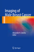
Imaging of Male Breast Cancer
by Alexander N. SenchaThis book is devoted to the diagnosis and treatment of male breast pathology. It provides a comprehensive overview of current strategies for the diagnosis of breast malignancies in men, with a focus on imaging modalities. Ultrasound of male breast abnormalities is discussed in particular depth, but the roles of other imaging techniques, genetic tests, and interventional diagnostic modalities are also carefully covered. Special attention is paid to differential diagnosis of malignant and benign lesions, and the most important benign breast diseases are described and illustrated with high-quality images. A special chapter analyzes treatment strategy in men with breast malignancies and principles of follow-up after breast surgery. Individual chapters are also devoted to the diagnosis of recurrent cancer and cancer metastases. This up-to-date and richly illustrated book will interest and assist specialists in ultrasound diagnostics, radiologists, oncologists, and surgeons.
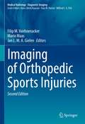
Imaging of Orthopedic Sports Injuries (Medical Radiology)
by Filip M. Vanhoenacker Mario Maas Jan L. M. A. GielenThis volume provides an updated review of imaging abnormalities in orthopedic sports injuries. The first part of the book contains background information on relevant basic science and general imaging principles in sports traumatology. The second part comprises a topographic discussion of sports injuries. Each chapter highlights the merits of different imaging techniques, focused on a specific clinical problem. In the third part, natural history, monitoring and follow-up imaging are discussed.
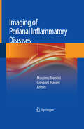
Imaging of Perianal Inflammatory Diseases
by Massimo Tonolini Giovanni MaconiThis practical volume is intended for all radiologists, gastroenterologists, and surgeons who are responsible for, or interested in, the diagnosis and care of patients with diseases of the anal and perianal region. After an introductory section focusing on surgical, MRI, and US anatomy, up-to-date clinical and therapeutic information is provided on the full range of perianal inflammatory conditions, with special attention to Crohn's disease, ulcerative colitis, and sexually transmitted diseases. Physical and surgical examination is carefully discussed, with consideration of limitations, results, clinical needs, and the questions likely to be posed of imaging. The subsequent three sections provide detailed information on the different imaging modalities: transanal and transperineal ultrasound, contrast-enhanced MRI, and CT. Techniques, diagnostic accuracy, common and unusual disease patterns, artifacts, and pitfalls are clearly presented.
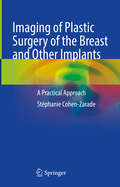
Imaging of Plastic Surgery of the Breast and Other Implants: A Practical Approach
by Stéphanie Cohen-ZaradeIn this comprehensive book, the author describes in a didactic and concise manner the sometimes misunderstood post-surgical aesthetic radiological aspects that can be misleading.It is intended for radiologists, whether or not specialized in breast imaging, cosmetic surgeons, gynecologists, and other specialists who are interested in aesthetics, in full development.In this book, all techniques of breast augmentation-reduction, butt augmentation, calf augmentation, pectus excavatum correction, are thoroughly described: surgical procedures, post-surgical radiological aspects or complications.For each technique, the imaging protocol to be performed is detailed.A rich and specific collection of radiological pictures facilitates understanding.This is an essential book that can be used for training purposes.
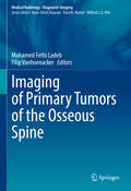
Imaging of Primary Tumors of the Osseous Spine (Medical Radiology)
by Mohamed Fethi Ladeb Filip VanhoenackerThis comprehensive book provides a detailed description of imaging techniques and findings in the detection, characterization, local staging and post-treatment tumors of the osseous spine. In the first part of the book, the epidemiology, classification, pathology, genetics and molecular biology are discussed. The second part gives an overview of the basic semiology, imaging modalities including CT, MRI, PET and PET-CT, and staging. The third part of the book discusses the imaging of primary bone tumors of the spine and its mimics. In the fourth part, the treatment of tumors and tumor-like conditions of the spine is reviewed. The final part of the book summarizes post-treatment evaluation and assessment. Thus, the book will be a useful reference for general and musculoskeletal radiologists and residents, orthopaedic surgeons, oncologists and histopathologists.
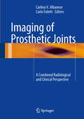
Imaging of Prosthetic Joints
by Carlina V. Albanese Carlo FalettiAn increasing percentage of the population has at least one prosthetic joint. Imaging is required for both initial assessment and routine follow-up of the implant, and this book is intended as an informative and up-to-date guide to the subject. After an introductory section covering a range of background topics, the level of information offered by different imaging techniques in presurgical planning and postimplantation assessment is analyzed. The application of imaging to different joints is then carefully explored in chapters devoted to the foot and ankle, hip, knee, shoulder, and elbow, wrist and hand. In addition, two innovative chapters focus on periprosthetic DXA as the gold standard in monitoring implant survival and on the role of drug therapy in helping to ensure the durability of the prosthesis. A central feature of the book is its combination of the clinical and radiological perspectives; it will be of value to radiologists and orthopedic specialists, as well as residents in these disciplines.
Imaging of Small Bowel, Colon and Rectum
by Pasquale Paolantonio Clarisse DromainThe new series A-Z Notes in Radiological Practice and Reporting provides practical guides for residents and general radiologists, organized alphabetically, primarily according to disease or condition. All booklets are designed so as to cover a large spectrum of topics referring to different anatomical regions of interest. Entries typically include a short description of pathological and clinical characteristics, guidance on selection of the most appropriate imaging technique, a schematic review of potential diagnostic clues, and useful tips and tricks. The present booklet, enriched by illustrations and schemes, is devoted to gastrointestinal imaging. Major topics in CT and MR imaging of the small bowel, colon, rectum, and anus are treated concisely in alphabetical order. For each topic a brief review of clinical features and pathology is presented, followed by a short description of imaging technique and an accurate review of imaging findings and signs which are useful in the differential diagnosis of gastrointestinal disease.
Imaging of Soft Tissue Tumors
by Filip M. Vanhoenacker Paul M. Parizel Jan L. GielenThis richly illustrated book, in an extensively revised new edition, provides a comprehensive survey of the role of medical imaging studies in the detection, staging, grading, tissue characterization, and post-treatment follow-up of soft tissue tumors. The indications for and relative merits of various imaging modalities are fully described, with particular emphasis on the role of advanced MRI techniques that can improve diagnostic accuracy and evaluation of treatment response. The most recent version of the WHO Classification of Soft Tissue Tumors is introduced, and individual chapters are devoted to imaging of each of the tumor groups in that classification as well as other soft tissue masses. Numerous new illustrations of both common and rare tumors are included, providing a rich pictorial database of soft tissue masses. In addition, imaging findings are correlated with clinical, epidemiologic, and histologic data. Imaging of Soft Tissue Tumors will be of value in daily practice not only for radiologists but also for orthopedic surgeons, oncologists, and pathologists.
Imaging of Spinal Infection (Medical Radiology)
by Wilfred C. G. Peh Mohamed Fethi LadebThis book examines all aspects of the imaging of spinal infection. The diagnosis of spinal infection has been a challenge for many years. In addition to clinical and laboratory findings and histopathological examination, imaging has a major role in aiding and expediting the correct diagnosis. This book comprehensively addresses how imaging can help in localizing the site and specifying the extent of a variety of spinal infections.After introductory chapters on the epidemiology and pathophysiology of spinal infection, the different imaging techniques are discussed in detail. The bulk of the book addresses different specific spinal infections caused by various pathogens. These comprise chapters on hematogeneous pyogenic spondylodiscitis, iatrogenic spinal infection, pyogenic epidural abscess, spinal brucellosis, salmonella spondylodiscitis, spinal tuberculosis, spinal hydatidosis, and fungal spondylodiscitis. The last chapter describes diagnostic algorithm of spinal infection.The book is written by Tunisian, Asian and European experts and will be a valuable resource for all medical practitioners who deal with spinal infection, including radiologists, rheumatologists and orthopedic surgeons.
Imaging of Synovial Tumors and Tumor-like Conditions (Medical Radiology)
by Filip M. Vanhoenacker Mohamed Fethi LadebThis vastly illustrated book offers a comprehensive survey of the role of imaging in the diagnosis of synovial tumors and tumor-like conditions. It gives an overview of the anatomy and histology of the synovium and discusses the classification, pathology, genetics and molecular biology of synovial tumors. The book presents the benefits of various imaging modalities in the diagnosis of these masses, which is important to provide appropriate treatment.Imaging of Synovial Tumors and Tumor-like Conditions will be a valuable resource not only for radiologists but also for orthopedic surgeons, oncologists, radiotherapists, and pathologists.
Imaging of Trabecular Microfracture and Bone Marrow Edema and Hemorrhage
by Yong-Whee BahkThis excellently illustrated monograph summarizes the updated fresh information on the theoretical, practical, and rapidly extended facets of gamma correction 99mTc-hydroxymethylene diphosphonate pinhole bone scan (99mTc-GCPBS) to MRI, CR, and MDCT. Basically, 99mTc-GCPBS is able to precisely visualize and quantitate callused trabecular microfracture (CTMF) which is as little as 200 μm in size. The extended gamma correction images can very neatly demonstrate CTMF on MRI, conventional radiography (CR), and multidetector computed tomography (MDCT). Whenever appropriate, histological correlation is provided in conjunction with fine gamma corrected images. In this setting, ACDSee 10 gamma correction MRI and CR have been found to offer a highly useful option that deserves wider clinical interest. In practice, gamma correction MRI, CR, and MDCT can distinctly visualize CTMF so that CTMF can be precisely measured simply using an optic lens. By comparison, the naïve MRI, for example, shows CTMF which measures as big as 2 mm in size. Furthermore, 99mTc-GCPBS can now differentiate bone marrow edema from hemorrhage using the visuospatial mathematic method, which includes the ImageJ of the NIH.
Imaging of Tuberculosis (Medical Radiology)
by Wilfred C. G. Peh Mohamed Fethi LadebThis book provides an extensive overview of the role of imaging in the detection, diagnosis, management, and follow-up of tuberculosis. Chapters cover the disease's epidemiology and pathophysiology, microbiological diagnosis and pathology relevant to radiologists, and the distinct aspects of imaging tuberculosis at various locations and body systems. This book discusses recent advances in imaging pertaining to tuberculosis, and addresses the approach to patients with tuberculosis and HIV co-infection. The final chapter offers an algorithmic approach to the diagnosis and management steps of tuberculosis. Imaging of Tuberculosis is an updated and comprehensive reference source that covers imaging of tuberculosis in a structured fashion and is valuable for radiologists.
Imaging of Ulcerative Colitis
by Massimo TonoliniDuring the past decade, the medical and surgical treatment of ulcerative colitis has undergone dramatic advances, including the widespread use of immunomodulators, biological drugs, and restorative proctocolectomy. In order to correctly balance the risks and benefits of medical therapies and surgical procedures, there is a need for improved diagnosis of colonic disease, acute complications, extraintestinal manifestations, and early and delayed postoperative complications. Cross-sectional imaging techniques are therefore playing an increasing role in the assessment of ulcerative colitis and provide an essential complement to clinical data and endoscopy. This practical, illustrated volume on the role of cross-sectional imaging is aimed at radiologists, gastroenterologists, and surgeons who are engaged or interested in the diagnosis and care of patients with inflammatory bowel disease, particularly ulcerative colitis. After an overview of diagnostic imaging techniques, state-of-the-art assessment of colorectal inflammatory disease with CT colonography using water enema is discussed, followed by description of the plain radiographic and CT findings in patients with acute exacerbations and surgical complications. Subsequent chapters review the diagnostic findings and role of cross-sectional imaging in the assessment of sclerosing cholangitis (with emphasis on MR cholangiopancreatography), vascular complications (particularly portal and mesenteric thrombosis), associated neoplasms, such as colorectal cancer and abdominal desmoids, and perianal inflammatory disease. Normal postoperative appearances and early and delayed complications in patients treated with proctocolectomy and ileal pouch-anal anastomosis are also comprehensively reviewed.
Imaging of Urinary Tract Diverticula
by Vladimir M. BuilovThis monograph covers all aspects of the radiologic diagnosis of urinary tract diverticula, including calyceal, ureteral, bladder and urethral diverticula. Characteristic and subtle diagnostic features are identified with the aid of numerous high-quality ultrasound, X-ray and magnetic resonance images, the vast majority of which are drawn from the author's personal clinical practice. In addition, issues relating to terminology, classification, statistics, etiology, pathogenesis, clinical presentation and differential diagnosis are discussed. The text is complemented by two helpful appendices that document the latest recommendations of the European Society of Urogenital Radiology regarding use of contrast media and the European Medicines Agency on minimizing the risk of nephrogenic systemic fibrosis when using gadolinium-containing contrast agents. This book will be of value for specialists in radiology and urology and also trainees and medical students.
Imaging of Vertebral Trauma
by Richard H. DaffnerThe imaging methods used to evaluate patients with suspected vertebral injuries have undergone radical changes in the past decade, the most significant being the ascendancy of computed tomography (CT) to become the primary investigative modality. Issues such as high radiation dose associated with CT studies and health care reform and cost containment also have a significant impact on clinical decision-making. This new edition of the classic landmark text for vertebral trauma imaging provides an in-depth discussion on the indications and methods of imaging the spine based on currently available clinical evidence. Every chapter has been extensively revised and the illustrations represent state-of-the-art imaging. A completely new chapter on pediatric injuries has been added. Imaging of Vertebral Trauma, third edition, is an invaluable and essential tool in the assessment of any patient with suspected vertebral or spinal cord injury.

Imaging of Vertebral Trauma (3rd Edition)
by Richard H. DaffnerThe imaging methods used to evaluate patients with suspected vertebral injuries have undergone radical changes in the past decade, the most significant being the ascendancy of computed tomography (CT) to become the primary investigative modality. Issues such as high radiation dose associated with CT studies and health care reform and cost containment also have a significant impact on clinical decision-making. This new edition of the classic landmark text for vertebral trauma imaging provides an in-depth discussion on the indications and methods of imaging the spine based on currently available clinical evidence. Every chapter has been extensively revised and the illustrations represent state-of-the-art imaging. A completely new chapter on pediatric injuries has been added. Imaging of Vertebral Trauma, third edition, is an invaluable and essential tool in the assessment of any patient with suspected vertebral or spinal cord injury.
Imaging of the Aorta: A Practical Guide for Radiologists
by Iacopo Carbone Davide Farina Pier Giorgio Nardis Davide BelliniThis book provides younger radiologists with a practical guide to aortic imaging, from acquisition to reporting. It discusses the pros and cons of the different available techniques and offers tips for the optimization of acquisition protocols and it covers the most frequent aortic pathologies, from a diagnostic and interventional point of view. Report building is discussed starting from standardized terminology; for each pathology, the book lists, in a simple and schematic way, the key points that must be included in the report of a cross-sectional study. Checklists used by clinicians and radiologists in routine practice are also included, and iconography is enriched by numerous stand-alone images. The Authors have long-standing experience in cardiovascular imaging and interventional procedures and with this publication they are providing a useful guide in everyday clinical practice.
Imaging of the Cardiovascular System, Thorax, and Abdomen
by Luca SabaMagnetic resonance imaging (MRI) is a technique used in biomedical imaging and radiology to visualize internal structures of the body. Because MRI provides excellent contrast between different soft tissues, the technique is especially useful for diagnostic imaging of the brain, muscles, and heart. <P><P>In the past 20 years, MRI technology has improved significantly with the introduction of systems up to 7 Tesla (7 T) and with the development of numerous post-processing algorithms such as diffusion tensor imaging (DTI), functional MRI (fMRI), and spectroscopic imaging. From these developments, the diagnostic potentialities of MRI have improved impressively with an exceptional spatial resolution and the possibility of analyzing the morphology and function of several kinds of pathology. <P><P>Given these exciting developments, the Magnetic Resonance Imaging Handbook: Imaging of the Cardiovascular System, Thorax, and Abdomen is a timely addition to the growing body of literature in the field. Offering comprehensive coverage of cutting-edge imaging modalities, this book: <Li>Discusses MRI of the heart, blood vessels, lungs, breasts, diaphragm, liver, gallbladder, spleen, pancreas, adrenal glands, and gastrointestinal tract <Li>Explains how MRI can be used in vascular, posttraumatic, postsurgical, and computer-aided diagnostic (CAD) applications <Li>Highlights each organ’s anatomy and pathological processes with high-quality images <Li>Examines the protocols and potentialities of advanced MRI scanners such as 7 T systems <Li>Includes extensive references at the end of each chapter to enhance further study <P><P>Thus, the Magnetic Resonance Imaging Handbook: Imaging of the Cardiovascular System, Thorax, and Abdomen provides radiologists and imaging specialists with a valuable, state-of-the-art reference on MRI.
Imaging of the Cervical Spine in Children
by Leonard E. SwischukDr. Leonard Swischuk has revised his outstanding work on imaging the cervical spine in children. He draws upon his extensive experience to provide practitioners with an insightful approach to pediatric cervical spine injuries. The text covers developmental anatomy, normal variants, congenital anomalies, abnormalities of the dens, trauma, and miscellaneous abnormalities of the cervical spine. The book has several strengths that appeal to radiology residents, such as its succinct overview of the topic and helpful reference lists that guide readers to additional resources. Dr. Swischuk illustrates conditions he discusses with excellent plain film examples that help residents identify cases they are likely to encounter during board exams and in practice. Accompanying CT and MR images clarify and qualify the findings. Dr. Swischuk's direct writing style makes the complex content highly accessible, providing imaging residents with an invaluable introduction to pediatric cervical spine radiology.
Imaging of the Foot and Ankle: Techniques and Applications (Medical Radiology)
by Steven James Mark Davies Rajesh BotchuUp-to-date and comprehensive textbook on imaging of the foot and ankle. In the first part, the various techniques and procedures are discussed in detail. Individual chapters are devoted to: radiography, arthrography and tenography, computed tomography and CT arthrography, magnetic resonance imaging and MR arthrography, ultrasonography, and intra-articular injections. The second part documents the application of these techniques to diverse clinical problems and diseases, including: congenital and developmental disorders, trauma, tendon and ligament pathology, compressive neuropathies, infection, and the diabetic foot. Each chapter is written by an acknowledged expert, and a wealth of illustrative material is included.
Imaging of the Hand and Wrist
by A. Mark Davies Andrew J. Grainger Steven J. JamesIn the past, radiographs of the hand have been described as the "skeleton's calling card", showing manifestations of many different diseases. As hand and wrist imaging has become increasingly sophisticated, this observation has become more true than ever. This is a comprehensive, up-to-date textbook on imaging of the hand and wrist. In the first part of the book, the various imaging techniques are discussed in detail. Individual chapters are devoted to radiography, ultrasound, CT, MRI and nuclear medicine. The second part of the book gives an authoritative review of the various pathologies that may be encountered in the hand and wrist, encompassing congenital and developmental abnormalities, trauma, and the full range of localized and systemic disorders. Each chapter is written by an acknowledged expert in the field, and a wealth of illustrative material is included. This book will be of great value to musculoskeletal and general radiologists, orthopaedic surgeons and rheumatologists.
Imaging of the Hip & Bony Pelvis: Techniques and Applications (Medical Radiology)
by Mark Davies Rajesh Botchu Karthikeyan P. IyengarThis volume provides an up-to-date and comprehensive review of imaging of the hip. In the first part of the book, the various techniques employed when imaging the hip are discussed in detail. Individual chapters are devoted to radiography, computed tomography, ultrasound and MRI. The second part then documents the application of these techniques to the diverse application and diseases encountered in the hip. Among the many topics addressed are congenital and developmental abnormalities, trauma, metabolic bone disease, infection, arthritis and tumours. Each chapter is written by an acknowledged expert in the field and a wealth of illustrative material is included. This book will be of great value to radiologists, orthopedic surgeons and other clinicians with an interest in the hip pathology.
Imaging of the Knee: Techniques and Applications (Medical Radiology)
by Steven James Mark Davies Rajesh BotchuAn up-to-date and comprehensive review of the discipline of imaging of the knee. The first part discusses the various techniques employed when imaging the knee. Individual chapters are devoted to radiography, arthrography, computed tomography and CT arthrography, magnetic resonance imaging and MR arthrography, and ultrasonography. The second part then documents the application of these techniques to the diverse clinical problems and diseases encountered in the knee. Among the many topics addressed are: congenital and developmental abnormalities, trauma, meniscal pathology, and others. Each chapter is written by an acknowledged expert in the field.