- Table View
- List View
Imaging of Urinary Tract Diverticula
by Vladimir M. BuilovThis monograph covers all aspects of the radiologic diagnosis of urinary tract diverticula, including calyceal, ureteral, bladder and urethral diverticula. Characteristic and subtle diagnostic features are identified with the aid of numerous high-quality ultrasound, X-ray and magnetic resonance images, the vast majority of which are drawn from the author's personal clinical practice. In addition, issues relating to terminology, classification, statistics, etiology, pathogenesis, clinical presentation and differential diagnosis are discussed. The text is complemented by two helpful appendices that document the latest recommendations of the European Society of Urogenital Radiology regarding use of contrast media and the European Medicines Agency on minimizing the risk of nephrogenic systemic fibrosis when using gadolinium-containing contrast agents. This book will be of value for specialists in radiology and urology and also trainees and medical students.
Imaging of Vertebral Trauma
by Richard H. DaffnerThe imaging methods used to evaluate patients with suspected vertebral injuries have undergone radical changes in the past decade, the most significant being the ascendancy of computed tomography (CT) to become the primary investigative modality. Issues such as high radiation dose associated with CT studies and health care reform and cost containment also have a significant impact on clinical decision-making. This new edition of the classic landmark text for vertebral trauma imaging provides an in-depth discussion on the indications and methods of imaging the spine based on currently available clinical evidence. Every chapter has been extensively revised and the illustrations represent state-of-the-art imaging. A completely new chapter on pediatric injuries has been added. Imaging of Vertebral Trauma, third edition, is an invaluable and essential tool in the assessment of any patient with suspected vertebral or spinal cord injury.

Imaging of Vertebral Trauma (3rd Edition)
by Richard H. DaffnerThe imaging methods used to evaluate patients with suspected vertebral injuries have undergone radical changes in the past decade, the most significant being the ascendancy of computed tomography (CT) to become the primary investigative modality. Issues such as high radiation dose associated with CT studies and health care reform and cost containment also have a significant impact on clinical decision-making. This new edition of the classic landmark text for vertebral trauma imaging provides an in-depth discussion on the indications and methods of imaging the spine based on currently available clinical evidence. Every chapter has been extensively revised and the illustrations represent state-of-the-art imaging. A completely new chapter on pediatric injuries has been added. Imaging of Vertebral Trauma, third edition, is an invaluable and essential tool in the assessment of any patient with suspected vertebral or spinal cord injury.
Imaging of the Aorta: A Practical Guide for Radiologists
by Iacopo Carbone Davide Farina Pier Giorgio Nardis Davide BelliniThis book provides younger radiologists with a practical guide to aortic imaging, from acquisition to reporting. It discusses the pros and cons of the different available techniques and offers tips for the optimization of acquisition protocols and it covers the most frequent aortic pathologies, from a diagnostic and interventional point of view. Report building is discussed starting from standardized terminology; for each pathology, the book lists, in a simple and schematic way, the key points that must be included in the report of a cross-sectional study. Checklists used by clinicians and radiologists in routine practice are also included, and iconography is enriched by numerous stand-alone images. The Authors have long-standing experience in cardiovascular imaging and interventional procedures and with this publication they are providing a useful guide in everyday clinical practice.
Imaging of the Cardiovascular System, Thorax, and Abdomen
by Luca SabaMagnetic resonance imaging (MRI) is a technique used in biomedical imaging and radiology to visualize internal structures of the body. Because MRI provides excellent contrast between different soft tissues, the technique is especially useful for diagnostic imaging of the brain, muscles, and heart. <P><P>In the past 20 years, MRI technology has improved significantly with the introduction of systems up to 7 Tesla (7 T) and with the development of numerous post-processing algorithms such as diffusion tensor imaging (DTI), functional MRI (fMRI), and spectroscopic imaging. From these developments, the diagnostic potentialities of MRI have improved impressively with an exceptional spatial resolution and the possibility of analyzing the morphology and function of several kinds of pathology. <P><P>Given these exciting developments, the Magnetic Resonance Imaging Handbook: Imaging of the Cardiovascular System, Thorax, and Abdomen is a timely addition to the growing body of literature in the field. Offering comprehensive coverage of cutting-edge imaging modalities, this book: <Li>Discusses MRI of the heart, blood vessels, lungs, breasts, diaphragm, liver, gallbladder, spleen, pancreas, adrenal glands, and gastrointestinal tract <Li>Explains how MRI can be used in vascular, posttraumatic, postsurgical, and computer-aided diagnostic (CAD) applications <Li>Highlights each organ’s anatomy and pathological processes with high-quality images <Li>Examines the protocols and potentialities of advanced MRI scanners such as 7 T systems <Li>Includes extensive references at the end of each chapter to enhance further study <P><P>Thus, the Magnetic Resonance Imaging Handbook: Imaging of the Cardiovascular System, Thorax, and Abdomen provides radiologists and imaging specialists with a valuable, state-of-the-art reference on MRI.
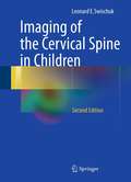
Imaging of the Cervical Spine in Children
by Leonard E. SwischukDr. Leonard Swischuk has revised his outstanding work on imaging the cervical spine in children. He draws upon his extensive experience to provide practitioners with an insightful approach to pediatric cervical spine injuries. The text covers developmental anatomy, normal variants, congenital anomalies, abnormalities of the dens, trauma, and miscellaneous abnormalities of the cervical spine. The book has several strengths that appeal to radiology residents, such as its succinct overview of the topic and helpful reference lists that guide readers to additional resources. Dr. Swischuk illustrates conditions he discusses with excellent plain film examples that help residents identify cases they are likely to encounter during board exams and in practice. Accompanying CT and MR images clarify and qualify the findings. Dr. Swischuk's direct writing style makes the complex content highly accessible, providing imaging residents with an invaluable introduction to pediatric cervical spine radiology.
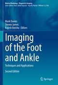
Imaging of the Foot and Ankle: Techniques and Applications (Medical Radiology)
by Steven James Mark Davies Rajesh BotchuUp-to-date and comprehensive textbook on imaging of the foot and ankle. In the first part, the various techniques and procedures are discussed in detail. Individual chapters are devoted to: radiography, arthrography and tenography, computed tomography and CT arthrography, magnetic resonance imaging and MR arthrography, ultrasonography, and intra-articular injections. The second part documents the application of these techniques to diverse clinical problems and diseases, including: congenital and developmental disorders, trauma, tendon and ligament pathology, compressive neuropathies, infection, and the diabetic foot. Each chapter is written by an acknowledged expert, and a wealth of illustrative material is included.
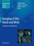
Imaging of the Hand and Wrist
by A. Mark Davies Andrew J. Grainger Steven J. JamesIn the past, radiographs of the hand have been described as the "skeleton's calling card", showing manifestations of many different diseases. As hand and wrist imaging has become increasingly sophisticated, this observation has become more true than ever. This is a comprehensive, up-to-date textbook on imaging of the hand and wrist. In the first part of the book, the various imaging techniques are discussed in detail. Individual chapters are devoted to radiography, ultrasound, CT, MRI and nuclear medicine. The second part of the book gives an authoritative review of the various pathologies that may be encountered in the hand and wrist, encompassing congenital and developmental abnormalities, trauma, and the full range of localized and systemic disorders. Each chapter is written by an acknowledged expert in the field, and a wealth of illustrative material is included. This book will be of great value to musculoskeletal and general radiologists, orthopaedic surgeons and rheumatologists.
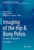
Imaging of the Hip & Bony Pelvis: Techniques and Applications (Medical Radiology)
by Mark Davies Rajesh Botchu Karthikeyan P. IyengarThis volume provides an up-to-date and comprehensive review of imaging of the hip. In the first part of the book, the various techniques employed when imaging the hip are discussed in detail. Individual chapters are devoted to radiography, computed tomography, ultrasound and MRI. The second part then documents the application of these techniques to the diverse application and diseases encountered in the hip. Among the many topics addressed are congenital and developmental abnormalities, trauma, metabolic bone disease, infection, arthritis and tumours. Each chapter is written by an acknowledged expert in the field and a wealth of illustrative material is included. This book will be of great value to radiologists, orthopedic surgeons and other clinicians with an interest in the hip pathology.
Imaging of the Knee: Techniques and Applications (Medical Radiology)
by Steven James Mark Davies Rajesh BotchuAn up-to-date and comprehensive review of the discipline of imaging of the knee. The first part discusses the various techniques employed when imaging the knee. Individual chapters are devoted to radiography, arthrography, computed tomography and CT arthrography, magnetic resonance imaging and MR arthrography, and ultrasonography. The second part then documents the application of these techniques to the diverse clinical problems and diseases encountered in the knee. Among the many topics addressed are: congenital and developmental abnormalities, trauma, meniscal pathology, and others. Each chapter is written by an acknowledged expert in the field.
Imaging of the Liver and Intra-hepatic Biliary Tract: Volume 1: Imaging Techniques and Non-tumoral Pathologies (Medical Radiology)
by Emilio QuaiaThis is the first of two volumes that together provide a comprehensive analysis of the embryology, normal anatomy, and pathology of the liver and intrahepatic biliary tract as seen on modern diagnostic imaging techniques. In this volume, readers will find fundamental information on embryology, radiological anatomy, and anatomic variants. A thorough introduction is then provided to each imaging technique, including ultrasound, computed tomography, magnetic resonance imaging, nuclear medicine techniques, angiography, and interventional radiology. The remainder of the volume is devoted to non-tumoral pathology of the liver and intra-hepatic biliary tract. For each disease, readers will find full description of the roles of individual imaging modalities and extensive illustration of the imaging appearances. The authors are world-leading experts in the field, and the book will be an ideal reference for all members of the radiology community, from residents to experts. It will also aid clinicians during their daily practice.
Imaging of the Liver and Intra-hepatic Biliary Tract: Volume 2: Tumoral Pathologies (Medical Radiology)
by Emilio QuaiaThis is the second of two volumes that together provide a comprehensive analysis of the embryology, normal anatomy, and pathology of the liver and intrahepatic biliary tract as seen on modern diagnostic imaging techniques. In this second volume, readers will find comprehensive description and illustration of the imaging appearances of tumoral pathologies, both in the “normal liver” and in the context of chronic liver disease and liver cirrhosis. In addition, the imaging findings in relation to different treatment approaches are presented, with extensive coverage of imaging of tumor response and post-treatment changes. The authors are world-leading experts in the field, and the book will be an ideal reference for all members of the radiology community, from residents to experts. It will also aid clinicians during their daily practice.
Imaging of the Newborn
by Haresh Kirpalani Monica Epelman John Richard Mernagh Haresh Kirpalani Monica EpelmanThis fully revised new edition of a popular practical guide provides a concise introduction to radiology in neonates, covering the full range of problems likely to be encountered in the neonatal ICU. The material is presented in atlas format, with concise text descriptions to provide a quick overview of the indications, utility, appearances and interpretation of images of common neonatal pathology. Numerous high-quality images enable easy 'matching' with clinical cases faced by the reader. New to this edition: • Images updated throughout to reflect improvements in equipment and scanning techniques • Expanded chapters on cardiovascular problems, bone and prenatal ultrasound • New chapters on clinical utility of procedures, metabolic and inborn errors of metabolism, and antenatal diagnosis of common abnormalities Concise and practical, this is an essential training resource for all those who work in the neonatal ICU, including pediatric residents and trainees, junior radiologists and nurse practitioners.
Imaging of the Pelvis, Musculoskeletal System, and Special Applications to CAD
by Luca SabaMagnetic resonance imaging (MRI) is a technique used in biomedical imaging and radiology to visualize internal structures of the body. Because MRI provides excellent contrast between different soft tissues, the technique is especially useful for diagnostic imaging of the brain, muscles, and heart.In the past 20 years, MRI technology has improved si
Imaging of the Scalp and Calvarium
by Philippe DemaerelThis richly illustrated book provides a comprehensive account of the imaging of scalp and calvarial lesions. It discusses essential facts such as the anatomy and pathology of the scalp and calvarium, imaging findings in CT and MRI, differential diagnosis, and selected references. The author presents the key information on the left and illustrations on the right side of the book. While the book shows the most common radiological examples, it also includes less typical cases.The uniform design and easy-to-use structure make the book a valuable reference guide for (neuro)radiology, neurosurgery, and dermatology specialists.
Imaging of the Shoulder: Techniques and Applications (Medical Radiology)
by Mark Davies Rajesh Botchu Karthikeyan. P. IyengarThis volume provides an up-to-date and comprehensive review of Imaging of the Shoulder. In the first part of the book, the various techniques employed when imaging the shoulder are discussed in detail. Individual chapters are devoted to radiography, computed tomography, ultrasound and MRI. The second part then highlights the application of these techniques to the diverse diseases encountered in the shoulder region. Among the many topics addressed are congenital and developmental abnormalities, trauma, metabolic bone disease, infection, arthritis and tumors. Each chapter is written by an acknowledged expert in the field and a wealth of illustrative material is included. This book will be of great value to radiologists, orthopedic surgeons and other clinicians with an interest in the shoulder pathology.
Imaging the Brain in Autism
by Jasjit S. Suri Ayman S. El-Baz Manuel F. CasanovaData compiled by the Center for Disease Control and Prevention indicates an alarming and continuing increase in the prevalence of autism. Despite intensive research during the last few decades, autism remains a behavioral defined syndrome wherein diagnostic criteria lack in construct validity. And, contrary to other conditions like diabetes and hypertension, there are no biomarkers for autism. However, new imaging methods are changing the way we think about autism, bringing us closer to a falsifiable definition for the condition, identifying affected individuals earlier in life, and recognizing different subtypes of autism. The imaging modalities discussed in this book emphasize the power of new technology to uncover important clues about the condition with the hope of developing effective interventions. Imaging the Brain in Autism was created to examine autism from a unique perspective that would emphasize results from different imaging technologies. These techniques show brain abnormalities in a significant percentage of patients, abnormalities that translate into aberrant functioning and significant clinical symptomatology. It is our hope that this newfound understanding will make the field work collaborative and provide a path that minimizes technical impediments.
Imaging the Brain with Optical Methods
by Anna W. RoeThe technology of detecting and interpreting patterns of reflected light has reached a remarkable degree of maturity that now permits high spatial and temporal resolution visualization at both the systems and cellular levels. There now exist several optical imaging methodologies, based on either hemodynamic changes in nervous tissue or neurally-induced light scattering changes, that can be used to measure ongoing activity in the brain. In two parts, Imaging the Brain with Optical Methods discusses the history of optical imaging and its use in the study of brain function, and the rapidly developing optical technologies and their applications that have recently developed. These include intrinsic signal optical imaging, near-infrared optical imaging, fast optical imaging based on scattered light, optical imaging with voltage sensitive dyes, and 2 photon imaging of hemodynamic signals. In total, this volume will encapsulate the current state of optical imaging methodologies and their contribution towards understanding the spatial and temporal organization of cerebral cortical function.
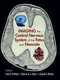
Imaging the Central Nervous System of the Fetus and Neonate
by Paul D. Griffiths Martyn N. J. Paley Elspeth H. WhitbyThis reference provides an authoritative overview of the role of ultrasonography and MR imaging technologies in the examination and assessment of the central nervous system of the fetus and neonate. Spanning advancements in fetal ultrasound, in-utero MR, the imaging of the neonatal brain, and the analysis of normal and abnormal brain development, t
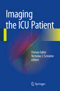
Imaging the ICU Patient
by Florian Falter Nicholas J. ScreatonThe critical care unit is an intense clinical environment with huge responsibilities on the professionals caring for these patients. Imaging is a key source of diagnostic information, but the conditions in which diagnostic imaging has to be performed are often extremely challenging and significantly different to imaging in the non acute setting. Imaging the ICU Patient reviews imaging procedures on the ICU in a highly practical and memorable manner. Swift and efficient clinical decision-making is rewarded on the ICU and this book serves as a practical handbook.
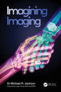
Imagining Imaging
by Michael R. JacksonFrom Roentgen to Rembrandt, Hounsfield to Hollywood and Vesalius to videogames, Imagining Imaging explores the deeply entwined relationship between art (and visual-based culture) and radiology / medical imaging. Including artworks from numerous historical eras representing varied geographic locations and visual traditions, alongside a diverse range of contemporary artists, Dr Jackson argues that the foundations of medical image construction and interpretation were laid down in artistic innovations dating back hundreds and thousands of years. Since the discovery of X-rays, artists and moviemakers have, in turn, drawn rich inspiration from radiographic imagery and concepts, but the process of cross-pollination between art and science has continued, with creative endeavour continuing to mould medical imaging examinations to this day.Blending a unique mix of art, science and medical history, together with aspects of visual neurophysiology and psychology, Imagining Imaging is essential reading for radiologists, radiographers and artists alike. Peppered with familiar TV and film references, personal insights into the business of image interpretation, and delivered in an accessible and humorous style, the book will also appeal to anyone who enjoys looking at pictures.Key features: Engaging synthesis of art and medical history, combined with anecdotes and experiences from a working clinical radiologist Diverse range of visual reference points including astronomy, botany and cartography, alongside comprehensive discussion of medical imaging modalities including plain radiography, ultrasound, CT and MRI 200 full colour illustrations
Imagining Welfare Futures (Social Policy: Welfare, Power and Diversity)
by Gordon HughesImagining Welfare Futures explores possible futures of welfare by considering different types of relationship between the public and the state through which social welfare may be organized beyond the millennium. By drawing on contemporary debates about the 'citizen', 'the community' and 'the consumer', the book explores what each of these imaginary figures might mean for the next generation of welfare users.
Imagining the Past, Constructing the Future
by Brady Wagoner Maria C. D. P. Lyra Alicia BarreiroThis book takes a sociocultural, developmental and dialogical perspective to explore the constructive and interconnected nature of remembering and imagining. Conceived as cognitive-affective processes, both emerge at the border of the person and his or her socio-cultural world. Memory is approached as a functional adaption to the environment using the resources of the past in preparation for action in the present. Imagination is tightly related to memory in that both aim to escape the confines of the concrete here-and-now situation; however, while memory is primarily oriented to the past, imagination looks to the future. Both are embedded in the exchanges with the social and cultural milieu, and thus theorizing them has relied on key ideas from Lev Vygotsky, Frederic Bartlett and Mikhail Bakhtin. Thus, this book aims to integrate theories of remembering and imagining, through rich empirical studies in diverse cultural settings and concerning the development of self and identity. These two groups of studies compose the subparts that organize the book.
Imagistic Care: Growing Old in a Precarious World (Thinking from Elsewhere)
by Cheryl MattinglyImagistic Care explores ethnographically how images function in our concepts, our writing, our fieldwork, and our lives. With contributions from anthropologists, philosophers and an artist, the volume asks: How can imagistic inquiries help us understand the complex entanglements of self and other, dependence and independency, frailty and charisma, notions of good and bad aging, and norms and practices of care in old age? And how can imagistic inquiries offer grounds for critique? Cutting between ethnography, phenomenology and art, this volume offers a powerful contribution to understandings of growing old. The images created in words and drawings are used to complicate rather than simplify the world. The contributors advance an understanding of care, and of aging itself, marked by alterity, spectral presences and uncertainty.Contributors: Rasmus Dyring, Harmandeep Kaur Gill, Lone Grøn, Maria Louw, Cheryl Mattingly, Lotte Meinert, Maria Speyer, Helle S. Wentzer, Susan Reynolds Whyte
Imitation from Infancy Through Early Childhood: Typical and Atypical Development
by Mikael HeimannThis book summarizes more than four decades of research on imitation in infancy and its relation to early learning and sociocognitive development in typically and atypically developing children. The studies were carried out in a Scandinavian context and thus provide important cultural validation of the central developmental processes. The book is divided into three parts: Part one focuses on the social and cognitive aspects of imitation, discussing links to early parent-infant interaction, and developmental meaning. It addresses evidence for an imitative capacity at birth for typical and atypical infants. Also covered are early individual differences in imitation, the role of imitation as a social and cognitive learning mechanism in early development, and possible links between imitation and temperament. Part two presents unique longitudinal studies on early memory development using deferred imitation as the key method. It discusses the biological basis of memory and explores the idea that deferred imitation is an indicator of an infant’s ability to understand intentions. Part three focuses on imitation in young children with autism and with Down syndrome. It examines the role of imitation as a “deficit” as well as a vehicle for change when used interactively in early interventions for children with autism. Imitation from Infancy Through Early Childhood is an essential resource for researchers, professors, and graduate students as well as clinicians and other professionals in developmental psychology, cognitive development, psycholinguistics, child psychiatry, and developmental neuroscience.