- Table View
- List View
Radiobioassays (Routledge Revivals #1)
by Fuad S. AshkarFirst Published in 1983, this book offers a full comprehensive guide to the examination of radiation in the body. Carefully compiled and filled with a vast repertoire of notes, diagrams, and references this book serves as a useful reference for radiologists and other practitioners in their respective fields.
Radiobiology Textbook
by Sarah BaatoutThis open access textbook focuses on the various aspects of radiobiology. The goal of radiobiological research is to better understand the effects of radiation exposure at the cellular and molecular levels in order to determine the impact on health. This book offers a unique perspective, by covering not only radiation biology but also radiation physics, radiation oncology, radiotherapy, radiochemistry, radiopharmacy, nuclear medicine, space radiation biology & physics, environmental and human radiation protection, nuclear emergency planning, molecular biology and bioinformatics, as well as the ethical, legal and social considerations related to radiobiology. This range of disciplines contributes to making radiobiology a broad and rather complex topic. This textbook is intended to provide a solid foundation to those interested in the basics and practice of radiobiological science. It is a learning resource, meeting the needs of students, scientists and medical staff with an interest in this rapidly evolving discipline, as well as a teaching tool, with accompanying teaching material to help educators.
Radiobiology and Environmental Security
by Carmel E Mothersill Colin B. Seymour Victoria KorogodinaThis volume - like the NATO Advanced Research Workshop on which it is based - addresses the fundamental science that contributes to our understanding of the potential risks from ecological terrorism, i.e. dirty bombs, atomic explosions, intentional release of radionuclides into water or air. Both effects on human health (DNA and systemic effects) and on ecosystems are detailed, with particular focus on environmentally relevant low-dose ranges. The state-of-the-art contributions to the book are authored by leading experts; they tackle the relevant questions from the perspectives of radiation genetics, radiobiology, radioecology, radiation epidemiology and risk assessment.
Radiobiology for the Radiologist
by Eric J. Hall Amato J. GiacciaPublisher's Note: Products purchased from 3rd Party sellers are not guaranteed by the Publisher for quality, authenticity, or access to any online entitlements included with the product. Considered the “gold standard” in the field for over 45 years, Radiobiology for the Radiologist combines traditional and molecular radiation biology principles and appeals to students, residents, and veteran clinical practitioners. This edition continues the two-part format of previous editions and features brand-new chapters, thoroughly updated content, and hundreds of figures that provide a visual context to the information in each chapter.
Radiochromic Film: Role and Applications in Radiation Dosimetry (Imaging in Medical Diagnosis and Therapy)
by Indra J. DasThis book provides a first authoritative text on radiochromic film, covering the basic principles, technology advances, practical methods, and applications. It focuses on practical uses of radiochromic film in radiation dosimetry for diagnostic x-rays, brachytherapy, radiosurgery, external beam therapies (photon, electron, protons), stereotactic body radiotherapy, intensity-modulated radiotherapy, and other emerging radiation technologies. <P><P>The expert authors address basic concepts, advantages, and the main applications including kilovoltage, brachytherapy, megavoltage, electron beam, proton beam, skin dose, in vivo dosimetry, postal and clinical trial dosimetry. The final chapters discuss the state of the art in microbeam, synchrotron radiation, and ultraviolet radiation dosimetry.
Radiofrequency Ablation for Small Hepatocellular Carcinoma
by Minshan Chen Yaojun Zhang Wan Y. LauThis book provides a comprehensive guide to the treatment of small hepatocellular carcinoma (sHCC) using a minimally invasive technique: radiofrequency ablation (RFA). RFA has emerged as a new treatment modality and become the main modality of locoregional therapy. Extensive clinical research indicates that RFA is as effective as surgical resection for sHCC, and it has the advantage of being less invasive. However, the outcomes after RFA are largely dependent on the operators' experience- known as the "learning curve". This book presents the characteristics of sHCC and discusses why sHCC is the best candidate for RFA. Then it introduces all the commercially available RFA systems, and their working principles, advantages, disadvantages and so on. It goes on to demonstrate how to perform RFA under the guidance of ultrasound, CT, laparoscopy, or during open operation. Finally, it discusses the radiologic assessment and followup after RFA, as well as adjuvant therapies and clinical trials on RFA. The authors are experts from the fields of pathology, radiology, surgery, and gastroenterology, as well as manufacturers. With this book, readers gain have a clear idea of when and how to do RFA. It aims to standardize and generalize the procedure of RFA, which will be very helpful in improving the outcome of RFA for sHCC.
Radiofrequency Treatments on the Spine
by Luca Saba Stefano MarciaThis book describes the principles and applications of radiofrequency treatments for various spinal indications, including disc herniation, discogenic and radicular pain, facet joint arthropathy, and benign and malignant lesions of the vertebral column. The aim is to provide a handy guide that will acquaint readers with all aspects of radiofrequency neurotomy at different levels of the spine, enabling them to carry out treatments effectively and safely. Radiofrequency neurotomy, or radiofrequency ablation, is a minimally invasive procedure that is associated with a reduction in complications, side effects, and risks of anesthesia as well as with lower costs. This book, written by world-renowned authorities in the field, fills a significant gap in the literature by specifically focusing on the use of radiofrequency for spinal conditions. It will be of value to a range of specialists, including interventional neuroradiologists and radiologists, neurosurgeons, and orthopedists.
Radiofrequenztherapie in der Kopf-Hals-Chirurgie: Ein klinischer Leitfaden
by Claudia Lill Klaus StelterDieses Buch beschreibt die breiten Einsatzmöglichkeiten und die professionelle Durchführung der Radiofrequenztherapie in der Hals-Nasen-Ohren- Heilkunde. Eine der bekanntesten Indikationen stellt die Vergrößerung der Nasenmuschel dar. Bei der Radiofrequenztherapie handelt es sich um einen schonenden, minimal-invasiven Eingriff, der wenig Risiken birgt. Schon nach kurzer Zeit kann der Patient die Praxis verlassen. Ähnliches gilt für Schnarchoperationen.Das vorliegende Werk gibt dem Hals-Nasen-Ohren- Arzt, aber auch dem plastischen Chirurgen eine übersichtliche, praxisorientierte Information und zeigt die vielfältigen Möglichkeiten der Radiofrequenzanwendung im Kopf-Hals-Bereich auf.
Radiography PREP: Program Review and Exam PREP
by D. A. SaiaHailed by Doody’s Review Service as “the gold standard among instructors and students,” Radiography PREP delivers a concise summary of the entire radiography curriculum in a readable narrative. Written by an experienced program director, this is a true “must read” for certification or recertification. The book includes more than 900 ARRT-style review questions with detailed answer explanations for correct and incorrect answers, a practice exam, hundreds of illustrations and radiographic images, and powerful learning aids such as summary boxes and a glossary.
Radiography in Veterinary Technology 4th Ed
by Lisa M. LavinWritten by a veterinary technician for veterinary technicians, students, and veterinary practice application, this concise, step-by-step text will help users consistently produce excellent radiographic images. It covers the physics of radiography, the origin of film artifacts, and positioning and restraint of small, large, avian, and exotic animals. It discusses everything from patient preparation, handling, and positioning to technical evaluation of the finished product.
Radiography of the Dog and Cat
by S. K. Kneller M. C. MuhlbauerRadiography of the Dog and Cat: Guide to Making and Interpreting Radiographs offers a comprehensive guide to producing high-quality radiographs and evaluating radiographic findings. Equally useful as a quick reference or for more in-depth information on specific diseases and disorders, the book is logically organized into sections describing how to make high-quality radiographs, normal radiographic anatomy, and interpretation of radiographic abnormalities. It is packed with checklists for systematic evaluation, numerous figures and line drawings, and exhaustive lists of differential diagnoses, resulting in an especially practical guide for the radiographic procedures performed in everyday practice.Written in a streamlined, easy-to-read style, the book offers a simple and fresh approach to radiography of the dog and cat, correlating physics, physiology, and pathology. Coverage includes patient positioning, contrast radiography, normal and abnormal radiographic findings, and differential diagnoses as they pertain to musculoskeletal, thoracic, and abdominal structures. Radiography of the Dog and Cat: Guide to Making and Interpreting Radiographs is a one-stop reference for improving the quality and diagnostic yield of radiographs in your clinical practice.
Radiography of the Dog and Cat: Guide to Making and Interpreting Radiographs
by M. C. Muhlbauer S. K. KnellerRadiography of the Dog and Cat A convenient and authoritative quick-reference guide to help you get the most from radiography of dogs and cats. In the newly revised second edition of Radiography of the Dog and Cat: Guide to Making and Interpreting Radiographs, the authors deliver a thorough update to a celebrated reference manual for all veterinary personnel, student to specialist, involved with canine and feline radiography. The book takes a straightforward approach to the fundamentals of radiography and provides easy-to-follow explanations of key points and concepts. Hundreds of new images have been added covering normal radiographic anatomy and numerous diseases and disorders. Readers of the book will also find: An expanded positioning guide along with images of properly positioned radiographs. Numerous examples of radiographic artifacts with explanations of their causes and remedies. Detailed explanations of many contrast radiography procedures, including indications, contraindications, and common pitfalls. Comprehensive treatments of Musculoskeletal, Thoracic, and Abdominal body parts, including both normal and abnormal radiographic appearances and variations in body types. Perfect for veterinary practitioners and students, the second edition of Radiography of the Dog and Cat: Guide to Making and Interpreting Radiographs is also a valuable handbook for veterinary technical staff seeking a one-stop reference for dog and cat radiography.
Radioguided Surgery
by Ken Herrmann Omgo E. Nieweg Stephen P. PovoskiThis book offers a comprehensive overviewof the state of the art in the practice of radioguided surgery. The openingsection is devoted to the basic physics principles for detection and imaging,radiation detection device technology, principles of surgical navigation,radionuclides and radiopharmaceuticals, and radiation safety. A series ofchapters then address the clinical application of radioguided surgery for avariety of malignancies, including breast cancer, melanoma and other cutaneousmalignancies, gynecologic malignancies, head and neck malignancies, thyroidcancer, urologic malignancies, colon cancer, gastroesophageal cancer, lungcancer, bone tumors, parathyroid adenomas, and neuroendocrine tumors. For eachapplication, the recommended methodological approaches are discussed and theavailable cumulative clinical experiences of investigators from across theglobe are reviewed. A conscious effort is made to highlight recent developmentsand innovative multidisciplinary approaches within each clinical area. Interesting issues and novel approaches are further explored through acollection of selected case reports at the end of the book. The contributingauthors are all experts in their own fields, ensuring that the book will holdwide appeal for surgeons, surgical technologists, nuclear medicine physicians,nuclear medicine technologists, and various trainees.
Radiologic Guide to Orthopedic Devices
by Hunter Tim B. Taljanovic Mihra S. Wild Jason R.Orthopedic devices improve the quality of life of millions of people, and show up on radiographs and cross-sectional imaging studies daily. This text will familiarise radiologists with the indications, applications, potential complications, and radiologic evaluation of many medical devices. The book offers a complete discussion of fracture fixation, joint arthroplasty, and orthopedic apparatus of the neck and spine, including the cervical, thoracic, and lumbar spine. It also provides detailed overviews of devices used for common dental disease, covers the general principles applicable to complications of orthopedic devices, foreign body ingestions, insertions and injuries, and details quality assurance issues concerning the manufacture and distribution of devices. Featuring a large gallery of apparatus for reference, an extensive glossary of terms and a list of manufacturers, Radiologic Guide to Orthopedic Devices is an essential resource for radiologists, orthopedists and emergency medicine physicians. Regular updates to the topics covered will be available on http://www. medapparatus. com.
Radiologic Physics: The Essentials
by Zhihua Qi Robert D. WissmanPerfect for residents to use during rotations, or as a quick review for practicing radiologists and fellows, Radiologic Physics: The Essentials is a complete, concise overview of the most important knowledge in this complex field. Each chapter begins with learning objectives and ends with board-style questions that help you focus your learning. A self-assessment examination at the end of the book tests your mastery of the content and prepares you for exams.
Radiologic Science for Technologists: Physics, Biology and Protection (10th Edition)
by Stewart Carlyle BushongThis textbook provides a solid presentation of radiologic science, including the fundamentals of radiologic physics, diagnostic imaging, radio biology, and radiation management.
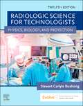
Radiologic Science for Technologists: Physics, Biology, and Protection
by Stewart Carlyle BushongDevelop the skills you need to produce diagnostic-quality medical images! Radiologic Science for Technologists: Physics, Biology, and Protection, 12th Edition provides a solid foundation in the concepts of medical imaging and digital radiography. Featuring hundreds of radiographs and illustrations, this comprehensive text helps you make informed decisions regarding technical factors, image quality, and radiation safety for both patients and providers. New to this edition are all-digital images and the latest radiation protection standards and units of measurement. Written by noted educator Stewart Carlyle Bushong, this text will prepare you for success on the ARRT® certification exam and in imaging practice.
Radiologic Technology Clinical Manual
by Robert J. ParelliThe Radiologic Technology Clinical Manual is designed to guide students through all aspects of clinical training in the area of radiological sciences. This practical workbook contains student self-evaluation forms, course outlines, instructional objectives, and all the procedures and work assignments necessary for training students in the clinical side of radiologic technology. It can be used as a supplement to any radiologic sciences program.When used as part of an occupational training course in radiologic technology, the Radiologic Technology Clinical Manual will help students qualify for examination by the American Registry of Radiologic Technologists (ARRT). The book contains valuable record keeping materials for clinical experience hours, background on the profession as a whole, and evaluation forms for quarterly periods of clinical training. Time sheets, attendance forms, and clinical log forms are also included.
Radiological Anatomy for FRCR Part 1
by Philip Borg Abdul Rahman AlviThe new FRCR part 1 Anatomy examination comprises 20 cases/images, with five questions about each. The cases are labelled 01 to 20 and the five questions are labelled (a) to (e). The authors have set out to emulate this format by gathering 200 cases which, from their experience, are representative of the cases on which candidates will be tested. The book consists of 10 tests with 20 cases each, and 5 stem questions each. The answers, along with an explanation and tips, accompany each test at the end of the chapter. This will help candidates to identify the level of anatomical knowledge expected by the Royal College of Radiologists. The aim of this book is not to replace the already available literature in radiological anatomy, but to complement it as a revision guide. Whereas radiological anatomy atlases and textbooks provide images with labels for every possible identifiable structure in an investigation, the cases in this book have only 5 labels, simulating the exam.
Radiological Anatomy for FRCR Part 1
by Philip Borg Abdul Rahman J. Alvi Nicholas T. Skipper Christopher S. JohnsThree years after the publication of the first edition, this book remains the best seller in its category based on its faithful representation of the FRCR Part 1 exam. The second edition is designed to reflect the change in exam format introduced in spring 2013. It includes two new chapters as well as some new cases in the remaining chapters and tests. Under the new exam format, candidates will be presented with 100 cases, with a single question per case and a single mark for the correct answer. This book covers all core topics addressed by the exam in a series of tests and includes chapters focussing specifically on paediatric cases and normal anatomical variants. The answers to questions, along with explanations and tips, are supplied at the end of each chapter. Care has been taken throughout to simulate the exam itself, so providing an excellent revision guide that will help candidates to identify the level of anatomical knowledge expected by the Royal College of Radiologists.
Radiological Anatomy for Radiation and Particle Therapy
by Thankamma Ajithkumar Sara Upponi Nicholas CarrollThis book is exceptional in addressing the common radiological anatomical challenges of target volume delineation faced by clinicians on a daily basis. The clear guidance that it provides on how to improve target volume delineation will help readers to obtain the best possible clinical outcomes in response to radiation and particle therapy. The first section of the book presents the fundamentals of the different imaging techniques used for radiation and proton therapy, explains the optimal integration of images for target volume delineation, and describes the role of functional imaging in treatment planning.The extensive second section then discusses site-specific challenges. Here, each chapter illustrates normal anatomy, tumor-related changes in anatomy, potential areas of natural spread that need to be included in the target volume, postoperative changes, and variations following systemic therapy.The final section is devoted to the anatomicalchallenges of treatment verification. The book is of value for radiation and clinical oncologists at all stages of their careers, as well as radiotherapy radiographers and trainees.
Radiological Atlas of Child Abuse: A Complete Resource for MCQs, v. 1
by Christine Hall Amaka OffiahThe radiological abnormalities associated with suspected child abuse can be extremely subtle. If missed, a baby or child may be returned to an environment where episodes of abuse may escalate. Similarly, a wrongful diagnosis can lead to an infant being removed from loving carers. This atlas will be of particular use to radiologists (both in training and at consultant level), and also to other doctors who may be first in line to encounter suspected abuse, including paediatricians, accident and emergency doctors, orthopaedic surgeons and pathologists. It uses numerous radiographs from Professor Hall's collection amassed over three decades, including many examples of the sorts of difficult cases and normal variants that are found in day to day practice. It offers assistance with the initial interpretation of what are often difficult and subtle findings in the emotionally charged environment that frequently exists when child abuse is suspected.
Radiological Evaluation Of The Spinal Cord (Routledge Revivals #2)
by Milosh PerovitchFirst Published in 1981, this book offers a full insight into the process of examination of the spine using radiological equipment. Carefully compiled and filled with a vast repertoire of notes, diagrams, and references this book serves as a useful reference for students of medicine and other practitioners in their respective fields.
Radiological Imaging of the Digestive Tract in Infants and Children
by Samuel Stafrace Johan G. G. Hans BlickmanThis book comprehensively reviews imaging of the pediatric gastrointestinal tract and accessory digestive organs from a practical approach. Starting with a brief discussion on techniques this is followed by a couple of comprehensive chapters covering emergency/acute pediatric abdominal imaging. A series of traditional anatomically structured chapters on the oesophagus, stomach, small bowel, colon and accessory organs then follow. Each chapter carefully considers the role of the currently available imaging techniques and discusses and illustrates diagnostic dilemmas. The closing chapter focuses on pediatric interventional procedures performed with imaging guidance. Since the first edition, the text has been fully updated and new illustrations included. Against the background of rapid advances in imaging technology and the distinctive aspects of gastrointestinal imaging in children and infants, this volume will serve as an essential reference for general and pediatric radiologists as well as for radiologists in training, neonatologists and pediatricians.
Radiological Imaging of the Kidney
by Emilio QuaiaThis book provides a unique and comprehensive analysis of the normal anatomy and pathology of the kidney and upper urinary tract from the modern diagnostic imaging point of view. The first part is dedicated to the normal radiological anatomy of the kidney and normal anatomic variants. The second part presents in detail all of the imaging modalities which can be employed to assess the kidney and the upper urinary tract, with careful descriptions of patient preparation, investigation protocols, and principal fields of application of each imaging modality. The entire spectrum of kidney pathologies is then presented with the aid of a large set of images, many of which are in color. The latest innovations in interventional radiology, biopsy procedures, and parametric and molecular imaging are also described. This book should be of great interest to all radiologists, oncologists, and urologists who are involved in the management of kidney pathologies in their daily clinical practice.
