- Table View
- List View
Radiology for Anaesthesia and Intensive Care
by Richard Hopkins Carol Peden Sanjay GandhiThe advent of small, affordable ultrasound machines and the widespread use of PACS systems have made imaging more accessible to anaesthetists and intensivists than ever before. This concise, highly illustrated text discusses the key aspects of radiology, examining all imaging modalities and body regions. Introductory sections review the imaging knowledge required for the FRCA exams and the role of imaging in the Pre-Operative Assessment. These are followed by chapters on each imaging modality and body region, each containing numerous illustrations, practical advice on diagnosis, and many case illustrations. Each modality chapter contains a concise introductory section on the principles of image formation. Containing over 300 scans and illustrations, and written by a multidisciplinary team of radiologists and anaesthetists, Radiology for Anaesthesia and Intensive Care, second edition, is an invaluable aid for all anaesthetists and intensivists.
Radiology for Medical Finals: A case-based guide
by Edward Sellon David HowlettThis book is an informed, educational and abundantly illustrated guide to the imaging knowledge that medical students in the clinical years of their undergraduate studies will be required to get to know, understand and recall in order to negotiate successfully their finals exams. Via the popular and instructive case-based format, readers are guided through 100 cases chosen specifically to reflect what the authors consider is necessary knowledge for finals, and imaging modalities that students can reasonably expect to encounter with a resulting emphasis on plain film with some CT and MR.
Radiology for PET/CT Reporting
by Stefano Fanti Cristina Nanni Lucia ZanoniReading PET/CT scans is sometimes challenging. Not infrequently, abnormal findings on CT images are functionally silent and therefore difficult for nuclear medicine practitioners to interpret. Furthermore, in general only a low-dose CT scan is produced as part of the combined PET/CT study, and the resulting CT images may prove suboptimal for image interpretation. This atlas is designed to enable nuclear medicine practitioners who routinely read PET/CT scans to recognize the most common CT abnormalities. Slice-by-slice descriptions are provided of anatomical structures as visualized on CT scans obtained in PET/CT studies. The CT findings that may be detected while reviewing PET/CT scans of various body regions and conditions are then illustrated and fully described. The concluding section of the book is devoted to the principal MRI findings in diseases which cannot be evaluated using PETs/CTs.
Radiology for PET/CT Reporting
by Stefano Fanti Cristina Nanni Lucia Zanoni Rita GolfieriThis atlas is intended to enable nuclear medicine practitioners who routinely read PET/CT scans to recognize the most common CT abnormalities. Reading PET/CT scans can sometimes be challenging. It is not infrequent, in fact, to encounter abnormal findings in CT images (not related to the neoplastic disease under evaluation) that are functionally silent and therefore difficult to interpret for nuclear medicine practitioners. Frequently, these findings are clinically relevant and should be reported, interpreted and compared to previous scans. This may also have an impact on patient management, since expensive tests like PET/CT are expected to provide the highest level of diagnostic information. Generally, CT images associated with a PET scan are acquired in a low-dose modality, and therefore prove to be sub-optimal for CT image interpretation. Sometimes a comparison with a full-resolution and contrast-enhanced CT atlas may be difficult. Low-dose CT slices are thicker than diagnostic CT and offer less anatomical detail, which can affect accuracy in terms of recognizing both anatomical structures and pathological findings.Today it is becoming increasingly common to acquire a standard PET/CT by combining the administration of contrast media and a diagnostic CT; here, too, basic CT reporting skills are needed in clinical practice. This atlas features a chapter on “normal anatomy” (with and without contrast media) that is based on low-dose and full-dose CT images from PET/CT standard acquisition, and which identifies all the relevant anatomical structures. Other chapters (focusing on the thorax, abdomen, pelvis, and musculoskeletal system) present cases with common and uncommon anatomical abnormalities. The addition of new cases with ceCT in this revised second edition rounds out the coverage of PET/CT reporting. Given its scope, the book will be of interest to nuclear medicine physicians, radiologists, and oncologists alike.
Radiology for Undergraduate Finals and Foundation Years: Key Topics and Question Types (MasterPass)
by Ashley Shaw Tristan Barrett Nadeem ShaidaBasic knowledge of radiology is essential for medical students regardless of the specialty they plan to enter. Hospital patients increasingly undergo some form of imaging, ranging from plain film through to CT and MRI. As technologies and techniques advance and radiology grows in scope, medical school curricula are reflecting its increased importance. This book provides a mixture of case-based teaching, structured questions, and self-assessment techniques relevant to the evolving modern curriculum. It covers critical areas including knowledge of when to investigate a patient, which modality best answers a specific clinical question and how to interpret chest and abdominal x-rays. Along with final year medical students, this book will also benefit postgraduate FY1 and FY2 junior doctors and those in the earlier clinical years who wish to expland their radiology knowledge. It also provides a useful basic radiology primer for the early MRCP and MRCS examinations. 'It is a great honour to be asked to provide a foreword for this excellent and unusual text. There is an eminently practical range of topics covered in this book and this reflects the commonsense approach by the authors. The images are good and the explanatory text educationally valuable and very much to the point.' - From the Foreword by Professor Adrian K. Dixon
Radiology for the Dental Professional (Ninth Edition)
by Herbert H. Frommer Jeanine J. Stabulas-SavageA complete guide to radiology principles and techniques, Radiology for the Dental Professional, 9th Edition helps you develop imaging skills through practical application. Detailed step-by-step procedures demonstrate proper techniques; photos and illustrations improve comprehension and readability. Written by Herbert H. Frommer, DDS, and Jeanine J. Stabulas, RDH, BS, MPH, this book will help you interpret radiographs, and troubleshoot and prevent common errors. For students, it's an ideal introduction to radiology; for dental hygiene/assisting professionals, it's a great review! A logical organization starts with the basics and makes it easier to progress through the material. Procedures boxes show detailed radiography procedures with illustrations and photos to demonstrate proper techniques. Common Errors boxes explain mistakes in radiographic techniques and describe how they can be resolved. Advantages/Disadvantages boxes compare and contrast the good and bad elements of radiographic techniques. Detailed outlines and educational objectives at the beginning of each chapter identify the information that you are expected to learn. Key terms are listed at the beginning of each chapter and highlighted upon first mention in the text. Expanded coverage of digital imaging techniques. Patient Management and Special Problems chapter improves coverage of nervous patients, patients with special needs, pediatric patients, and specific problems such as endodontic issues and third molars. New illustrations depict techniques and show the latest technology.
Radiology in Forensic Medicine: From Identification to Post-mortem Imaging
by Giuseppe Lo Re Massimo Midiri Antonina Argo Cristina CattaneoThis book offers a comprehensive overview of the forensic and radiological aspects of pathological findings, focusing on the most relevant medico-legal issues, such as virtual autopsy (virtopsy), anthropometric identification, post-mortem decomposition features and the latest radiological applications used in forensic investigations. Forensic medicine and radiology are becoming increasingly relevant in the international medical and legal field as they offer essential techniques for determining cause of death and for anthropometric identification. This is highly topical in light of public safety and economic concerns arising as a result of mass migration and international tensions. The book discusses the latest technologies applied in the forensic field, in particular computed tomography and magnetic resonance, which are continuously being updated. Radiological techniques are fundamental in rapidly providing a full description of the damage inflicted to add to witness and medical testimonies, and forensic/radiological anthropology supplies valuable evidence in cases of violence and abuse.Written by international experts, it is of interest to students and residents in forensic medicine and radiology. It also presents a new approach to forensic investigation for lawyers and police special corps as well as law enforcement agencies.
Radiology in Global Health
by Daniel J. Mollura Matthew P. LungrenThe World Health Organization stated that approximately two-thirds of the world's population lacks adequate access to medical imaging. The scarcity of imaging services in developing regions contributes to a widening disparity of health care and limits global public health programs that require imaging. Radiology is an important component of many global health programs, including those that address tuberculosis, AIDS-related disease, trauma, occupational and environmental exposures, breast cancer screening, and maternal-infant health care. There is a growing need for medical imaging in global health efforts and humanitarian outreach, particularly as an increasing number of academic, government, and non-governmental organizations expand delivery of health care to disadvantaged people worldwide. To systematically deploy clinical imaging services to low-resource settings requires contributions from a variety of disciplines such as clinical radiology, epidemiology, public health, finance, radiation physics, information technology, engineering, and others. This book will review critical concepts for those interested in managing, establishing, or participating in a medical imaging program for resource-limited environments and diverse cross-cultural contexts undergoing imaging technology adaptation.
Radiology in Global Health: Strategies, Implementation, and Applications
by Daniel J. Mollura Anne-Marie Lugossy Lauren Fuller KulinskiThis third edition provides a definitive, comprehensive, and multidisciplinary reference for a diverse healthcare audience to navigate the complexities of radiology service planning, implementation, and diagnostics in low and middle-income countries (LMICs) and resource-limited medically underserved regions. This book integrates clinical approaches with concepts of project planning to assist radiology professionals, public health specialists, and students to implement medical imaging in under-served and resource-constrained contexts to improve global health outcomes and optimize the use of radiologic technologies. The first section of the book introduces the general role of radiology in international public health, to help readers (i) understand the impact of medical imaging on health disparity (ii) learn how to assess radiologic service infrastructure in low-resource environments, and (iii) learn how to plan projects aiming to address health care shortages. The second section of the book presents clinical contexts for deploying radiology such as pediatrics, trauma, infectious disease, and cancer control. Themes from the two sections are interwoven so that the learner can integrate concepts of project development with clinical objectives. For example, the chapter on information technology (IT) reinforces the chapter on cancer control by addressing IT solutions applicable to cancer management in medically underserved areas. All chapters have been fully updated and this edition adds chapters on artificial intelligence, COVID, and breast imaging, as well as others. This is an ideal guide for radiologists and medical professionals working in LMICs and resource-limited medically underserved regions.
Radiology in Global Health: Strategies, Implementation, and Applications
by Daniel J. Mollura Matthew P. Lungren Melissa P. CulpExploring the question as to why more than half the world continues to have little or no access to medical imaging and radiology, this important second edition, fully revised and expanded, offers not only answers but practical solutions, providing new tools, ideas, and strategies for bringing vital radiology to low-resource areas. Based on RAD-AID’s ten years of work (2008-2018) serving indigent communities around the world, the book’s interdisciplinary approach offers the synthesis of business management, government policy formulation, clinical methods, and engineering in order to integrate economic development, technology innovation, clinical model planning, educational strategies, and public health measures. The gold-standard title in the field, Radiology in Global Health, 2nd Edition is intended for a broad audience, including physicians (especially radiologists and radiology residents), radiology technologists, radiology nurses, sonographers, hardware/software engineers, policy-makers, business leaders, researchers, and public health specialists at all levels who use or implement health care services for underserved populations. In addition, as health care providers use radiology in the process of clinical decision-making, this title is also designed for clinical physicians, nurses, nurse-practitioners, physician assistants, and paramedical personnel. Administrators and public health personnel will also be interested, as the planning of radiology services for health care systems at both the facility level and at the population level requires a clear understanding of the technological challenges and management opportunities.
Radiology of HIV/AIDS
by Hongjun LiThis book provides a comprehensive overview of modern methodology, diagnostic methods and techniques of HIV/AIDS imaging. It starts with a general review of HIV/AIDS imaging and evidence based medicine (EBM). In the following chapters, authors introduce 13 of most common types of AIDS/HIV-associated diseases separately. High-quality examples combined with computed tomography (CT), Magnetic resonance imaging (MRI), gross anatomy and pathology images are clearly illustrated. Moreover, the use of functional imaging in HIV/AIDS diagnosis and differential diagnosis is an essential part that diagnostic doctors will find to be useful.
Radiology of Infectious Diseases: Volume 2
by Hongjun LiThis book provides a comprehensive overview of diagnostic imaging in infectious diseases. It starts with a general review of infection diseases, including their classification, characteristics and epidemiology. In separate chapters, the authors then introduce the radionuclide imaging of 50 kinds of infectious diseases. Volume 1 covers 21 viral infections. Volume 2 has 29 chapters discussing 24 bacterial infections and 5 parasitic infections. Each disease is clearly illustrated using cases combined with high-quality computed tomography (CT) and magnetic resonance imaging (MRI). The book provides a valuable reference source for radiologists and doctors working in the area of infectious diseases.
Radiology of Infectious and Inflammatory Diseases - Volume 1: Brain and Spinal Cord
by Jian Wang Hongjun Li Xuening ZhangThis book provides a comprehensive overview of diagnostic imaging in infectious and inflammatory diseases in central nervous system and spine. It starts with basic theory of infectious diseases, including pathological basis, laboratory diagnostic methods, and clinical imaging techniques. In following chapters, the authors firstly introduce the central nervous system infections related to respiratory, gastrointestinal, and contagious diseases. Then viral, bacterial, fungal, parasitic and special infections in central nervous system and spine are presented with clinical cases. Each disease is clearly illustrated using cases combined with high-quality CT and MRI. The book provides a valuable reference source for radiologists and doctors working in the area of infectious and inflammatory diseases.
Radiology of Infectious and Inflammatory Diseases - Volume 2: Head and Neck
by Hongjun Li Shuang Xia Yubo LyuThis book provides a comprehensive overview of state-of-the-art imaging in infectious and inflammatory diseases in head and neck. It starts with a brief introduction of infectious diseases in head and neck, including normal anatomy, classification, and laboratory diagnostic methods. In separate parts of eye, ear, nose, pharynx, larynx, and maxillofacial region, the common imaging techniques and imaging anatomy is firstly introduced, and then typical infectious and inflammatory diseases is presented with clinical cases. Each disease is clearly illustrated with PET and MR images and key diagnostic points. The book provides a valuable reference source for radiologists and doctors working in the area of infectious and inflammatory diseases.
Radiology of Infectious and Inflammatory Diseases - Volume 3: Heart and Chest
by Li Li Hongjun Li Jingzhe LiuThis user-friendly book provides a comprehensive overview of diagnostic imaging in infectious and inflammatory diseases in heart and chest. It starts with a general review of infectious diseases, including pathogenesis and pathology-radiology correlations. In separate chapters, the authors then introduce clinical images of typical bacterial, viral, fungal, and parasitic infections in a wide range of clinical conditions and circumstances. Each disease is clearly illustrated using cases combined with high-quality CT and MRI for readers to correctly identify causes of abnormalities. The book provides a valuable reference source for radiologists and doctors working in the area of infectious and inflammatory diseases.
Radiology of Infectious and Inflammatory Diseases - Volume 5: Musculoskeletal system
by Jun Zhou Hongjun Li Shinong PanThis book offers a systematic coverage of diagnostic imaging in infectious and inflammatory diseases in musculoskeletal system. The first part is devoted to a general review of infectious diseases in musculoskeletal system, as well as pathogenic classification, imaging techniques, pathogenic and imaging characteristics. In the following parts, imaging interpretation of typical infectious and inflammatory diseases affecting bone, joint, and soft tissue is described. Each disease is clearly illustrated using cases combined with high-resolution CT, MRI and PET. The book provides a valuable reference source for radiologists and doctors working in the area of infectious and inflammatory diseases.
Radiology of Influenza
by Hongjun LiThis book presents the theory of influenza and its imaging characteristics. The first section introduces readers to the fundamental theory of influenza, including the etiology, epidemiology and pathogenesis of influenza, influenza-related diseases, and imaging technologies. The second section is divided into 5 chapters and includes extensive DR, CT and MRI images on H1N1 influenza, H5N1, H7N9, H5N6 and H10N8 avian influenza.
Radiology of Influenza A (H1N1)
by Hongjun Li Ning LiA novel strain of influenza A (H1N1) virus emerged and an outbreak of influenza A (H1N1) occurred worldwide in 2009 and 2010, posing a severe threat to the health of human beings. The two Editors work together in Beijing You An Hospital, to combat this type of influenza. This book, Radiology of Influenza A (H1N1), is intended to provide first-hand clinical data and its scientific analysis, thus supporting sound clinical practice for influenza A (H1N1) prevention and treatment as well as related scientific research. The authors have collected over 100 critical influenza A (H1N1) virus-infected cases. For cases of influenza A (H1N1) that resulted in death, autopsies were performed for 8 cases after receiving their families' informed consent. Over 200 reports on pathological and histological analysis as well as immunohistochemical and electroscopic analysis were brought together. This book was then compiled by comparing the published data with the present first-hand data. This book includes a wealth of content, including DR, CT and MRI images, pathological and histological figures, and immunohistochemical and electroscopic figures. Related research, clinical and educational needs can be readily satisfied by its detailed data, texts and figures, as well as typical case reports. Li Hongjun, is professor and director of the diagnostic radiology department in Beijing You An Hospital, affiliated with Capital Medical University, Beijing, China. Li Ning, is professor and president of Beijing You An Hospital, affiliated with Capital Medical University, Beijing, China.
Radiology of Non-Spinal Pain Procedures
by Azim Shaikh Mubin I. SyedThis handy, well-illustrated manual has been designed to meet the need of interventional pain physicians to understand the radiologic imaging involved in the performance of non-spinal pain procedures. It provides information on such topics as radiologic anatomy, the radiologic manifestations of indications and contraindications to interventional procedures, and the radiologic appearance of complications that may arise from these procedures. In addition, it will be useful for the diagnostic radiologist, who may be unaware of many of the interventional pain procedures. The chosen format will ensure that the reader is quickly able to reference any given procedure. As this is a guidebook, it does not encompass every pathologic entity that may be encountered; however, the commonly performed non-spinal pain procedures are included. This text will prove essential for any interventionalist who does not have easy access to a radiologist and vice versa.
Radiology of Orthopedic Implants
by Sanjeev Agarwal Gaurav Jyoti BansalThere is an ever-expanding range of implants used in Orthopaedic Surgery. Nearly 200,000 joint replacement procedures are done in UK every year. The performance of these implants is assessed on radiographs. This is of interest to Orthopaedic surgeons and Radiologists alike. Information on interpretation of these radiographs is not readily available in an easily readable format. This book will assist both trainees and practicing orthopedic surgeons and radiologists in assessing the radiologic appearance of implants and their potential for future performance.
Radiology of Parasitic Diseases
by Hongjun LiThis book primarily summarizes the imaging characteristics and theory of parasitic disease, offering a clinical guide and practical approach to understanding, preventing and diagnosing parasitic disease. Through imaging diagnosis and cross-research of imaging with autopsy and pathology, it highlights the chief characteristics and evolution of imaging, which provides primary scientific data for the understanding and research of parasitic disease. Divided into 9 chapters, this volume is fully illustrated with DR, CT and MRI images of different kinds of parasitic diseases, providing a valuable resource for radiologists in this field.
Radiology of the Abdomen and Pelvis: A Case-Based Guide
by Swati GoyalA succinct account of various routinely experienced pathologies and suitable images has been presented as approximately162 case studies. The cases are structured into thematic chapters with an integrated approach to basic learning. Each case study follows a similar format with a brief clinical presentation, relevant imaging findings, discussion with differential diagnosis, management, and suggested readings. This book focuses on the pointwise description of cases routinely encountered in abdominopelvic imaging that help students, trainees and radiologists to write certificate examinations. Key Features: Presents chapters in the form of case studies, along with a brief illustrative description of normal anatomy and abnormal findings. Uses image-based quizzes for easy comprehension for trainees, residents, and practicing radiologists. Incorporates pivotal cases from the hepatobiliary, pancreatic, genitourinary, and gastrointestinal systems in a single book.
Radiology of the Chest and Related Conditions
by F W WrightThe book presents a comprehensive overview of the various disease processes affecting the chest and related abnormalities. It discusses biopsy and bronchography, as well as a variety of imaging techniques including radiography, fluoroscopy, tomography, and ultrasound.
Radiology of the Post Surgical Abdomen
by Damian J.M. Tolan John BrittendenA comprehensive description of the most common abdominal operations involving the gastrointestinal tract, pancreas, liver and genitourinary systems, illustrated with artists' drawings and images of normal post operative anatomy. The complications associated with each procedure will be in table format consisting of text alongside imaging examples. There will also be teaching points included. The book will be divided into nine chapters.
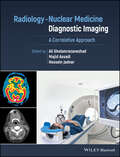
Radiology-Nuclear Medicine Diagnostic Imaging: A Correlative Approach
by Hossein Jadvar Majid Assadi Ali GholamrezanezhadRadiology-Nuclear Medicine Diagnostic Imaging Radiology-Nuclear Medicine Diagnostic Imaging: A Correlative Approach provides in-depth guidance on applying the principles of radiologic-nuclear medicine correlation to the interpretation of imaging for diagnostic, prognostic, and predictive indications. Describing the clinical implications of all major imaging modalities, this comprehensive professional reference offers one-stop coverage of the common diagnostic applications encountered by nuclear medicine physicians and radiologists in day-to-day practice. The book develops the nuclear diagnostic skills necessary to interpret combined imaging modalities and correlate radiologic findings using a disease and organ-based approach to radiologic interpretation. Thematically organized sections explore a variety of pathologies including diseases of the head and neck, gastrointestinal tract, and pulmonary, endocrine, and central nervous system. Written by internationally recognized experts, this important resource: Helps physicians better understand the clinical and treatment implications of diseases with characteristic radiologic appearances Includes detailed descriptions of nuclear medicine presentations of diseases of most organ systems combined with radiologic correlation Explains refinement of differential diagnoses in various organ systems based on specific imaging features Demonstrates how to correlate scintigraphy and PET images with radiography, CT, MRI, and other imaging techniques Includes a timely review of the application of nuclear medicine-radiology correlative imaging in research Features practical, hands-on clinical imaging references, and more than 600 color illustrations and high-resolution images throughout Radiology-Nuclear Medicine Diagnostic Imaging: A Correlative Approach is a must-have for both trainee and experienced radiologists, nuclear medicine physicians, and specialist nurses.