- Table View
- List View
Atlas of Cardiac Catheterization for Congenital Heart Disease
by Massimo Chessa Gianfranco Butera Andreas Eicken John D. ThomsonThis atlas depicts and describes catheter-based interventions across the entire pediatric age range, from fetal life through to early adulthood, with the aim of providing an illustrated step-by-step guide that will help the reader to master these techniques and apply them in everyday practice. Clear instruction is offered on a wide range of procedures, including vascular access, fetal interventions, valve dilatation, angioplasty, stent implantation, defect closure, defect creation, valve implantation, hybrid approaches, and other miscellaneous procedures. The atlas complements the previously published handbook, Cardiac Catheterization for Congenital Heart Disease, by presenting a wealth of photographs, images, and drawings selected or designed to facilitate the planning, performance, and evaluation of diagnostic and interventional procedures in the field of congenital heart disease. It will assist in the safe, efficient performance of these procedures, in decision making, and in the recognition and treatment of complications.
Atlas of Cardiac Innervation
by Vasken Dilsizian Jagat NarulaThis atlas elucidates the role of the neuronal component in normal cardiovascular function and cardiac disorders and discusses the currently available imaging targets and probes. It provides a foundation in cardiac neuronal imaging and image processing and serves as a guide in the effective utilization of these techniques in clinical and research settings. The atlas discusses the principles of autonomic control in the regulation of cardiac function and disease; chemistry and biology of radiotracers designed to target changes in the myocardial sympathetic and parasympathetic innervation as a function of disease or treatment; neuronal imaging in heart failure and reverse remodeling and the use of PET imaging in quantification of cardiac innervation; cardiac sympathetic innervation in ventricular arrhythmias and device therapy; conditions that affect the autonomic nervous system; and the role of myocardial blood flow and cardiac neuronal imaging in denervation and reinnervation in cardiac transplant recipients. Featuring full-color illustrations, schematic diagrams, and diagnostic algorithms, Atlas of Cardiac Innervation is a valuable resource for cardiologists, radiologists, nuclear medicine physicians, and electrophysiologists.
Atlas of Cardiac Surgery (Springer Surgery Atlas Series)
by Francis C. WellsThis atlas provides a comprehensive overview of the current techniques practised in a leading cardiac surgical institution. It offers an up-to-date review of procedures, authored by foremost surgeons in each specialist area. Richly illustrated, its aim is to serve as an appealing “go-to” text for training and practising cardiac surgeons.
Atlas of Cardiometabolic Risk
by William T. Cefalu Christopher P. CannonThe first atlas devoted specifically to cardiometabolic risk, this book will provide a concise visual primer on the pathophysiology, epidemiology, diagnosis, treatment, and clinical and radiologic features of this disorder. Describing recent care strategies and current practices in patient assessment, this source will allow clinicians to accuratel
Atlas of Cardiovascular Computed Tomography (2nd Edition): Includes Narrated Videos
by Jagat Narula Matthew J. Budoff Stephan S. Achenbach Harvey S. HechtIncludes nearly 1000 high quality images in step with the latest developments.<P><P> Contributors include world-renowned authors from a variety of disciplines.<P> Includes high-quality hand-drawn illustrations.<P> Offers in-depth explanation for each image.<P> This atlas is a comprehensive visual reference for the use of cardiovascular computed tomography (CT) containing photomicrographs, anatomic illustrations, tables, and charts paired with extensive legends and explanations that are supplemented by extensive research, peer-reviewed articles, and textbooks.<P> In addition to providing historical perspective and current direction for CT, this new edition of Atlas of Cardiovascular Computed Tomography 2e focuses on research involving coronary artery diseases and anomalies, congestive heart failure, atherosclerotic plaques and asymptomatic disease, as well as imaging techniques, including preparation, acquisition, and processing, involving the great vessels and carotids, the peripheral vasculature, and coronary and pulmonary veins. The increasing role of CT in the emergency room and in private cardiology practice is also reviewed thoroughly, making this an essential read for all involved in cardiac imaging, cardiology and emergency medicine.
Atlas of Cell Organelles Fluorescence
by Elli Kohen Rene Santus Joseph G. Hirschberg Nuri OzkutukContaining over 150 original photomicrographs accompanied by protocol information, Atlas of Cell Organelles Fluorescence delineates organelles structures, interaction, and organization into complexes. It provides a collection that shows living cells under physiopathological conditions and in the context of treatment with carcinogens, xenobi
Atlas of Chest Imaging in COVID-19 Patients
by Jinxin Liu Xiaoping Tang Chunliang LeiThis book presents X-ray and CT findings of patients with 2019 Novel Coronavirus (2019-nCoV) pneumonia in early, progressive, critical, and recover stage. It starts with a general review of CT features of new coronavirus pneumonia. In the following chapters, imaging manifestations in different groups and stages are presented, especially CT findings of asymptomatic COVID-19 patients and those first nucleic tests for the novel coronavirus are negative, but turn positive in one day or two. In addition, imaging and pathological analysis of COVID-19 death are summarized in the last chapter. The book provides a valuable reference source for radiologists and doctors working in the area of coronavirus pneumonia.
Atlas of Cilia Bioengineering and Biocomputing
by Richard Mayne Jaap Den ToonderCilia are microscopic finger-like cell-surface organelles possessed by a great many eukaryotic organisms, including humans, whose purposes include generating local fluid movements via rhythmic whip-like beating and environmental sensing. Despite intense research efforts since their discovery by van Leeuwenhoek in the 1670's, several key questions regarding ciliary functions, experimental manipulation and in silico imitation remain unanswered. Major justifications for cilia research lie in their involvement in various forms of human disease (ciliopathies) and their ability to instantiate decentralised, asynchronous sensorial-actuation of adjacent matter through modulation of beating characteristics. Further elucidation of these characteristics, which is a problem requiring the combined expertise of mathematicians, computer scientists, engineers and life scientists, will lead to novel biomedical therapies, creation of `smart' actuating surfaces for microfluidics/lab-on-chip applications and a greater understanding of fluid mechanics in real-world scenarios. This lavishly-illustrated anthology presents recent advances in the fields of ciliary investigation, manipulation, emulation, mimesis and modelling from key researchers in their fields: its goal is to explain the state-of-the-art in cilia bioengineering and bio-computation in a uniquely creative, accessible manner, towards encouraging further transdisciplinary work in the field as well as educating a broad spectrum of scientists and lay people. The volume is split into three distinct but interwoven themes:Biology: Biological preliminaries for the study of cilia; the state-of-the-art in genetic engineering of ciliated cells for biomedical purposes; reprogramming of cilia dynamics in live cells.Engineering: Creation of macro cilia robots for object sorting applications; pneumatic cilia for the optimization of fluid motion; electrostatic, magnetic and MEMS cilia for microfluidic mixing; reviews in artificial cilia fabrication, actuation and flow induction methods.Numerical and computational modelling. Analyses of thin film cilia for `lab on chip' microfluidic mixing applications; modelling of gel-based artificial cilia towards simulating dynamic behaviors of responsive cilia layers in complex fluids across a wide range of potential applications.
Atlas of Clear Cell Carcinoma of the Ovary: A Pathological Guide
by Junzo Kigawa Tsunehisa Kaku Toru Sugiyama Steven G. SilverbergClear cell carcinoma (CCC) of the ovary with its unique clinical and biological features has attracted great attention, and calls for the publication of a specialized book on the subject. This is the first atlas that has narrowed its focus to CCC. Revealed here are the typical and variable histological features of CCC and related tumors. Hundreds of high-quality photographs help the reader to recognize the pathological features and clinical manifestations of CCC and to formulate diagnoses confidently and accurately. Data are based on the international central pathological review of the JGOG/GCIG3017 clinical trial in which experts around the world participated. Using a virtual slide system, interesting and significant features of CCC were discovered in the review of 652 cases. This book provides the newest information on the categorization and classification of CCC: growth patterns (papillary, tubocystic, solid, and adenofibromatous), cell types (classical hobnail and clear cell, eosinophilic, oxyphilic, and oncocytic), stromal changes (hyalinized, necrotic, hemorrhagic, lymphocytic infiltrative, luteinized, and psammomatous calcification), presence of endometriosis and atypical endometriosis, and borderline/atypical proliferative tumors. The Atlas of Clear Cell Carcinoma of the Ovary is an invaluable diagnostic resource for pathologists and gynecologic oncologists.
Atlas of Cleft Lip and Palate & Facial Deformity Surgery
by Jian-Min Yao Jing-Hong XuThis book consists the basic theories and comprehensive skills of surgery, plastic surgery and chin surgery. The book contains more than 1900 color surgical photos, which mainly from lip and palate surgery in “Operation Smile” and the accumulation in decades of clinical practice. It briefly introduces various surgical methods of cleft lip and palate and alternative methods, procedures and steps of recommended repair surgeries by the way of pictures and graphic. It also emphasizes on describing common and practical cleft lip and palate surgery, and explores innovative surgical methods, operative techniques and key points. By means of schematic diagrams and surgical photos, various surgical procedures for cleft lip and palate are introduced. It is a useful reference book for surgeons in plastic surgery, oral surgery, cosmetic surgery and otolaryngology.
Atlas of Clinical Cases in Rhinoplasty: Volume I
by Motaz H. ShafyThis atlas discusses a variety of common through to complex clinical presentations in Rhinoplasty. Simple methods of handling different types of noses including large, bulky, crooked, pediatric, deviated and oblique are covered throughout the book. It contains over 2,000 full-colour surgical photographs of both rare and routine cases in Rhinoplasty with all aesthetic challenges examined in detail. The display of the pre- and post-operative photographs is made simple and easy for the eyes to see. Pre-operative photographic assessment, surgical planning and post-operative analysis is included in each case in an easy to follow format so that photographs and texts can be seen and read at a glance. A well-informed, clear guide is written at the beginning of the Atlas.Atlas of Clinical Cases in Rhinoplasty: Volume I is aimed at those who are planning to take Rhinoplasty as a career and experienced surgeons who wish to expand their knowledge and skills. It will be an essential resource for all those seeking a clear, highly visual guide to new developments in Rhinoplasty.
Atlas of Clinical Cases in Rhinoplasty: Volume II
by Motaz H. ShafyThis atlas discusses a variety of common through to complex clinical presentations in Rhinoplasty. Simple methods of handling fractured, finesse, secondary, ethnic and special clinical cases are covered throughout the book. It contains over 2,000 full-colour surgical photographs of both rare and routine cases in Rhinoplasty with all aesthetic challenges examined in detail. The display of the pre- and post-operative photographs is made simple and easy for the eyes to see. Pre-operative photographic assessment, surgical planning and post-operative analysis is included in each case in an easy to follow format so that photographs and texts can be seen and read at a glance. A well-informed, clear guide is written at the beginning of the Atlas. Atlas of Clinical Cases in Rhinoplasty: Volume 2 is aimed at those who are planning to take Rhinoplasty as a career and experienced surgeons who wish to expand their knowledge and skills. It will be an essential resource for all those seeking a clear, highly visual guide to new developments in Rhinoplasty.
Atlas of Clinical Cases on Brain Tumor Imaging
by Yelda Özsunar Utku ŞenolThis book presents and analyzes clinical cases of brain tumors and follows the classification provided by the WHO in 2016. After introductory chapters reviewing the international literature on the topic, the advances made in all imaging modalities (especially Magnetic Resonance and Computed Tomography) are examined.All radiological findings are supplemented with a wealth of images and brief explanations. The clinical information is given as part of the case discussion, as are the characteristics and differential diagnosis of the tumors. Radiologic-pathologic correlations round out the description of each clinical case.Intended as a quick and illustrative reference guide for radiology residents and medical students, this atlas represents the most up-to-date, practice-oriented reference book in the field of Brain Tumor Imaging.
Atlas of Clinical Dermatology in Coloured Skin: A Morphological Approach
by Piyush Kumar Pc DasThis Atlas of Dermatology has a unique clinical orientation and lays emphasis on morphology. The disease entities are arranged according to the morphology of lesions, not etiologically. This pattern of placement is more useful for both teaching and learning and serves as an important aid in making a diagnosis as this approach is based on the way patients present themselves in out-patient departments. The key diagnostic clinical pointers too have been provided with images, which helps one suspect the diagnosis and exclude clinical differentials. This Atlas would serve as a ready reckoner for residents and practitioners. Key Features ● An atlas and text covering skin, mucosa, hair and nail diseases in skin of color ● Emphasis on key diagnostic clinical pointers ● 3000+ clinical images--multiple images of a disease to cover the clinical spectrum ● Dedicated section on Regional Dermatology and Skin changes in systemic diseases ● Clinician’s desk reference for OPD practice
Atlas of Clinical Dermatopathology: Infectious and Parasitic Dermatoses
by Werner Kempf Heinz Kutzner Josef Feit Günter Burg Ram Chandra AdhikariATLAS OF CLINICAL DERMATOPATHOLOGY INFECTIOUS AND PARASITIC DERMATOSES Differential diagnosis is at its most accurate and efficient when clinical presentation and histopathological features are considered in correlation with one another. With that being so, the expert team behind this innovative atlas has integrated both perspectives to provide all those working in dermatologic healthcare with a complete guide to infectious and parasitic dermatoses in their many forms. More than 600 high-quality images demonstrate the common presentation of a wide range of bacterial, viral, and fungal infections, as well those of parasitic conditions of various kinds. Accompanying these are direct and easily understood descriptions of key features and diagnostic clues, making this new text an essential quick-reference tool for trainees and practicing clinicians alike. The Atlas of Clinical Dermatopathology: Infectious and Parasitic Dermatoses includes: A straightforward, pattern-based approach to dermatologic diagnosis Full-color illustrations and clear descriptions for easy reference Combined clinical and histopathological perspectives Handy diagnostic tips throughout Featuring all this and more, this invaluable atlas offers a uniquely balanced, clear, and comprehensive guide to what can be a difficult process, and will be of tremendous assistance to students, dermatologists, dermatopathologists, and pathologists everywhere.
Atlas of Clinical Endocrinology and Metabolism
by Pauline CamachoThis updated text is a pictorial atlas of endocrine and metabolic disorders. Each chapter focuses on providing multiple illustrations as well as a thorough discussion of the diagnosis and management of a variety of endocrine diseases with their appropriate treatment plans. Using updated guidelines, it provides a comprehensive discussion of the latest therapies, including diabetes technology. Presenting a large number of clinical images, including imaging of thyroid ultrasounds, DXA images, bone scans, and new technologies in diabetes mellitus, this atlas aims to provide the reader with the information needed to make accurate diagnoses, making it an updated source of highly illustrated information for endocrinologists, clinicians, residents, fellows and trainees. With new chapters on transgender medicine and obesity, this textbook is a valuable resource for the contemporary endocrine practitioner. • Features new chapters such as transgender medicine and inborn errors of metabolism • Aims to be an invaluable aid for endocrinologists, internal medicine specialists, family practice clinicians, residents, fellows, and trainees • Explores diabetes technology with updated guidelines
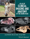
Atlas of Clinical Imaging and Anatomy of the Equine Head
by Larry Kimberlin Lynn Ruoff Alex Zur LindenAtlas of Clinical Imaging and Anatomy of the Equine Head presents a clear and complete view of the complex anatomy of the equine head using cross-sectional imaging. Provides a comprehensive comparative atlas to structures of the equine head Pairs gross anatomy with radiographs, CT, and MRI images Presents an image-based reference for understanding anatomy and pathology Covers radiography, computed tomography, and magnetic resonance imaging
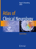
Atlas of Clinical Neurology
by Roger N. RosenbergLike its preceding editions, this atlas is an indispensable guide to the field of neurology, featuring the most clinically essential images and figures. Chapters offer insight and research written by deeply practiced, knowledgeable neurologists that is supplemented with detailed imagery, tables, algorithms, and delineative drawings. Topics covered include developmental and genetic diseases, neuroendocrine disorders, critical care neurology, cerebrovascular disease, dementias, behavioral neurology, neuro-oncology, movement disorders, epilepsy, neuromuscular diseases, infectious diseases, neuroimmunology, neurotoxic disorders, and headache. The authors also delve into specific issues currently prevalent in neuroscientific research, including Alzheimer’s disease, dementia, Machado-Joseph disease, Huntington’s disease, and brain scanning with PET and fMRI. The Atlas of Clinical Neurology, 4th Edition serves as a comprehensive and premier visual resource for neurologists.
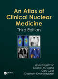
Atlas of Clinical Nuclear Medicine
by Gopinath Gnanasegaran Ignac Fogelman Gary Cook Susan E. M. ClarkeThis long-awaited Third Edition has been revised and updated to encapsulate the developments in the field since the previous edition was published nearly two decades ago. The successful style of the previous editions has been built upon and expanded to provide the ultimate guide for beginners, those in training, and experienced practitioners. Each section contains comprehensive cases with first-class examples of correlative/hybrid imaging (SPECT and PET/CT) included where appropriate. This atlas contains superb illustrative cases and valuable supportive information, together with highlighted teaching points aiding all clinicians in routine practice.
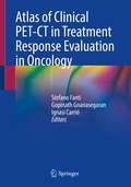
Atlas of Clinical PET-CT in Treatment Response Evaluation in Oncology
by Stefano Fanti Gopinath Gnanasegaran Ignasi CarriÓThis atlas is a superb guide to the use of PET-CT for the evaluation of treatment response in oncology patients based on its ability to assess tumor metabolic status. The first part of the book explains the role of PET-CT in response evaluation in different treatment settings. For comparison, overviews of the value and limitations of CT alone, PET alone, and anatomical and functional MRI are included. Guidance is also provided on the reporting of PET-CT scans in post-therapy scenarios. The second part of the book describes and illustrates the use of PET-CT with FDG and other tracers to assess the treatment response of malignancies at different anatomic sites. Featuring a wealth of images, informative case-based discussion, and evidence-based teaching points, these disease-specific chapters clearly demonstrate the key role that PET-CT can play in distinguishing early responders from patients who are non-responders or are resistant to treatment. Prompt and accurate evaluation of treatment response is vital as we enter the era of individualized medicine, and this atlas will persuade readers of the considerable advantages of PET-CT over conventional radiological and clinical methods.
Atlas of Common and Rare Genodermatoses
by Nayera Moftah May El Samahy Nadia Abd El Wadood Monira WaseefThe management of cutaneous findings in genodermatoses is challenging to the practicing clinician, especially when all hues of skin and skin types are contemplated, and first-line therapies often inadequate. This carefully crafted Atlas includes all sorts of different ethnic skin types, while featuring a wealth of over 1000 commented images collected in more than a decade, including 200 rare genodermatoses, and providing the reader with key points for correct dermatological diagnosis. The contents start with disorders of keratinization and pigmentation, as well as bullous, acantholytic, dermal, hair and nail diseases. Successively, immunodeficiency syndromes and their skin related symptoms, hamartoneoplastic syndromes and defective excision repair syndromes are presented. The addition of vascular disorders, as well as syndromes with premature aging grant full topic coverage. This Atlas of Common and Rare Genodermatoses will be a valued companion to dermatologists and paediatric specialists in their daily practice.
Atlas of Comparative Diagnostic and Experimental Hematology
by Alfred Jarecki Clifford SmithA vital resource on blood and bone marrow cell morphology in laboratory animal medicine. This fully revised new edition is an essential reference for clinical pathologists in diagnostic laboratories, and medical or veterinary research. The atlas contains over 400 color images of cells from the peripheral blood and bone marrow from a variety of animals encountered in laboratory animal medicine, in health and disease. Key features: New chapter on flow cytometry and its application in terms of routine analyses as a means of identifying abnormalities in cell marker expression, which is of particular relevance for pre-clinical safety assessment Covers the most recent developments in laboratory animal hematology, including parameters measured by the latest generation of analyzers Coverage of a wide range of laboratory animal species, as well as those used in clinical veterinary trials Photomicrographs present normal and abnormal blood cells from a variety of hematological conditions along with descriptive text
Atlas of Conducted Electrical Weapon Wounds and Forensic Analysis
by Jeffrey D. Ho Mark W. Kroll Donald M. DawesAtlas of Conducted Electrical Weapon Wounds and Forensic Analysis provides a comprehensive publication on the subject of Conducted Electrical Weapon (CEW) wounds and signature markings created by this class of weapon. This volume will serve as a very useful resource for all professions tasked with assisting persons that have allegedly been subjected to a CEW exposure. The volume provides an introduction to basic CEW technology and the types of CEWs currently available. It also serves as a comprehensive pictorial atlas of signature markings that CEW exposures make in the immediate and more remote post-exposure periods. Also, it discusses the ability of forensic specialty examinations of the CEW itself to aid in the determination of whether the alleged CEW exposure is consistent with the objective evidence and the subjective statements. Finally, this text addresses the important and growing area of factitious CEW markings that will be useful for consideration by investigators and litigators. Atlas of Conducted Electrical Weapon Wounds and Forensic Analysis provides an objective atlas of evidence for reference that will benefit those professionals who often must make diagnostic, treatment or legal judgments on these cases including Emergency and Primary-Care Physicians, Medical Examiners, Forensic Pathologists, Coroners, Law Enforcement Investigators, and Attorneys.
Atlas of Cone Beam Computed Tomography
by Ghassem Ansari Yaser Safi Mitra Ghazizadeh Ahsaie Ingrid Różyło-Kalinowska Mahsima Tayefi Nasrabadi Maryam FazlalipourA comprehensive collection of oral and maxillofacial cases using cone beam CT imaging Atlas of Cone Beam Computed Tomography delivers a robust collection of cases using this advanced method of imaging for oral and maxillofacial radiology. The book features over 1,500 high-quality CBCT scans with succinct descriptions covering a wide range of maxillofacial region conditions, including normal anatomy, anomalies, inflammatory diseases, and degenerative diseases. Easy to navigate and featuring multiple images of normal variation and pathologies, the book offers readers guidance on the diagnostic values of CBCT, as well as CBCT images of the inferior alveolar nerve canal, dental implants, temporomandibular joint evaluations, and surgical interventions. The book also includes: A thorough introduction to cone beam computed tomography, including in vivo and in vitro preparation and evaluation, indications in dentistry, and indications in medicine Comprehensive explorations of cone beam computed tomography artefacts and anatomic landmarks Practical discussions of cone beam computed tomography of dental structure, including normal anatomy, anomalies, and the difficulties of eruption In-depth examinations of cone beam computed tomography of pathological growth and development, including maxillofacial congenital and developmental anomalies Perfect for graduate dental students and postgraduate dental students in oral and maxillofacial radiology, Atlas of Cone Beam Computed Tomography is also useful to general dentists, oral and maxillofacial radiologists, head and neck maxillofacial surgeons, head and neck radiologists, general radiologists, and ENT surgeons.