- Table View
- List View
Atlas of Ear Diseases of the Dog and Cat
by Sue Paterson Karen M. TobiasBringing together a wealth of images of normal and diseased dog and cat ears, this is an indispensible diagnostic tool for the small animal veterinary practitioner seeing ear cases on a regular basis. This fully illustrated atlas covers the anatomy of the canine and feline ear, diagnostic techniques, a range of commonly seen diseases, and ear surgery.Atlas of Ear Diseases of the Dog and Cat is one of the most complete picture references for this rapidly expanding branch of small animal medicine and surgery. It is an invaluable aid for general practitioners, as well as those specialising in dermatology, and serves as an effective revision aid for veterinary students and those studying for further qualifications in veterinary dermatology.Includes over 400 high quality colour clinical images and clear line drawingsImages are accompanied by clear explanatory text throughoutEnables veterinarians to match cases seen in practice with photos supplied to aid diagnosisWritten by highly qualified specialist veterinary dermatologist and veterinary surgeon
Atlas of Echocardiography in Pediatrics and Congenital Heart Diseases
by Azin Alizadehasl Maryam MoradianThis atlas provides a practical guide to the diagnosis of congenital heart disease using echocardiography in both adults and children. A plethora of high-quality echocardiography images provide practical examples of how to diagnose a range of conditions correctly, including aortic stenosis, tricuspid atresia, coronary artery fistula and hypoplastic left heart syndrome. Atlas of Echocardiography in Pediatrics and Congenital Heart Diseases describes the diagnostic management of a range of congenital heart diseases successfully in both adults and children. Therefore it provides a valuable resource for both practicing cardiologists who regularly treat these patients and for trainees looking to develop their diagnostic skills using echocardiography.
Atlas of Elastosonography: Clinical Applications with Imaging Correlations
by Emilio Quaia Dirk-André Clevert Mirko D’onofrioWith the aid of an extensive series of high-quality images, this atlas describes all the potential applications of elastosonography in routine clinical practice. After a brief introduction on technical and physical aspects, the diagnostic benefits of elastosonography in a range of settings are illustrated with the aid of numerous representative cases. The coverage encompasses pathologies of the liver, spleen, pancreas, kidney, breast, thyroid, testis, musculoskeletal system, and vascular system. In addition, helpful correlations with imaging appearances on ultrasound and computed tomography are included. Elastosonography is a powerful, relatively new diagnostic technique that assesses the elasticity of tissues as an indicator of malignancy. Readers will find this book to be an excellent aid to use and interpretation of the technique.
Atlas of Elbow Surgery
by Andrea Celli Luigi CelliThis richly illustrated atlas is entirely devoted to the most modern surgical techniques of the elbow, offering a comprehensive, step-by-step and detailed examination of all the technical aspects of the surgical exposure of this joint. With the help of over 100 original drawings and photographs, it offers readers a unique understanding of elbow anatomy and surgical approaches, providing precise indications and surgical timing for clinical practice. An entire section focuses on the surgical management of the most common diseases of the elbow: the surgical exposures are related to pathologies affecting the lateral medial, anterior and posterior compartments of the elbow. This atlas will be of great value both to trainees and to specialists who manage disorders of the elbow, including orthopedic surgeons, traumatologists, and sports physicians, as well as anatomists.
Atlas of Electroencephalography in Sleep Medicine
by Nidhi S Undevia Hrayr P. AttarianSleep Medicine is a field that attracts physicians from a variety of clinical backgrounds. As a result, the majority of sleep specialists who interpret sleep studies (PSG) do not have specialized training in neurophysiology and electroencephalography (EEG) interpretation. Given this and the fact that PSGs usually are run at a third of the speed of EEGs and that they usually have a limited array of electrodes, waveforms frequently appear different on the PSGs compared to the EEGs. This can lead to challenges interpreting certain unusual looking activity that may or may not be pathological. This Atlas of Electroencephalograpy in Sleep Medicine is extensively illustrated and provides an array of examples of normal waveforms commonly seen on PSG, in addition to normal variants, epileptiform and non-epileptiform abnormalities and common artifacts. This resource is divided into five main sections with a range of topics and chapters per section. The sections cover Normal Sleep Stages; Normal Variants; Epileptiform Abnormalities; Non-epileptiform Abnormalities; and Artifacts. Each example includes a brief description of each EEG together with its clinical significance, if any. Setting the book apart from others in the field is the following feature: Each EEG discussed consists of three views of the same page -- one at a full EEG montage with 30mm/sec paper speed, the same montage at 10mm/sec (PSG speed) and a third showing the same thing at 10 mm/sec, but with the abbreviated PSG montage. Unique and the first resource of its kind in sleep medicine, the Atlas of Electroencephalograpy in Sleep Medicine will greatly assist those physicians and sleep specialists who read PSGs to identify common and unusual waveforms on EEG as they may appear during a sleep study and serve as a reference for them in that capacity.
Atlas of Emergency Imaging from Head-to-Toe
by Mariano Scaglione Douglas S. Katz Michael N. PatlasThis reference work provides a comprehensive and modern approach to the imaging of numerous non-traumatic and traumatic emergency conditions affecting the human body. It reviews the latest imaging techniques, related clinical literature, and appropriateness criteria/guidelines, while also discussing current controversies in the imaging of acutely ill patients. The first chapters outline an evidence-based approach to imaging interpretation for patients with acute non-traumatic and traumatic conditions, explain the role of Artificial Intelligence in emergency radiology, and offer guidance on when to consult an interventional radiologist in vascular as well as non-vascular emergencies. The next chapters describe specific applications of Ultrasound, Magnetic Resonance Imaging, radiography, Multi-Detector Computed Tomography (MDCT), and Dual-Energy Computed Tomography for the imaging of common and less common acute brain, spine, thoracic, abdominal, pelvic and musculoskeletal conditions, including the unique challenges of imaging pregnant, bariatric and pediatric patients. Written by a group of leading North American and European Emergency and Trauma Radiology experts, this book will be of value to emergency and general radiologists, to emergency department physicians and related personnel, to obstetricians and gynecologists, to general and trauma surgeons, as well as trainees in all of these specialties.
Atlas of Emergency Imaging from Head-to-Toe
by Mariano Scaglione Douglas S. Katz Michael N. PatlasThis new reference work provides a comprehensive and modern approach to the imaging of numerous non-traumatic and traumatic emergency conditions affecting the human body. It reviews the latest imaging techniques, related clinical literature, and appropriateness criteria/guidelines, while also discussing current controversies in the imaging of acutely ill patients. The first chapters outline an evidence-based approach to imaging interpretation for patients with acute non-traumatic and traumatic conditions, explain the role of Artificial Intelligence in emergency radiology, and offer guidance on when to consult an interventional radiologist in vascular as well as non-vascular emergencies. The next chapters describe specific applications of Ultrasound, Magnetic Resonance Imaging, radiography, Multi-Detector Computed Tomography (MDCT), and Dual-Energy Computed Tomography for the imaging of common and less common acute brain, spine, thoracic, abdominal, pelvic and musculoskeletal conditions, including the unique challenges of imaging pregnant, bariatric and pediatric patients. There are two new sections for 2nd edition. One section is devoted to imaging of emergency conditions in geriatric patients. The second section covers special considerations in emergency imaging including imaging of intimate partner violence and emergencies in transplant patients. Written by a group of leading North American and European Emergency and Trauma Radiology experts, this book will be of value to emergency and general radiologists, to emergency department physicians and related personnel, to obstetricians and gynecologists, to general and trauma surgeons, as well as trainees in all of these specialties.
Atlas of Emergency Medicine Procedures
by Latha GantiThis full-color atlas is a step-by-step, visual guide to the most common procedures in emergency medicine. Procedures are described on a single page, or two-page spreads, so that the physician can quickly access and review the procedure at hand. The atlas contains more than 600 diagnostic algorithms, schematic diagrams and photographic illustrations to highlight the breadth and depth of emergency medicine. Topics are logically arranged by anatomic location or by type of procedure and all procedures are based on the most current and evidence-based practices known.
Atlas of Emergency Medicine Procedures
by Latha GantiThe significantly expanded second edition of this full-color atlas provides a step-by-step, visual guide to the most common procedures in emergency medicine. Completely revised, it also includes new procedures such as REBOA, the HINTS test, sphenopalatine ganglion block, occipital nerve block, and lung ultrasonography. Procedures are described on a single page, or two-page spreads, so that the physician can quickly access and review the procedure at hand. The atlas contains more than 700 diagnostic algorithms, schematic diagrams, and photographic illustrations to highlight the breadth and depth of emergency medicine. Topics are logically arranged by anatomic location or by type of procedure, and all procedures are based on the most current and evidence-based practices. Atlas of Emergency Medicine Procedures, Second Edition is an essential resource for physicians and advanced practice professionals, residents, medical students, and nurses in emergency medicine, urgent care, and pediatrics.
Atlas of Emergency Neurovascular Imaging
by Yang TangIschemic and hemorrhagic strokes are common neurological emergencies. In recent years, endovascular intervention has become a standard of care in treating acute ischemic stroke, aneurysms, and vascular malformations. As a result, noninvasive CT- and MRI-based techniques have been increasingly used in emergency settings. In this context, neurovascular imaging has become an essential part of the curriculum for training emergency radiologists, stroke neurologists, and vascular neurosurgeons. This book provides a comprehensive review of the entire spectrum of emergent neurovascular imaging, with the emphasis on noninvasive CT angiography (CTA), MR angiography (MRA), and perfusion techniques. It is organized into 11 chapters. The first three chapters address the topics of acute stroke imaging, including algorithms based on recent clinical trials and updated American Heart Association stroke guideline, vascular territories, and stroke mimics. These are followed by discussions of cerebrovenous thrombosis, vasculopathies, aneurysms, and vascular malformations. Remaining chapters are devoted to the traumatic neurovascular injury, as well as the relatively rare albeit important topics of head and neck vascular emergencies and spinal vascular diseases. The book has an image-rich format, including more than 300 selected CT, MRI, or digital subtraction angiography (DSA) images. Atlas of Emergency Neurovascular Imaging is an essential resource for physicians and related professionals, residents, and fellows in emergency medicine, neuroradiology, emergency imaging, neurology, and vascular neurosurgery and can successfully serve as a primary learning tool or a quick reference guide.
Atlas of Emergency Radiology: Vascular System, Chest, Abdomen and Pelvis, and Reproductive System
by Rita AgarwalaThis book presents a vast collection of radiologic images of cases seen in a very busy emergency room. It encompasses common and very unusual pathology and every imaging modality. The book is divided into four parts on pathology of the vascular system, chest, abdomen and pelvis and reproductive organs. Images obtained with the modalities that best depict the abnormality in question are presented, with marking of the salient pathology and explanation of the abnormal imaging features in concise captions. Whenever possible, differential diagnosis is covered using further images and guidance is also provided on selection of additional modalities to confirm the diagnosis. The book will help residents to analyze different diseases and relate pathophysiology to imaging and assist students in appreciating what is abnormal. It will be a useful guide for the busy practicing radiologist and aid clinicians in understanding the complexity of these cases and delivering better focused treatment. p>

Atlas of Emergency Ultrasound
by John Christian FoxEmergency medicine specialists explain how to use the new mobile ultrasound systems at bedside to help with diagnosis in emergency departments. They consider such topics as the focused assessment of sonography in trauma, ultrasound of the lung, pelvic ultrasound, ultrasound-guided procedures, and venous ultrasound. Annotation ©2012 Book News, Inc. , Portland, OR (booknews. com)
Atlas of Endocrine Pathology (Atlas of Anatomic Pathology)
by Lori A. EricksonAtlas of Endocrine Pathology provides a comprehensive compendium of photomicrographs of common and uncommon entities in endocrine pathology. The volume includes histologic features of normal features, reactive conditions, hyperplasia and tumors. The most helpful diagnostic features are illustrated to provide direction and clues to the diagnosis of endocrine tumors. Furthermore, photomicrographs highlight the most pertinent diagnostic features in problematic diagnoses in endocrine pathology. Authored by a nationally and internationally recognized pathologist, Atlas of Endocrine Pathology is an important learning tool for those becoming familiar with the diverse entities encountered in endocrine pathology and a valuable reference for practicing pathologists faced with challenging diagnoses in endocrine pathology.
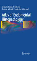
Atlas of Endometrial Histopathology
by Dietmar Schmidt Gisela Dallenbach-Hellweg Friederike DallenbachThe prime purpose of this atlas is to help the pathologist find, classify and differentially diagnose the changes he is observing. Information on the daily changes during the menstrual cycle and how to date them is essential for recognizing changes caused by abnormal endogenic and iatrogenic hormonal stimuli in functional endometrial disturbances. To provide their patients with the proper hormonal therapy, gynecologists cannot rely on blood samples only because of the constantly fluctuating hormonal levels. A precise functional diagnosis of the endometrial biopsy is the most accurate and best means for assessing hormonal action. Numerous microphotographs explain in detail how to recognize the normal and pathological changes that can develop in the endometrium. The subject of endometritis and the complex endometrial neoplasms and their precursors, with their differential diagnosis as well as their clinical prognosis are covered in detail. To complete the endometrial histopathology, gestational changes are reviewed and illustrated in the last chapter.
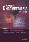
Atlas of Endometriosis (Encyclopedia of Visual Medicine Series)
by Colin Davis Robert W. Shaw Caroline Overton Lindsay McMillanEndometriosis affects women in the reproductive years, is associated with pelvic pain and infertility, and - although not life threatening - can seriously impair health, with huge economic and social consequences. It is arguably the most frequent problem encountered in contemporary More...gynecology and is the subject of much ongoing research and innovation in management. This beautifully and comprehensively illustrated Atlas, now in its third edition, provides a useful educational tool for trainees and general obstetricians and gynecologists who may not be up-to-date with the most important recent research on the diagnosis and management of the condition; particularly expanded for this edition are the chapters on ultrasound imaging and the nutritional aspects of the subject.
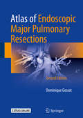
Atlas of Endoscopic Major Pulmonary Resections
by Dominique GossotIt is my greatest honor to be asked to write this foreword for the first edition of the Atlas of Endoscopic Major Pulmonary Resections by Dr Dominique Gossot. I have known Dr Gossot for over 15 years and have worked with him for many workshops and thoracic meetings. He is a pioneer in video-assisted thoracic surgery, and one of the most innovative thoracic surgeons I have known. Minimally invasive surgery has set a new standard of care for all surgical disciplines. Video-assisted thoracic surgery (VATS) offers a much kinder approach to the management of a wide variety of surgical conditions c- pared with conventional thoracotomy for these patients. Anatomical or major lung resections are a complex set of procedures commonly performed by thoracic s- geons. The adoption of the VATS approach for these procedures has received increasing acceptance by the thoracic surgical community, our pulmonologist and oncology colleagues, as well as the patients over the past two decades. There is now a growing body of evidence in the literature showing that the VATS approach is safe, oncologically sound, and associated with much lower morbidity compared with its conventional counterparts in the management of early lung cancers and benign conditions. Although there have been other books and atlases on VATS, this volume distinguishes itself in two respects.
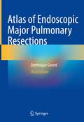
Atlas of Endoscopic Major Pulmonary Resections
by Dominique GossotThis third edition Atlas of Endoscopic Major Pulmonary Resections describes in detail the totally thoracoscopic approach to major pulmonary resections, in which only endoscopic instruments and monitor control are used. Pulmonary lobectomies and segmentectomies are presented step by step, using brief technical notes and high-quality, clearly labeled still images. Each chapter begins with information on the anatomical background, which is illustrated in three-dimensional reconstructions. In turn, technical ‘tricks’ and specific pitfalls are explained in pictograms. The technical descriptions presented here are based on the author’s own technique, which in some cases differs from other video-assisted approaches. The goal is for surgeons embarking on video-assisted major pulmonary resections – regardless of which approach they use – to find helpful hints and guidance on this totally new vision of pulmonary and mediastinal anatomy.The first edition of this atlas was released at a time when video-assisted major pulmonary resections were just emerging as a valid alternative to conventional techniques. In the second edition, chapters on sublobar resections, as a new alternative to lobectomy in selected patients, were added. In this third edition, as the interest in sublobar resections is growing, and because they are challenging, the technique is dealt with in depth. In particular, readers will be introduced to new imaging technologies to support these techniques.
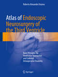
Atlas of Endoscopic Neurosurgery of the Third Ventricle: Basic Principles for Ventricular Approaches and Essential Intraoperative Anatomy
by Roberto Alexandre DezenaThis book describes in practical terms the endoscopic neurosurgery of the third ventricle and surrounding structures, emphasizing aspects of intraoperative endoscopic anatomy and ventricular approaches for main diseases, complemented by CT / MRI images. It is divided in two parts: Part I describes the evolution of the description of the ventricular system and traditional ventricular anatomy, besides the endoscopic neurosurgery evolution and current concepts, with images and schematic drawings, while Part II presents a collection of intraoperative images of endoscopic procedures, focusing in anatomy and main pathologies, complemented by schemes of the surgical approaches and CT / MRI images. The Atlas of Endoscopic Neurosurgery of the Third Ventricle offers a revealing guide to the subject, addressing the needs of medical students, neuroscientists, neurologists and especially neurosurgeons.
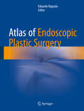
Atlas of Endoscopic Plastic Surgery
by Edoardo RaposioConcentrating on technique, which is explained and illustrated in detail, this book is written by worldwide experts and provides detailed, step-by-step instructions on how to perform state-of-the-art endoscopic surgical techniques in the complex Plastic Surgery field. More than 300 high-quality photos help clarify complex techniques throughout the book. Atlas of Endoscopic Plastic Surgery represents a comprehensive description of the current endoscopic techniques in the plastic, reconstructive an aesthetic field. It supplies surgeons with all the information necessary to successfully accomplish an endoscopic approach to vary plastic surgery procedures, from carpal and cubital tunnel release, breast augmentation and reconstruction, migraine surgery, hyperhidrosis management, to facial aesthetic surgery, flap and fascia lata harvesting, and mastectomy and abdominal wall surgery.
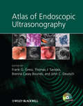
Atlas of Endoscopic Ultrasonography
by John C. Deutsch Frank Gress Thomas Savides Brenna C. BoundsThe Atlas of Endoscopic Ultrasonography provides readers with a large collection of excellent images obtained from both diagnostic and therapeutic procedures. The Atlas includes a DVD which will be an invaluable addition to the library of trainee and practising gastroenterologists with video clips and searchable database of images. Together the book and DVD offer a first class collection of images to give a highly integrated introduction to endoscopic ultrasonography. The Atlas is an ideal companion to Dr Gress et al’s Endoscopic Ultrasonography, Second Edition.
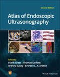
Atlas of Endoscopic Ultrasonography
by Frank Gress Thomas Savides Brenna Casey Everson L. A. ArtifonAtlas of Endoscopic Ultrasonography Atlas of Endoscopic Ultrasonography Atlas of Endoscopic Ultrasonography, Second Edition offers an outstanding visual guide to this very common diagnostic and therapeutic endoscopic tool. With contributions from noted experts in the field, the Atlas contains 400 high-quality color and black and white images obtained from real cases, each accompanied by detailed annotation to aid readers in their understanding of this popular technical procedure. In addition, there is a companion website featuring 50 video clips of real-life procedures in action, as well as the entire collection of images from within the book. Updated throughout to include the most recent advances in interventional Endoscopic Ultrasound (EUS) guided therapies Contains a large collection of color images obtained from both diagnostic and therapeutic procedures, also available on the companion website image bank Provides a highly integrated and accessible multimedia introduction to endoscopic ultrasonography Includes a companion website offering insightful videos Written for gastroenterologists, students, residents, and radiologists, Atlas of Endoscopic Ultrasonography, Second Edition is an essential introduction to endoscopic ultrasonography.
Atlas of Endoscopy with Narrow Band Imaging
by Manabu Muto Kenshi Yao Yasushi SanoWith its focus on narrow band imaging, this book is an excellent reference for new as well as experienced practitioners in the field of endoscopy. Narrow band imaging has brought about a revolutionary improvement in diagnostic endoscopy, enabling objective diagnosis and precise detection of lesions. It has enhanced the capability of endoscopy to facilitate qualitative diagnoses for the great benefit of patients who undergo endoscopic examinations. However, a standardized system of classification has not yet been established and many clinicians and researchers are not yet highly skilled in utilizing the technique or interpreting the images that are produced. This atlas addresses those issues, providing clear, simple and easy-to-understand descriptions illustrated with generous use of endoscopic images.
Atlas of Equine Ultrasonography
by Michele L. Frazer Kristina G. Lu Jessica A. KiddThe only visual guide to equine ultrasonography based on digital ultrasound technology. Atlas of Equine Ultrasonography provides comprehensive coverage of both musculoskeletal and non-musculoskeletal areas of the horse. Ideal for practitioners in first opinion or referral practices, each chapter features normal images for anatomical reference followed by abnormal images covering a broad range of recognised pathologies. The book is divided into musculoskeletal, reproductive and internal medicine sections and includes positioning diagrams demonstrating how to capture optimal images. With contributions from experts around the world, this book is the go-to reference for equine clinical ultrasonography.Key features include:Pictorially based with a wealth of digital ultrasound images covering both musculoskeletal and non-musculoskeletal areas and their associated pathologies.Each chapter begins with a discussion of normal anatomy and demonstrates how to obtain and interpret the images presented.A video library of over 50 ultrasound examinations is available at www.wiley.com/go/kidd/equine-ultrasonography for streaming or download and viewing on-the-go.
Atlas of Equine Ultrasonography
by Michele L. Frazer Kristina G. Lu Jessica A. KiddATLAS OF EQUINE ULTRASONOGRAPHY A THOROUGH EXPANSION TO THE FIRST ATLAS OF ULTRASONOGRAPHY IN THE HORSE, WITH NEW AND SIGNIFICANTLY IMPROVED IMAGES Ultrasonography is a vital diagnostic tool that can be applied in numerous functions in a veterinary practice. In conjunction with relevant clinical information—patient history and physical examination findings, for example—it can act as an important aid in the veterinarian’s decision-making process. Many vets in equine practice rely upon ultrasonography as a mainstay of equine diagnostic imaging on a wide range of structures and body systems. Ultrasonography is a useful procedure that is non-invasive and acts in complement to radiography to successfully diagnose the animal’s condition. This book’s aim is to encourage the clinician to rely further on the use of ultrasonography in their practice. The second edition of Atlas of Equine Ultrasonography provides an updated and expanded revision of the first atlas of ultrasonography in the horse. The first edition of this important resource was the first pictorially-based book to cover ultrasonography in the horse, and remains the only book currently available on the subject. The current version offers 450 additional images with greater clarity and precision in the images throughout and demonstrates how to obtain images in each body region while offering clinical ultrasonograms that show pathology. Atlas of Equine Ultrasonography readers will also find: High-quality clinical ultrasonograms for important musculoskeletal, reproductive, and medical conditions in the horse More than 1,500 images, with accompanying concise text describing the images A companion website that provides video clips showing dynamic ultrasound exams Atlas of Equine Ultrasonography is an invaluable reference to any veterinarian evaluating ultrasonograms in equine patients. As a result, this book will be of particular interest to equine specialists, veterinary radiologists, equine practitioners, and veterinary students.
Atlas of Esophageal Disease and Intervention: A Multidisciplinary Approach
by Shanda H. Blackmon Min P. Kim Karen J. DickinsonThis atlas provides a comprehensive, state-of-the-art review of all interventions that pertain to the esophagus. It includes a review of the current staging modalities, ablation technologies, resection and reconstruction techniques, and disease classification. Evidence-based guidelines regarding how each intervention is chosen are also included. With color illustrations and photographs for each surgery, the atlas details specific anatomic topics such as micro-anatomy of Barrett's and Dysplasia, EMR pathology, endoscopic ultrasound, and conventional surgical anatomy. Each intervention is presented in task format as a task list to be checked-off as each step is completed. Written by experts in the field, Atlas of Esophageal Disease and Intervention: A Multidisciplinary Approach serves as a valuable resource for any practitioner who performs esophageal intervention and will guide new surgeons and gastroenterologists into the hybrid multidisciplinary approach to this disease.