- Table View
- List View
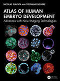
Atlas of Human Embryo Development: Advances with New Imaging Technologies
by Nicolas Plachta Stephanie BissiereThis important and exciting atlas shows with unprecedented resolution the development of the human embryo from fertilization to the end of pre-implantation development. Technology can now capture dynamic changes at the subcellular level that drive embryogenesis and development, allowing the whole team in reproductive medicine to move on from traditional imaging of fixed specimens or classic light microscopy to seeing, as if in 3D and real time, the process of fertilization and development of the human embryo – and of developmental defects. The human embryo research documented here will be of paramount importance for reproductive health, offering the chance to advance fertility treatments, genetic disease prevention, and our understanding of early human development.
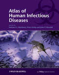
Atlas of Human Infectious Diseases
by Peter Horby John P. Woodall Heiman F.L.WertheimThe Atlas of Human Infectious Diseases provides a much needed practical and visual overview of the current distribution and determinants of major infectious diseases of humans. The comprehensive full-color maps show at a glance the areas with reported infections and outbreaks, and are accompanied by a concise summary of key information on the infectious agent and its clinical and epidemiological characteristics. Since infectious diseases are dynamic, the maps are presented in the context of a changing world, and how these changes are influencing the geographical distribution on human infections. This unique atlas: Contains more than 145 high quality full-color maps covering all major human infectious diseases Provides key information on the illustrated infectious diseases Has been compiled and reviewed by an editorial board of infectious disease experts from around the world The result is a concise atlas with a consistent format throughout, where material essential for understanding the global spatial distribution of infectious diseases has been thoughtfully assembled by international experts. Atlas of Human Infectious Diseases is an essential tool for infectious disease specialists, medical microbiologists, virologists, travel medicine specialists, and public health professionals. The Atlas of Human Infectious Diseases is accompanied by a FREE enhanced Wiley Desktop Edition - an interactive digital version of the book with downloadable images and text, highlighting and note-taking facilities, book-marking, cross-referencing, in-text searching, and linking to references and glossary terms.
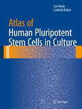
Atlas of Human Pluripotent Stem Cells in Culture
by Lyn Healy Ludmila RubanThis lavishly-illustrated, authoritative atlas explores the intricate art of culturing human pluripotent stem cells. Twelve chapters - containing more than 280 color illustrations - cover a variety of topics in pluripotent stem cell culturing including mouse and human fibroblasts, human embryonic stem cells and induced pluripotent stem cells, characteristic staining patterns, and abnormal cultures, among others. Atlas of Human Pluripotent Stem Cells in Culture is a comprehensive collection of illustrated techniques complemented by informative and educational captions examining what good quality cells look like and how they behave in various environments. Examples of perfect cultures are compared side-by-side to less-than-perfect and unacceptable examples of human embryonic and induced pluripotent stem cell colonies. This detailed and thorough atlas is an invaluable resource for researchers, teachers, and students who are interested in or working with stem cell culturing.
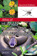
Atlas of Human Poisoning and Envenoming
by James H. DiazClinicians undergoing competency testing, certification, and periodic recertification are frequently faced with computer-based exams designed to evaluate clinical acumen and judgment. Test questions often include an image or radiograph followed by a vignette of the clinical encounter and a series of questions. Designed to better prepare practitione
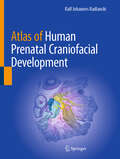
Atlas of Human Prenatal Craniofacial Development
by Ralf Johannes RadlanskiThis comprehensive atlas captures the prenatal formation of the human head with precision and detail, helping the reader discover the intricate world of craniofacial development. It serves as both a rich visual reference and a foundation for further research on how molecular signaling shapes the unique features of the craniofacial structure. Authored by a leading researcher with more than four decades in prenatal morphogenesis, this atlas goes beyond existing texts by offering an in-depth look at embryonic head formation through high-quality histology and advanced 3D reconstructions. More than 400 meticulously curated, vivid color images include detailed illustrations and 3D models that trace each crucial stage in the development of the human head's anatomical structures. An invaluable resource for scientists, medical professionals, educators, and anyone studying human development, the Atlas of Human Prenatal Craniofacial Development provides insights that lay the groundwork for understanding not only human morphology but also the roots of congenital anomalies.
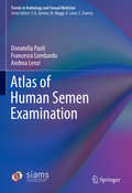
Atlas of Human Semen Examination (Trends in Andrology and Sexual Medicine)
by Andrea Lenzi Francesco Lombardo Donatella PaoliThis atlas provides valuable information on crucial aspects of sperm examination as well numerous meaningful color illustrations. It discusses successful evaluation of the sperm morphology and the cellular elements other than spermatozoa, enabling readers to unambiguously interpret seminal cytologic images and compare reports for diagnostic, therapeutic and prognostic purposes. With its extensive collection of colored images, the book is intended as a reference resource for students and technicians in the field of andrology as well as practitioners and clinicians in andrology, urology, pathology, IVF and other ART programs
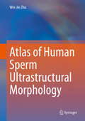
Atlas of Human Sperm Ultrastructural Morphology
by Wei-Jie ZhuThis atlas provides ultrastructural morphological images of human spermatozoa. Sperm morphology plays an essential role in sperm-oocyte interactions and early embryonic development, and human sperm ultrastructural morphology offers a valuable reference tool for assessing certain etiologies of male infertility and reproductive failure. However, the ultrastructural morphology of human sperm has not been systematically evaluated or thoroughly described in the literature. Using 470 original and unpublished images, the book shows various ultrastructural morphological phenotypes; defects of the sperm head, neck, middle piece, principal piece, and terminal piece; as well as artefacts of sperm ultrastructural morphology and phenomena related to inadequate preparation, demonstrating many sperm phenotypes and surface structural appearances for the first time. As such, it helps researchers and practitioners in andrology, reproductive medicine, and reproductive pathology gain a better understanding of human sperm ultrastructural morphology.
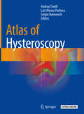
Atlas of Hysteroscopy
by Andrea Tinelli Luis Alonso Pacheco Sergio HaimovichThis book provides a wealth of detailed hysteroscopic images, illustrating the various gynecological pathologies that can be directly diagnosed by hysteroscopy. Providing comprehensive coverage, the book examines each pathology on the basis of concrete images, offering readers support with the diagnostic evaluation. This is especially important when there are recrudescent pathologies, such as TBC, which can often confuse physicians in the context of diagnosis, and with regard to oncological, infectious and infertility issues. Written by prominent experts in the field, this atlas offers an extremely useful and comprehensive tool for all gynecologists, which can support them to overcome any diagnostic doubts and arrive at an accurate diagnosis.
Atlas of Ileoscopy: A Collection of Clinical Cases
by Antonello TreccaThis atlas presents high-quality images of clinical cases of small bowel diseases, with a focus on the importance of terminal ileoscopy as the first step in detecting this group of rare pathologies. Among the conditions covered are celiac disease, inflammatory bowel disease, neoplasms, infections, and pediatric disease. The multidisciplinary approach to the presentation of clinical cases, with contributions from histology, radiology, capsule endoscopy, enteroscopy, and surgery, ensures that the atlas will provide an accurate learning course for beginners and also an effective update for specialists.
Atlas of Imaging Anatomy
by Lucio OlivettiThis book is designed to meet the needs of radiologists and radiographers by clearly depicting the anatomy that is generally visible on imaging studies. It presents the normal appearances on the most frequently used imaging techniques, including conventional radiology, ultrasound, computed tomography, and magnetic resonance imaging. Similarly, all relevant body regions are covered: brain, spine, head and neck, chest, mediastinum and heart, abdomen, gastrointestinal tract, liver, biliary tract, pancreas, urinary tract, and musculoskeletal system. The text accompanying the images describes the normal anatomy in a straightforward way and provides the medical information required in order to understand why we see what we see on diagnostic images. Helpful correlative anatomic illustrations in color have been created by a team of medical illustrators to further facilitate understanding.
Atlas of Imaging in Cardio-Oncology: Case-Based Study Guide
by Richard M. Steingart Jennifer E. LiuThis atlas provides a practically applicable didatic case-based guide to the use of cardiac imaging in patient care during and after cancer therapy. It features detailed information on a variety of imaging modalities, including transesophageal echocardiography, magnetic resonance angiography and positron emission tomography. A range of actual patient presentations are included covering both diagnosis and management during curative and palliative surgery, as well as cancer pharmacotherapy with the use of drugs including Trastuzumab, Anthracycline, Cisplatin and Carboplatin. Topics relevant to survivorship are also described. Atlas of Imaging in Cardio-Oncology enables the reader to develop a deep understanding of how to utilise a variety of imaging modalities used in cardio-oncology in a range of scenarios. It provides a critical and timely resource for the trainee and experienced cardiologist, oncologist and radiologist.
Atlas of Imaging in Infertility: A Complete Guide Based in Key Images
by Luis Ronan Marquez Ferreira de Souza Ana Luisa Alencar De Nicola Harley De NicolaThe present Atlas intends to gather the most frequent findings of alterations that causes infertility, both in male and in female, and how they should be analyzed and described, to serve as a guide for physicians who will ask and will perform these imaging tests. There are a group of professionals that may be interested on further knowledge on infertility, since this is a highly prevalent condition: about 10% of married couples are infertile and males and females are equally affected. These professionals include endocrinologists, gynecologists and urologists. Moreover, these professionals shall make good use of additional tests, in order to properly treat their patients whenever they suspect of or face a case of infertility. Among these tests, the diagnostic imaging exams are essential. Thus, the Atlas of Imaging in Infertility intends to provide such knowledge, allowing physicians of different specialties to a manage of cases of infertility, both in males and in females.
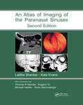
Atlas of Imaging of the Paranasal Sinuses, Second Edition
by Kate Evans, Thomas R. Marotta, Eugene Yu, Michael Hawke and Heinz StammbergerWith color illustrations, the Second Edition of this best-selling guide concentrates on the advances in technology that are now available to the clinical otolaryngologist. This reinforces the book's position as a classic guide, especially to the problems associated with endoscopic sinus surgery.
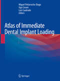
Atlas of Immediate Dental Implant Loading
by Miguel Peñarrocha-Diago Ugo Covani Luis CuadradoThis atlas, in which a wealth of illustrations are supported by clear explanatory text, offers an up-to-date and comprehensive overview of the immediate restoration of teeth and immediate functional loading when using different implant systems and surfaces in patients with single tooth loss or partial or complete edentulism. It provides guidance on all aspects of technique, including procedures for impression and measurement taking, and describes the surgical and prosthetic protocols applicable in various settings. The coverage encompasses the more advanced techniques used for immediate loading of implants placed in conjunction with grafting/augmentation procedures or in fresh extraction sockets, as well as immediate implant loading for mandibular and maxillary full-arch rehabilitation. This atlas will help dental students and practitioners to gain a sound understanding of immediate loading techniques, including their indications and limitations, and to apply them optimally in their practice. The atlas also shows and explains how to integrate a full digital workflow from the intraoral scanner to solve complex cases in a simple way.
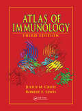
Atlas of Immunology
by Robert E. Lewis Julius M. Cruse MD PhDIn the 11 years since this atlas first published, the immunology field has experienced an exponential increase in information. Besides the unprecedented advances in knowledge of cell receptors and signal transduction pathways, an avalanche of new information has been gleaned from contemporary research concerning cytokines and chemokines, with speci
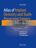
Atlas of Implant Dentistry and Tooth-Preserving Surgery: Prevention and Management of Complications
by Zoran StajčićWeighs the pros and cons of tooth-preserving surgical procedures against implant dentistry.<P> Offers extensive guidance on the etiology, prevention, and management of complications.<P> Includes numerous full color clinical photos of step-by-step surgical procedures.<P>This atlas is unique in comparing the two disciplines of dental implant surgery and tooth-preserving surgery with respect to common procedures, problems, and failures and in providing excellent guidance on the prevention and management of complications. The etiology of a wide variety of implant-related and non-implant-related complications and failures is carefully explained. Since many complications have their roots in oral and periodontal surgical maneuvers, these maneuvers are themselves discussed and extensively illustrated. The most frequently used tooth preservation procedures are also fully described, with emphasis on correct surgical technique as a means to avoid complications. The use of these procedures is constantly weighed against the effects of tooth removal and insertion of dental implants. The text is complemented by the inclusion of a substantial number of helpful references. While the atlas is intended primarily for dentists involved in outpatient implant dentistry and oral surgery, oral and maxillofacial surgeons will also find the descriptions of innovative techniques or maneuvers to be of interest, including those relating to the selection of incision and flap design and the sinus floor elevation techniques.
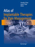
Atlas of Implantable Therapies for Pain Management
by Jason E. Pope Timothy R. DeerFrom reviews of the First Edition: "For the practitioner new to the field of implantable therapies, this book shows the scope of this particular branch of pain medicine. It is full of good advice on techniques and how to avoid complications. " British Journal of Clinical Pharmacology "All practicing pain physicians, whether their background is in anesthesia or neurosurgery, will find [this atlas] very useful. . . beautifully written and well-illustrated with x-ray images and pictures. . . a must-read for every pain fellow and a very useful one for every pain physician. " Doody's Review Service Now in its Second Edition, this atlas remains an essential guide to the treatment of pain using neuromodulation and is written for all implanters, from beginners to the most advanced practitioners. It has been significantly expanded with many brand new chapters and figures and thoroughly updated to address new techniques, targets, waveforms, and concepts that have emerged since publication of the last edition and now provides even more detailed coverage of patient safety, including infection control and reduction of bleeding risks. The new and returning physicians who have collaborated on the Second Edition are world-class in their research, clinical expertise, and ethics of practice. They have endeavored to make each segment of the atlas a great learning event through careful integration of extensive photographs, illustrations, and detailed instructions and to create a resource that helps to improve practice and enhance outcomes while complementing fellowship training, peer-to-peer experiences, and hands-on continuing medical education.
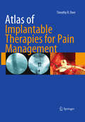
Atlas of Implantable Therapies for Pain Management
by Timothy R DeerThis Atlas serves as a guide to beginning implanters, intermediate implanters, and the most advanced practitioners. The author covers the process of implanting and managing spinal cord stimulators, peripheral nerve stimulators, and intrathecal pumps from the beginning of the process to long term management. The book also discusses the recognition, prevention, and management of complications. The Atlas is a must for any physician hoping to improve their skills in any segment of this important area of interventional pain treatment. The combination of instructional photographs and detailed instructions makes each segment a great learning event.
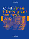
Atlas of Infections in Neurosurgery and Spinal Surgery
by Ali AkhaddarThis Atlas is the first reference Atlas covering exclusively all aspects of this multifaceted topic. It is designed to serve as a succinct appropriate resource for neurosurgeons, spinal surgeons, radiologists, neurologists, microbiologists, researchers and infectious disease specialists with an interest in cranio-cerebral and vertebro-medullary infections especially encountered in neurosurgery and spinal surgery. This Atlas is designed to deliver more information in less space than traditional texts, allowing for quick review of the essential facts of this complex infectious topic through pictures. Pertinent imaging and laboratory information are combined with intraoperative photographs and illustrations to help readers visualize variable presentations and enhance their perioperative management. The comprehensive content of this richly-illustrated book covers different infectious diseases seen on neurosurgical and spinal practices. The Atlas is divided into five sections, after a general introduction, the second section focuses on infections of the brain and its coverings. The third section focuses on vertebromedullary infections. The fourth section includes infections following cranial and spinal surgery, and the fifth section provides a description of the most important specific pathogens and other particular conditions. The format makes it easily accessible and includes a definition of each infection and its epidemiology, main clinical presentations, imaging features and laboratory findings, treatment options, and prognosis information. It will help the reader in choosing the most appropriate way to manage this multipart problem. In addition, the book supplies clinicians and investigators with both basic and more sophisticated information and procedures relating to the complications associated with neurosurgical and spinal infections.
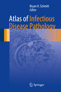
Atlas of Infectious Disease Pathology (Atlas of Anatomic Pathology)
by Bryan H. SchmittProvides a comprehensive overview of the histopathology of the majority of infectious diseases.<P><P> Richly illustrated Atlas with 418 full color photographs.<P> Written by experts in the field.<P> Infectious diseases may be encountered in nearly every aspect of pathology. This atlas provides an informative reference for the identification of the common and esoteric pathogens, presenting in a wide array of specimen types. The focus of the presented images is on the hematoxylin and eosin-stained appearances of these infections and highlight common special stains that can be used to aid in the diagnosis of the infectious agent. Where appropriate, commentary regarding additional testing such as immunohistochemistry and molecular-based methods is supplied. The Atlas of Infectious Disease Pathology is organized primarily by pathogen type followed by discussion of the various manifestations that may occur in individual organ systems. The reader will be provided with a comprehensive overview of the histopathology of the majority of infectious diseases encountered in general and subspecialty practice alike.
Atlas of Inflammatory Bowel Disease-Associated Intestinal Cancer: Examining the Macroscopic Images of Small and Large Intestine
by Toshiyuki Matsui Takayuki Matsumoto Akinori Iwashita Takashi Hisabe Kitaro Futami Hiroshi TanabeThis book is structured in three major parts and the first part is a comprehensive overview of IBD-related intestinal cancer. The second and third parts provide rare and important cases on ulcerative colitis (UD) and Crohn’s disease (CD) presenting the macroscopic image of the resected specimen and analyzing the endoscopic and the pathological image to explain how to find the IBD cancer. It also delivers a detailed analysis of the histological structure of cancers. This book tries to clarify the critical but yet to be answered questions such as “Why is it so difficult to find out?”, “Why so flat?” and “How to name superficial lesion with invisible margin?” and so on.Atlas of Inflammatory Bowel Disease-Associated Intestinal Cancer is designed to serve all endoscopists and gastroenterologists managing IBD patients. It also is valuable reading for pathologists who are diagnosing gastrointestinal lesions. This book is designed to assist these medical professionals to find cancers in the early stage and stay up-to-date on image-enhanced endoscopy techniques. This book is a translation of an original Japanese edition. This has been facilitated using machine translation (by the service DeepL.com) followed by editors and authors revising, editing and verifying the translated manuscript.
Atlas of Inflammatory Bowel Diseases
by Won Ho Kim Jae Hee CheonThis comprehensive atlas, containing a wealth of high-quality images, illustrates the complete spectrum of presentations of inflammatory bowel diseases (IBD). It focuses especially on the most recent developments in the use of endoscopy in IBD, providing detailed guidance on endoscopic indices of disease activity, diagnosis, and differential diagnosis. In addition to ileocolonoscopy, small bowel endoscopy, and esophagogastroduodenoscopy, chapters are included on the role of both established radiological techniques, such as CT, MRI, and abdominal ultrasonography, and the newest approaches, including high-resolution endoscopy, narrow band imaging, and confocal laser endomicroscopy. Extraintestinal manifestations and complications are addressed in separate chapters, and the book concludes by presenting surgical findings. The authors are international authorities with diverse expert knowledge who have collaborated to create an ideal tool for all who wish to master endoscopic evaluation in IBD.
Atlas of Inherited Metabolic Diseases
by William L Nyhan Georg F HoffmannIn a field where even experts may find that years have elapsed since they last encountered a child with a given disorder, it is essential for the clinician to have a comprehensive source of practical and highly illustrated information covering the whole spectrum of metabolic disease to refer to. The content is divided into sections of related disorders, including disorders of amino acid metabolism, lipid storage disorders, and mitochondrial diseases for ease of reference, with an introductory outline where appropriate summarizing the biochemical features and general management issues. Within the sections, each chapter deals with an individual disease, opening with a useful summary of major phenotypic expression including clear and helpful biochemical pathways, identifying for the reader exactly where the defect occurs. Throughout the book, plentiful photographs, often showing extremely rare disorders, are an invaluable aid to diagnosis. Key Features • Fully updated to incorporate all new developments in the field • Brand new chapters cover methylmalonic aciduria of ACSF3 deficiency, branched chain keto acid dehydrogenase deficiency, serine deficiencies, purine nucleoside phosphorylase deficiency, antiquitin deficiency, and others • Excellent and detailed clinical descriptions, with numerous valuable hints and suggestions for management • Helpful explanatory algorithms and decision trees, and high-quality illustrative material including biochemical pathways and an unrivaled photographic collection, which enhance clinical applicability The fourth edition of this highly regarded book, authored by two of the foremost authorities in pediatric metabolic medicine, continues to provide incomparable insight into the problems associated with metabolic diseases and remains invaluable to pediatricians, geneticists, and general clinicians worldwide.
Atlas of Inherited Retinal Diseases (Advances in Experimental Medicine and Biology #1467)
by Stephen H. Tsang Tarun Sharma Vlad DiaconitaThis atlas provides a thorough overview of various inherited retinal dystrophies with an emphasis on phenotype characteristics and how they relate to the most frequently encountered genes. It also meets the previously unmet needs of PhD students who will benefit from seeing the phenotypes of genes they work on and study. Further, because genetic-testing costs are quite high and spiraling higher, this atlas will help geneticists familiarize themselves with the candidate gene approach to test patients’ genomes, enabling more cost-efficient testing. This invaluable atlas is organized into eight sections starting with an introduction to the basic knowledge on retinal imaging, followed by diseases listed according to inheritance pattern and disorders with extraocular manifestations grouped by defining features. This structure will be intuitive to clinicians and students studying inherited retinal disorders. In this thoroughly updated new edition featuring new images and the latest research developments, new chapters on the history taking of IRD suspect patients. Updated chapters on mimics of IRD diseases, as well as a novel section on current available treatments.
Atlas of Inherited Retinal Diseases (Advances in Experimental Medicines and Biology #1085)
by Stephen H. Tsang Tarun SharmaThis Atlas of Inherited Retinal Disorders provides a thorough overview of various inherited retinal dystrophies with emphasis on phenotype characteristics and how they relate to the most frequently encountered genes. It also meets the previously unmet needs of PhD students who will benefit from seeing the phenotypes of genes they work on and study. Further, because genetic-testing costs are quite high and spiraling higher, this Atlas will help geneticists familiarize themselves with the candidate gene approach to test patients’ genomes, enabling more cost-efficient testing. This invaluable atlas is organized into eight sections starting with an introduction to the basic knowledge on retinal imaging, followed by diseases listed according to inheritance pattern and disorders with extraocular manifestations grouped by defining features. This structure will be intuitive to clinicians and students studying inherited retinal disorders.