- Table View
- List View
Atlas of Interstitial Cells of Cajal in the Gastrointestinal Tract
by Terumasa KomuroThis atlas will illustrate the distribution and morphological features of interstitial cells of Cajal (ICC) which are the key cells to understanding of the regulatory mechanism of gastrointestinal motility, since ICC act as both pacemaker and as intermediates in neural transmission, and since ICC show specific distribution patterns depending on their anatomical positions. All subtypes of ICC located in the different tissue layers and different levels of the gastrointestinal tract will be revealed by immunohistochemistry for Kit receptors and nerves by using mainly whole-mount stretch preparation of the guinea-pig tissues. Three-dimensional reconstruction of confocal images will particularly help the readers to understand the peculiar arrangement of ICC networks in situ and the correlation between ICC and nerves. Electron micrographs will help illustrate the characteristic features of ICC and their ultrastructural differences from fibroblasts, smooth muscles and other interstitial cells.
Atlas of Interstitial Lung Disease Pathology: Pathology With High Resolution Ct Correlations
by Andrew Churg Neston L. MullerWith a concise, image-rich format, Atlas of Interstitial Lung Disease Pathology, Second Edition, features new entities, classifications, molecular pathways, and other discoveries introduced to the field since the previous edition. Each chapter addresses radiological, clinical, and prognostic features, and boasts numerous illustrations of pathologic findings—all to make it easier for you to better understand and make an accurate diagnosis of ILD.
Atlas of Interstitial Lung Disease Pathology: With High Resolution CT Correlations
by Andrew Churg Nestor L. MüllerThis objective of this atlas is to serve as an updated diagnostic resource that allows pathologists to diagnose interstitial lung disease (ILD) when they encounter it in biopsies. Pathologists often struggle in this area due to the numerous different and sometimes overlapping entities encompassed by term &“ILD&”. ILD pathology is unique in that diagnostic accuracy always requires correlation with the clinical and particularly the radiologic findings. This is different from most areas of pathology in which only minimal clinical/radiologic information will allow an accurate diagnosis. By including CT findings, pathologists will know what to look for in radiology reports or when talking with radiologists. A further recent development has been the increasingly widespread use of transbronchial cryobiopsy for diagnosing interstitial lung disease. What is becoming evident, however, is that there is astonishingly little in the way of pathologic guidance for making diagnoses with cryobiopsies, and pathologists struggle with them. Advances in understanding the genetics of ILD have led to realization that there are major overlaps in the genetic abnormalities associated with a UIP pattern, even if the underlying disease is fibrotic hypersensitivity pneumonitis or connective tissue disease. A new idea is that of telomeropathy; i.e. the finding that patients with short telomeres are particularly likely to develop fibrotic lung disease, and, most importantly, should not be treated with immunosuppressive agents. Atlas of Interstitial Lung Disease: Pathology with High Resolution CT Correlations 3rd Edition is primarily directed toward non-expert pathologists who encounter interstitial lung disease specimens, as well as radiologists who want some understanding of the pathologic basis for the imaging findings. The book incorporates current clinical/radiological/pathologic guidelines and provides information on how to apply them to biopsy specimens.
Atlas of Interventional EUS: Case-based Strategies
by Anthony Y. B. Teoh Marc Giovaninni Mouen A. Khashab Takao ItoiThis atlas aims to systematically provide a case-by-case discussion on selected interventional endoscopic ultrasonography (EUS) procedures and how to approach such procedures on different patients. The atlas offers illustrations of the interventional procedures and several cases of the procedures are included. Within each case, a standardized report is provided including a brief history, imaging findings, a detailed discussion on treatment options, considerations in the EUS intervention, how to perform the procedures and final outcomes. EUS-guided drainages of the bile duct, pancreatic duct, gallbladder and abscesses are summarized. Ablative procedures including celiac plexus, pancreatic cyst and radiofrequency ablations are also covered. Other procedures guided by EUS, including gastroenterostomy, injected therapy, iodine beads insertion and fiducial marker insertion and endovascular interventions are further demonstrated. Gastroenterologists and surgeons can benefit from this atlas on learning strategies and preparing for these procedures.
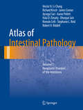
Atlas of Intestinal Pathology: Volume 1: Neoplastic Diseases of the Intestines (Atlas of Anatomic Pathology)
by Hector H. Li-Chang Richard Kirsch James Conner Aysegul Sari Aaron Pollett Hala El-Zimaity Dhanpat Jain Romulo Celli Stephanie L. Reid Robert H. RiddellIntestinal neoplasia comprises a large part of a surgical pathologist’s workload. Pathologists play a key role not only in the classification of malignancies but also in assisting screening programs, identifying incidental neoplasms, and guiding treatment by providing essential prognostic features for individual entities. A large variety of neoplasms affect the intestines, and there is ongoing discovery of new entities and prognostic features for known diseases. Pathologists and trainees should have a solid understanding of key morphologic features, pitfalls, and differential diagnoses. Importantly, pathologists should recognize and communicate features that help their clinical colleagues in making treatment decisions, with the ultimate goal of benefiting the patient first and foremost.The first volume of the Atlas of Intestinal Pathology provides a comprehensive yet concise, primarily visual review of intestinal neoplasms. It also serves as a useful resource primarily for pathologists and trainees in pathology by providing a concise yet comprehensive summary of morphology of intestinal neoplasia. Clinical practitioners and trainees also benefit from an understanding of the pathologic correlates to the diseases they manage.
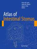
Atlas of Intestinal Stomas
by Victor W. Fazio James S. Wu James M. ChurchDesigned to provide a highly visual reference for surgeons and other members of the patient management team, Atlas of Intestinal Stomas is based on the 1967 gold standard text, Turnbull and Weakly's Atlas of Intestinal Stomas. Additions include chapters on anatomy and physiology, biliary stomas, pediatric ostomies, the continent ileostomy, urostomy, laparoscopic stoma construction, stomas in trauma surgery, stomas for antegrade continence enema, percutaneous ostomies, and quality of life. There are also sections on ileostomy, colostomy, enterostomal therapy and on the management of complications of stomas such as management of the high output ostomy, enterocutaneous fistula, parastomal hernia, prolapse, and skin conditions. The Cleveland Clinic pioneered the entire practice of ostomies, beginning in 1858 and continuing to this day as the world's leading academic and clinical center. The editors and contributors are all current or former Cleveland Clinic physicians and instructors. The fundamental focus of the book is not only how to install ostomies, but how to avoid complications and how to treat complications when they arise. Atlas of Intestinal Stomas will be of great value to colorectal and general surgeons, both in practice and in training.
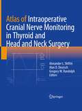
Atlas of Intraoperative Cranial Nerve Monitoring in Thyroid and Head and Neck Surgery
by Gregory W. Randolph Alexander L. Shifrin Alan D. DeutschThis comprehensive atlas is the modern, state-of-the-art guide for intraoperative neurophysiologic monitoring (IONM) and management of the recurrent laryngeal nerve, vagus nerve and other cranial nerves at risk during thyroid, parathyroid and modified radical neck dissection surgery. Based on real-time electrophysiologic images, it will assist the surgeon in the decision-making process by incorporating important information related to the identification of the nerves and their functional status, aiding in the interpretation and improvement of the quality of neural monitoring and reducing inappropriate variations in monitoring technique. Utilization of IONM enables the surgeon to interrogate nerve anatomy and function with immediate quantitative feedback, thereby augmenting surgical training, and importantly, surgical skills and sound anatomic knowledge remain prerequisite and are not supplanted by IONM use. Authored by experts in the field, Atlas of Intraoperative Cranial Nerve Monitoring in Thyroid and Head and Neck Surgery will be the gold-standard text for IONM for endocrine surgeons, otolaryngology surgeons, neurophysiologists, and head and neck surgeons, as well as fellows and residents in these areas.
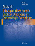
Atlas of Intraoperative Frozen Section Diagnosis in Gynecologic Pathology
by Pei Hui Natalia BuzaThis atlas is dedicated specifically to gynecologic frozen section diagnosis and addresses professional practice gaps such as high diagnostic error rate, slow turnaround time, and inefficient communication between surgeons and pathologists at the time of intraoperative frozen section consultation of gynecologic specimens. The format of the volume is a combination of concise text and high quality gross and frozen section microscopic images, meticulously selected from the superb collection of pathology specimens of gynecologic tumors provided at Yale-New Haven Hospital in the past decades. Frequent entities with diagnostic pitfalls are balanced with less common lesions. Strategies to recognize their diagnostic features are emphasized, in addition to the impact on optimal surgical treatment of patients with gynecologic cancer. The indications, limitations, morphologic diagnostic criteria and pitfalls of frozen section consultation in gynecologic pathology are thoroughly reviewed with an ultimate goal of avoiding patient mismanagement in real-time. High quality frozen section microscopic illustrations aid the recognition of morphologic patterns and serve as quick reference during intraoperative consultation. Written by experts in the field, Atlas of Intraoperative Frozen Section Diagnosis in Gynecologic Pathology is a valuable resource for pathologists at all career/expertise levels who are involved in intraoperative consultation in their daily clinical practice.
Atlas of Keystone Reconstructive Technique in Melanoma Management
by Felix BehanWorldwide, it is quoted that 85% of Melanoma cases are treated surgically. The reliability of the Keystone Perforator Island Flap (KPIF) as a reconstructive technique has wide applications. Its low complication rate is the key to its surgical success. The technique is clearly explained in this Atlas by the author, a renowned plastic and reconstructive surgeon, who established this reconstructive principle over 20 years. The illustrations range from simple to complex cases in an easy-to-follow format. Audio files, as a supplementary tool, amplify the technique to enhance the reader’s experience and foster clear understanding. Chapters focussing on anatomical regions contain carefully selected cases accompanied by images to demonstrate a step-by-step surgical technique. The basic design format for all Keystones are highlighted, all sitting within the dermatomal precincts. The KPIF must contain a fascial base for lining and the island outline is an essential pre-requisite. It is hypothesized that this islanding creates a sympathectomy effect resulting in hyperaemic blood flow which optimises healing. Undermining up to 2/3 is permissible as long as there is a deep attachment of 1/3 at the base of the flap to contain the random perforator vascular support. This design allows the rotation, advancement and transposition (ART) of the flap to facilitate the reconstructive closures. Presumably, there are somatic and autonomic neural support lines accompanying the vascular pathways of these random perforator flaps. The vascular perforator tree must not be skeletonised (as in propellor flaps) to allow preservation of such anatomical elements - somatic, autonomic and lymphatic pathways, all of which are an essential component in any surgical repair. This KPIF provides an alternative to microvascular procedures where the biggest drawback is tissue match. The KPIF addresses this problem admirably with a low pain component, an excellent aesthetic match with a low vascular complication rate and a performed in an expeditious manner – these all characterise the KPIF. Simple solutions solve problems and this KPIF Atlas for Melanoma becomes a welcome companion to any surgical speciality including plastic and reconstructive surgeons, surgical oncologists, general surgeons and dermatologists.
Atlas of Knee Arthroscopy
by Radu PrejbeanuThis atlas provides readers with a concise and accessible resource for performing knee arthroscopy, one of the most common orthopaedic procedures in the US and increasingly around the world. Illustrated with over 150 surgical images, residents, consultants and senior surgeons alike will find this atlas to be a key reference for improving knee arthroscopy procedures and outcomes for patients.
Atlas of Lacrimal Drainage Disorders
by Mohammad Javed AliWritten by an expert in the field, this book is a comprehensive and up-to-date guide to the evaluation and management of lacrimal drainage disorders. Lacrimal disorders are one of the most common conditions encountered not only by oculoplastic surgeons and general ophthalmologists, but also by otorhinolaryngologists in their daily practice. Consisting of 77 chapters, it addresses the basic anatomy and underlying pathology, patient evaluation, and the surgical procedures currently performed in managing various lacrimal disorders. Surgical modalities including the endoscopic approaches are thoroughly and succinctly captured in pictures with detailed legends to aid understanding and offer a visual treat. Since familiarity with a surgical technique is incomplete without the knowledge of risk factors and red flags, the book discusses in detail how to deal with surgical complications and failure. The Atlas of Lacrimal Drainage Disorders is an essential companion to the a uthor's previous work "Principles and Practice of Lacrimal Surgery". .
Atlas of Lacrimal Drainage Disorders
by Mohammad Javed AliThe second edition of this successful Atlas provides an updated and comprehensive guide to the evaluation and management of lacrimal drainage disorders with 4400+ images.. Lacrimal disorders are one of the most common conditions encountered not only by oculoplastic surgeons and general ophthalmologists, but also by otorhinolaryngologists in their daily practice. It is authored by a world renowned expert in the field. The 2nd edition consists of 103 chapters addressing the basic anatomy and underlying pathology, patient evaluation, and the surgical procedures currently performed in managing various lacrimal disorders. Surgical modalities including the endoscopic approaches are thoroughly and succinctly captured in pictures with detailed legends to aid understanding and offer a visual treat. The book discusses how to deal with surgical complications and failure in detail since familiarity with a surgical technique is incomplete without the knowledge of risk factors and redflags.The 2nd edition of the Atlas of Lacrimal Drainage Disorders is an essential companion to the author’s previous work ‘Principles and Practice of Lacrimal Surgery’.This detailed guide is an indispensable resource for practicing ophthalmologists, oculoplastic surgeons, dacryologists, ophthalmology residents, ophthalmology fellows, practicing otorhinolaryngologists, otolaryngology residents and rhinology fellows.
Atlas of Laparoscopic Gastrectomy for Gastric Cancer: High Resolution Image for New Surgical Technique
by Ping Li Chang-Ming Huang Chao-Hui Zheng Jian-Wei XieDue to recent advances in laparoscopic gastrectomy for gastric cancer, a high-resolution atlas in this field was felt necessary. This book describes the laparoscopic surgical procedure and precautions of lymph node (LN) dissection, comprehensively. The details of preoperative preparation, regional LN dissection, and digestive tract reconstruction are introduced based on a large numbers of clinical cases. Modified intracorporeal anastomosis procedure and clinical application of indo cyanine green (ICG) in LN dissection are included. This atlas will be of benefit to gastrointestinal surgeons, surgical oncologists, and minimally invasive surgeons.
Atlas of Laparoscopic Gynecological Anatomy
by Helizabet Salomão Ayroza Paulo Ayroza RibeiroThis book allows readers to gain a comprehensive understanding of gynecological surgical anatomy from a laparoscopic perspective. In recent years, with the growing number of gynecological surgical procedures performed by laparoscopy, many surgeons are faced with a “new” anatomy, not yet presented in traditional books. Addressing this gap in the literature and written in a colloquial style, this book presents much-needed information, especially regarding the spaces and the vessels, as well as numerous surgical tips and tricks. Focusing on retroperitoneal dissection, gynecological oncology and endometriosis, the book is intended for surgeons (gynecologists, urologists, general surgeons and others) interested in performing advanced pelvic surgery, offering them insights into how to transfer their knowledge of the traditional open surgery anatomy to the laparoscopic anatomy. Further, the book addresses 2D visualization and changes in the angle of visualization. The Atlas of Laparoscopic Gynecological Anatomy includes photos, surgical videos, drawings and figures to help readers quickly grasp the new concepts and to enhance the teaching power of the text.
Atlas of Laparoscopic Urologic Surgery
by Jan Roigas Serder Deger Stefan A Loening Andreas H WilleIdeal not only for the beginners in the field, but also for urologists looking to expand their knowledge, this clearly presented book adopts a useful step-by-step format to present established laparoscopic procedures. Easy and quick to use, the text includes a wealth of illustrations with important instructions, as well as tips and tricks described
Atlas of Laparoscopic and Robotic Single Site Surgery (Current Clinical Urology)
by Robert J. Stein Jihad H. Kaouk Georges-Pascal HaberThis text provides a broad and current review of this field and will serve as a valuable resource for trainees, academic and community surgeons, and members of industry with an interest in LESS. Due to the novelty and complexity of these procedures, the book focuses on detailed descriptions as well as pertinent illustrations for various upper and lower tract urologic procedures. The development of novel minimally invasive and robotic technology for more comfortable performance of these demanding procedures is covered. A complete description of instrumentation, platforms, and optics developed specifically for LESS is another primary focus of this text. Finally, a description of outcomes and complications as well as comparative data defining the status of LESS in relation to other current minimally invasive techniques is offered. Atlas of Laparoscopic and Robotic Single Site Surgery will provide a detailed summary of the current status of LESS that will help guide surgical decision making, encourage investigative efforts, and stimulate industry led technology development.
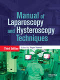
Atlas of Laparoscopy and Hysteroscopy Techniques
by Togas TulandiThis beautifully illustrated book provides a practical step-by-step guide to all the laparoscopic and hysteroscopic procedures performed by gynecologists. Each procedure is described in detail and fully illustrated with color photographs. The potential complications are described, and the circumstances in which a procedure is contraindicated are ex
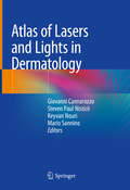
Atlas of Lasers and Lights in Dermatology
by Keyvan Nouri Giovanni Cannarozzo Steven Paul Nisticò Mario SanninoThis richly illustrated atlas written by a team of experts guides the reader to the applications of lasers and light technologies in dermatology. It is divided in two parts: the first reviews the physical and optical concepts related to lasers and light sources, and provides a detailed description of surgical (ablative and non-ablative), vascular and pigmentary laser devices. It also discusses difficult-to-treat conditions, such as melasma and scars. The second part of the atlas is more clinically-oriented, presenting reproducible parameters and high-resolution images of pre and post-treatment, and desired end points in order to achieve an optimal result. Enabling readers to gain an understanding of the various topics concerning lasers, it explores conventional, non-conventional and combined laser treatments in a wide range of indications, as well as practical aspects such as medicolegal issues, informed consent and management of complications. The increasing knowledge and growing expertise in lasers and light devices make it necessary for physicians to be aware of the latest developments in this quickly evolving field. As such, this book is of interest to all physicians working in dermatology, cosmetology and aesthetic medicine, as well as to physician assistants and nurses using lasers in their daily practice.
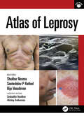
Atlas of Leprosy
by Shekhar Neema Biju Vasudevan Santoshdev P Rathod Senkadhir Vendhan Akshay AmbasanaAtlas of Leprosy comprehensively covers the varied manifestations, including common and uncommon presentations, of leprosy, supported by patient photographs and additional video content, to assist medical practitioners diagnose leprosy in the early stages.With a detailed guide to approaching bedside diagnostic tests, this book will help readers to visualise common and uncommon presentations of leprosy, enabling effective diagnosis and treatment in this pre-elimination era, where increasingly difficulties in identifying its early stages can lead to preventable morbidity and deformity.Clinicians, dermatologists and trainees alike will find the wealth of high-quality illustrations and video demonstrations a vital guide to better diagnose this mycobacterial disease and combat resistance, relapse, reactions and any other difficult challenges posed.
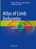
Atlas of Limb Deformity: Etiological Classification
by Sihe Qin Wen Tian Qianyu Zhuang Baofeng GuoThis book focuses on classification and category of etiology, clinical manifestations, diagnosis, and orthopedic principles of different types of limb deformities in China. The relativity of the definition of limb deformity, the cognitive history of human beings on limb deformity and disability for thousands of years, and the characteristics and classification methods of the diseases category of limb deformity in Chinese population are covered in the chapters, in which the dynamic nature of the occurrence and development of limb deformity from the perspective of biological evolution and auxology, is analyzed too. Written by experts with wealthy of experience, it will be an ideal reference for physicians involved in the diagnosis and treatment of limb deformity.
Atlas of Lip and Nose Plastic and Cosmetic Surgery
by Jianhua Liu Bing ShiThis Atlas covers various types of lip and nose deformities and introduces individualized surgical plan for patients. The surgical techniques are classified according to the various types of deformities. Schematic diagrams, pre-operative and post-operative photographs are supplemented by detailed explanations. It consists of four parts: anatomical structure of lips and nose; cosmetic surgery of lips and nose; plastic surgery of nasolabial congenital deformity; plastic surgery of acquired nasolabial defect and deformities.The management of nasolabial deformities and defects crosses many surgical disciplines, and therefore, this book recommended to be read by surgeons of different backgrounds i.e. oral and maxillofacial surgery, plastic surgery, cosmetic surgery, otorhinolaryngology, etc. It can also be used as a reference for anatomy, developmental biology, physiology, linguistics, aesthetics and other basic medical sciences.
Atlas of Liquid Biopsy: Circulating Tumor Cells and Other Rare Cells in Cancer Patients’ Blood
by Ludmilla Thomé Domingos ChinenThis book provides a wealth of images and extensive information on circulating tumor cells (CTCs) and other cells usually observed in blood from patients with cancer, such as giant macrophages, and circulating tumor microemboli (CTM). The field of “liquid biopsy” began with the analysis of CTCs in the early 2000s. In the beginning, molecular techniques were developed to detect these cells in the blood. However, it has since become clear that CTCs initially require a cytopathological analysis to be detected without false positive and negative results. After detection, molecular analysis can be subsequently performed. Cancer is an important cause of mortality, especially when detected in late stages. Even with all the advances that have been made in its treatment, cancer is still challenging, as many patients do not respond to any therapy. Many health agencies have considered early diagnosis as a feasible tool. In this context, it is of the utmost importance to know the morphology and characteristics of CTCs to determine a correct diagnosis. Currently much of the scientific community is committed to expanding our knowledge of CTCs, and this work makes a valuable contribution, presenting hundreds of cell images from patients with various types of cancer, in many different stages of disease, and after receiving various treatments. The Atlas of Liquid Biopsy: Circulating Tumor Cells and Other Rare Cells in Cancer Patients’ Blood is an essential reference guide for all physicians, biologists, biomedics, and professionals working at clinical and research laboratories, hospitals and research centers.
Atlas of Liver Pathology (Atlas of Anatomic Pathology)
by Anthony W. H. Chan Alberto Quaglia Beate Haugk Alastair BurtThe liver is a complex organ due to its unique microscopic structure, intricate metabolic functions and susceptibility to a wide variety of insults, manifesting in countless histological patterns. Atlas of Liver Pathology considers both changes seen in medical liver biopsies as well as lesional biopsies when the specimen has been taken from a mass. The book starts by reviewing normal structure and its variants and the optimal approaches for the preparation of histological sections for diagnostic liver pathology. The following chapters are dedicated to developmental, metabolic, infectious, drug related, autoimmune, biliary, vascular and neoplastic disorders. Two sections on liver pathology in pregnancy and transplantation conclude the work. Macroscopic illustrations are included where appropriate. All photographs are complemented by legends describing the picture and providing relevant related information. Authored by nationally and internationally recognized pathologists, Atlas of Liver Pathology is a valuable resource that serves as a quick reference guide for the diagnosis of usual and unusual diseases.
Atlas of Liver Pathology: A Pattern Based Approach To Neoplastic Biopsies
by Michael TorbensonPart of the new Pattern-Based Approach series, designed to teach pathology in a way that reflects actual sign-out, this new visually-oriented guidebook presents real-life cases and practical diagnostic tips. Disease processes are organized into ‘patterns’ of injury—the method by which pathologists approach their work—and self-assessment quizzes are provided for each chapter to give you experience with high-yield, board-style teaching topics.
Atlas of Liver Transplantation
by Daniel Azoulay Chetana Lim Chady SalloumThe Atlas of Liver Transplantation presents the principles and techniques of liver transplantation in a vividly illustrated, easy-to-understand format. Its primary objective is to portray common situations in liver transplantation: from graft harvesting to explantation of the native liver, portocaval anastomosis to biliary anastomosis. Featuring more than 350 color photographs organized into thirteen chapters, Atlas of Liver Transplantation is designed to be equally useful for students, surgical residents, trainees, surgeons seeking current evidence-based information in subspecialty areas relevant to their general surgical practice, and leading experts in the field.