- Table View
- List View
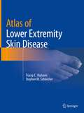
Atlas of Lower Extremity Skin Disease
by Tracey C. Vlahovic Stephen M. SchleicherLower extremity skin disorders are often overlooked by clinicians. Ailments such as eczema, psoriasis and tinea at times prove difficult to distinguish clinically, and misdiagnosis can lead to inappropriate therapy. Many practitioners are mystified when confronted with an abnormal appearing nail. Delay in recognizing skin cancer may adversely impact morbidity and mortality. This full-color atlas is a concise guide for medical professionals who deal with the lower extremities and will aid in both diagnosis and treatment. Topics featured in the Atlas include nail pathology, fungal and bacterial infections, xerotic and hyperkeratotic disorders, autoimmune diseases and vasculopathies, benign and malignant lesions, systemic diseases, and ulcerations. Each chapter contains vibrant photographic representative examples. Concluding chapters present a review of biopsy techniques as well as an overview of current dermatological therapies. The Atlas of Lower Extremity Skin Disease is a unique resource for podiatrists, dermatologists, and primary care physicians as well as nurse practitioners and physician assistants.
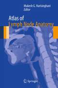
Atlas of Lymph Node Anatomy
by Mukesh G. HarisinghaniDetailed anatomic drawings and state-of-the-art radiologic images combine to produce this essential Atlas of Lymph Node Anatomy. Utilizing the most recent advances in medical imaging, this book illustrates the nodal drainage stations in the head and neck, chest, and abdomen and pelvis. Also featured are clinical cases depicting drainage pathways for common malignancies. 2-D and 3-D maps offer color-coordinated representations of the lymph nodes in correlation with the anatomic illustrations. This simple, straightforward approach makes this book a perfect daily resource for a wide spectrum of specialties and physicians at all levels who are looking to gain a better understanding of lymph node anatomy and drainage. Edited by Mukesh G. Harisinghani, MD, with chapter contributions from staff members of the Department of Radiology at Massachusetts General Hospital.
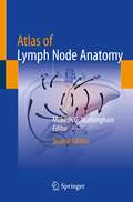
Atlas of Lymph Node Anatomy
by Mukesh G. HarisinghaniThis book is a comprehensive atlas on lymph node anatomy and drainage to aid in cancer staging and therapy. Nodal drainage is pertinent to all aspects of cancer staging and therapy and is used by radiation oncologists, surgeons, and medical oncologists to increase accuracy. The first edition of this text was the first comprehensive monograph on this topic, allowing physicians across various specialties to utilize this information and easily share that knowledge with residents, fellows, and junior faculty.Detailed anatomic drawings and state-of-the-art radiologic images combine to produce this essential Atlas of Lymph Node Anatomy. Utilizing the most recent advances in medical imaging, this book illustrates the nodal drainage stations in the head and neck, chest, abdomen, and pelvis. Also featured are clinical cases depicting drainage pathways for common malignancies. 2-D and 3-D maps offer color-coordinated representations of the lymph nodes in correlation with the anatomic illustrations. This simple, straightforward approach makes this book a perfect daily resource for a wide spectrum of specialties and physicians at all levels who are looking to gain a better understanding of lymph node anatomy and drainage.This new edition enables physicians to educate themselves on the location of various nodal stations, especially in the context of common primary tumors, so that they are able to detect, localize, and characterize nodes seen with novel new imaging methods and with an increased level of accuracy. Chapters now cover the significant strides made in the imaging realm, such as PET CT, conventional MRI, MRI with novel imaging agents, and multidetector CT, which allows visualization of lymph nodes in various anatomic compartments.
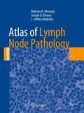
Atlas of Lymph Node Pathology (Atlas of Anatomic Pathology)
by Joseph D. Khoury L. Jeffrey Medeiros Roberto N. MirandaAtlas of Lymph Node Pathology reviews the histopathology of nodal diseases, illustrating the use of ancillary studies and includes concise discussions of pathogenesis, clinical settings and clinical significance of the pathologic diagnosis. The atlas features an overview of the benign reactive processes secondary to infectious, environmental or unknown insults, as well as relevant illustrations of virtually all primary and secondary neoplasms involving lymph nodes. The atlas also includes macroscopic images of some disorders, tables that help readers understand and comprehend diseases that look alike, and diagnostic algorithms for certain groups of diseases. Authored by highly experienced pathologists, Atlas of Lymph Node Pathology is a valuable resource that illustrates the vast majority of diseases practicing pathologists, clinicians and oncologists are likely to encounter in daily practice.
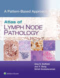
Atlas of Lymph Node Pathology: A Pattern Based Approach
by Amy S. Duffield Joo Y. Song Girish VenkataramanClosely mirroring the daily sign-out process, Atlas of Lymph Node Pathology: A Pattern Based Approach is a highly illustrated, efficient guide to accurate diagnosis. This practical reference uses a proven, pattern-based approach to clearly explain how to interpret challenging cases by highlighting red flags in the clinical chart and locating hidden clues in the slides. Useful as a daily “scope-side guide,” it features numerous clinical and educational features that help you find pertinent information, reach a correct diagnosis, and assemble a thorough and streamlined pathology report.
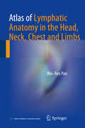
Atlas of Lymphatic Anatomy in the Head, Neck, Chest and Limbs
by Wei-Ren PanThis atlas provides detailed information on the human lymphatic system in the head, neck and chest regions as well as the extremities, with more than 400 photographs and radiographs, including micro and macro views of the morphology. Much of the content is presented for the first time, such as the individual differences in lymphatic distribution, especially in the head neck region; characteristics of the indirect precollecting lymph vessel in the scalp; the lymphatic ampulla and diverticulum; and the transparent lymph node. Providing insights into the lymphatic anatomy, the book is an essential resource for medical and science students as well as therapists, clinicians and researchers working in this field.
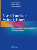
Atlas of Lymphatic System in Cancer: Sentinel Lymph Node, Lymphangiogenesis and Neolymphogenesis
by Shamil Gantsev Kamil Gantsev Shamil KzyrgalinThis atlas contains a large number of original photographs illustrating the anatomy, research methods, and structural features of the structure of the lymphatic apparatus in oncological diseases. The tissue complexes of the axillary region have been studied in detail. The lymphatic maps of the axillary region in cancer are presented, including those with manifestations of lymphangiogenesis; newly formed lymph nodes formed as a result of neolymphogenesis are also considered. Atlas of Lymphatic System in Cancer: Sentinel Lymph Node, Lymphangiogenesis and Neolymphogenesis is intended for oncologists, anatomists, morphologists and doctors of other specialties. The book is the result of ten years of research. The applied technology of ultrasonic isolation of anatomical structures made it possible to take a fresh look at the lymphatic apparatus in cancer and describe the phenomenon of neolymphogenesis.
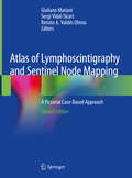
Atlas of Lymphoscintigraphy and Sentinel Node Mapping: A Pictorial Case-Based Approach
by Giuliano Mariani Sergi Vidal-Sicart Renato A. Valdés OlmosThe main goal of the second edition of this book is to update the content on the rapidly growing field of lymphoscintigraphy, a radionuclide-based imaging procedure that provides information on the functional status of the lymphatic system. Although the technique was originally introduced to identify the cause of peripheral edema (i.e., blockage of the venous or lymphatic circulation), more recent and widespread applications include radioguided biopsy of the sentinel lymph node in patients with solid cancers. This procedure is crucial for the adequate planning of oncologic surgery in a growing number of cancers, most notably breast cancer, cutaneous melanoma, head and neck cancers, penile cancer, and cervical cancer.The book focuses on the latest advances in lymphoscintigraphy techniques, including both novel tracers recently approved for clinical use (especially in the field of sentinel lymph node mapping) and the expanding role of hybrid imaging with SPECT/CT – and in sentinel node detection using hybrid tracers (radiolabeled and fluorescent) for dual-signature guidance. Each chapter addresses the clinical application of lymphoscintigraphy in different anatomic areas or disease conditions. After an introductory section concerning the pathophysiology of the specific site/disease, the clinical relevance and impact of lymphoscintigraphy is demonstrated by a collection of richly illustrated teaching cases describing the lymphoscintigraphic patterns most commonly observed, as well as anatomic variants and technical pitfalls. Emphasis is placed on tomographic multimodality imaging. The book gathers contributions by experts in nuclear oncology, who have revised their chapters by updating the didactic material and adding clinical cases. Regarding sentinel lymph node biopsy in particular, a major distinction of this text is the incorporation of the staging guidelines of the American Joint Committee on Cancer (8th edition) into the didactic material.
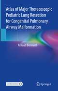
Atlas of Major Thoracoscopic Pediatric Lung Resection for Congenital Pulmonary Airway Malformation
by Arnaud BonnardThis atlas offers a step-by-step approach to Thoracoscopic surgery for Congenital Pulmonary Malformation (CPAM) in each major lung resection. This technique is still a matter of debate and is used approxmately by 50% of the surgeons in Europe. The apprehension of the thoracoscopic approach is complicated firstly by the small size of the child to whom it is proposed and secondly by the risks of perioperative or postoperative complications. Furthermore, the pulmonary malformations object of this investigation are rare, thus making this technique difficult to learn and reproduce The volume - the result of a surgical experience acquired since 2007 in this field - offers a step-by-step approach to the procedure in each major lung resection and is illustrated by numerous intraoperative photos, short video clips and even complete resection videos; tips and tricks are presented to facilitate surgery, to make it reproducible and accessible by all surgeons, whether they are young surgeons in training or experienced surgeons. This atlas aims to complement and not to replace the direct observation in the operating room, or the procedure learning by the reader. It offers essential guide for practitioners, trainees and thoracoscopic surgeons interested in CPAM.
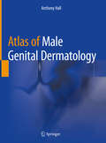
Atlas of Male Genital Dermatology
by Anthony HallThis atlas provides a practical guide applicable to the diagnosis and management of skin diseases affecting the male genital area. Chapters feature concise descriptions and advice on potential management strategies and cover sexually transmissible infections, candidal balano-posthitis, rare male genital malignancies, and pigmentary disorders. Every description is accompanied by a broad range of images of a particular disease or disorder, enabling the reader to develop a deeper understanding of both diagnostic and management aspects of skin diseases affecting male genitalia. Atlas of Male Genital Dermatology enables readers to quickly and successfully identify a variety of dermatological disorders that can affect the male genitalia. It provides instruction on how to effectively manage these conditions and is a valuable resource for any physician who encounters these conditions in daily practice.
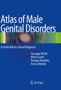
Atlas of Male Genital Disorders: A Useful Aid for Clinical Diagnosis
by Franco Dinotta Giuseppe Micali Marco Cusini Pompeo DonofrioMale genital disorders represent a common issue in medical practice, especially in the dermatological setting. Correct clinical evaluation of these disorders is essential when addressing the diagnosis, which in some cases may require histopathological confirmation. Depending on the disease, early diagnosis may be not only lifesaving, but also of fundamental importance to the planning of successful treatment. This atlas introduces the most common penile diseases, along with more rarely encountered ones. It provides invaluable guidance on clinical diagnosis by highlighting prominent clinical features and presenting particular videodermatoscopy findings when these are indicative of the diagnosis. In addition, for each condition the most appropriate treatment is proposed, taking into account recent therapeutic advances of proven benefit.
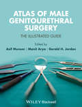
Atlas of Male Genitourethral Surgery
by Manit Arya Asif Muneer Gerald H. JordanMale genitourethral abnormalities are a source of great concern and distress to those affected. Surgery, when required, is very specialized and often extremely complex requiring expert surgical skills in order to achieve the best outcomes. Atlas of Male Genitourethral Surgery: The Illustrated Guideprovides urological surgeons, at all levels of experience from trainees to established specialists, with a full colour, highly illustrated and step-by-step approach to male genitourethral surgery, enabling complete mastery of surgical techniques in this difficult and challenging area. Full-color throughout and with over 430 high-quality images, this comprehensive atlas covers key areas of urologic surgery including:Surgery for penile curvatureUrethral reconstruction and artificial urinary sphinctersPenile and scrotal reconstructionSurgery for male infertilitySurgery for erectile dysfunctionSurgery for penile cancerEach chapter includes an introduction to the condition and its challenge, a step-by step guide to the surgical procedures applicable for that condition with surgical tips and tricks for improved techniques and outcomes. Brought to you by the world's leading experts in the field of genitourethral surgery, this outstanding book guides you through the most challenging of operations, helping you deliver high quality clinical care to your patients.
Atlas of Male Infertility Microsurgery
by Marc GoldsteinInfertility affects approximately 15% of couples attempting to conceive, and male factor is known to be either the primary or a contributing factor in at least half of all cases of couple infertility. More than half of male infertility is amenable to microsurgical treatment, but a majority of urology residency programs provide little or no training in male infertility or its microsurgical treatment. Textbooks typically include a chapter or two on surgery of male infertility, but often these chapters only cover basic concepts and simplified overviews and miss the many finer points that are only taught by observing or scrubbing directly in the operating room. Utilizing Dr. Marc Goldstein's extensive surgical experience, this atlas focuses primarily on microsurgical techniques for restoring and preserving male fertility. The chapters discuss surgical anatomy and diagnosis, microsurgery of the vas deferens and epididymis, varicocele and other fertility-related procedures. Topics and techniques include vasectomy, vasovasostomy, vasoepididymostomy, varicocelectomy, hydrocelectomy, and microsurgically assisted inguinal hernia repair. Equipment, instrumentation and training in microsurgery, including animal models, are also discussed. In each chapter, practical tips and tricks are highlighted. Enhanced by plentiful intraoperative photos and original illustrations, Atlas of Male Infertility Microsurgery is an essential resource for male reproductive surgeons, urologists, residents and fellows.
Atlas of Mandibular and Maxillary Reconstruction with the Fibula Flap: A step-by-step approach
by Giorgio De Santis Peter G. Cordeiro Luigi ChiariniThis atlas deals with the standard technique used for reconstructing the mandible and the maxilla - the fibula flap. The reader will find useful information on all issues that are important in the surgical procedure, including the use of CAD-CAM technology (Computer Assisted Technology), bone synthesis and flap modelling. The editors draw on their 30 years of experience to provide a step-by-step description of this surgical procedure. With the help of numerous illustrations, the reader will learn the technical, functional and aesthetic developments since 1989 when this technique was first described.Plastic surgeons, otorhinolaryngologists and oral- and maxillofacial surgeons will find this book a valuable guide to the sophisticated principles of jaw reconstruction and how to apply them in their everyday practice.
Atlas of Mediastinal Pathology (Atlas of Anatomic Pathology)
by Saul SusterThe mediastinum is a virtual compartment in the chest cavity that is the seat of several vital organs and structures that can be involved in a variety of pathologic processes, including congenital and developmental abnormalities, inflammatory conditions, and benign and malignant neoplasms. The Atlas of Mediastinal Pathology provides a pictorial survey of the major disease processes that can affect this anatomic compartment, including congenital and acquired cysts, benign hamartomatous processes, inflammatory processes involving the mediastinum, and benign and malignant neoplasms. The latter includes tumors of the thymus (thymoma and thymic carcinoma), neuroendocrine neoplasms, germ cell tumors, mesenchymal neoplasms, and hematolymphoid malignancies. The use of ancillary diagnostic methods is illustrated, where appropriate, providing assistance for pathologists in arriving at the correct diagnosis.
Atlas of Medical Renal Pathology (Atlas of Anatomic Pathology)
by Stephen M. BonsibThe kidney is an organ with complex organogenesis susceptible to numerous misadventures in development and is exposed to a diverse array of insults of hematogenous and lower urinary tract origin. This Atlas of Medical Renal Pathology provides an overview of the development, macroscopic and microscopic features of the normal kidney. This is followed by a comprehensive survey of developmental and cystic kidney diseases, vascular diseases and tubulointerstitial diseases. An emphasis is placed on gross diagnostic findings with detailed histological correlates. In addition, the histological, immunofluorescent, immunohistochemical and ultrastructural features of the major glomerular diseases and renal transplantation pathology are presented. This compendium of non-neoplastic kidney diseases illustrates the vast majority diseases you are likely to encounter in surgical and autopsy pathology.
Atlas of Mesotherapy in Skin Rejuvenation
by Antonella Tosti Maria Pia De PadovaThe injection of a combination of vitamins and medications into the middle layer of the skin has been practised in continental Europe for some fifty years now, but because the literature has hitherto not been published in English the topic is still surrounded by a great deal of ignorance and prejudice. This atlas from a renowned authorship will doc
Atlas of Minimally Invasive Facelift: Facial Rejuvenation with Volumetric Lipofilling
by Jose Maria Serra-Renom Jose Maria Serra-MestreThis atlas provides a concise overview of the principle of fat grafting and its clinical applications to treat aging in the central areas of the face in a minimally invasive manner. The authors start with a short description of the anatomical basis of facial aging and by presenting current facial rejuvenation techniques. Here the emphasis is on the process of harvesting and preparing macro-, micro-, intradermal and nano-fat for injection. Thanks to the book's numerous illustrations and short descriptions, readers will find valuable information on where and how to inject the fat combined with the authors' individual techniques and refinements. Aging is a major issue in today's society. Facial rejuvenation procedures seek to enhance the final outcome and durability of surgery and to minimize drawbacks. The current trend is to use less invasive methods requiring fewer incisions, which produce more natural-looking results. This atlas shows the authors' personal techniques to help achieve that goal.
Atlas of Minimally Invasive Hand and Wrist Surgery (Minimally Invasive Procedures in Orthopaedic Surgery)
by John T. Capo Virak TanHand and wrist surgery is evolving rapidly and often, advances are directed at developing procedures that are less invasive, with smaller incisions and shorter rehabilitation times. Minimally Invasive Hand and Wrist Surgery is the only book devoted exclusively to these exciting new percutaneous and minimal access techniques for the treatment of chr
Atlas of Minimally Invasive Surgery for Lung and Esophageal Cancer
by Mark K. Ferguson Jun WangThis Atlas presents a state-of-the-art review of VATS and robotic approaches to managing lung and esophageal cancers. It discusses cancer staging, physiological evaluation of patients, and patient selection for minimally invasive surgery and offers detailed descriptions of individual operations accompanied by anatomic drawings, intraoperative images, and 3-dimensional anatomic reconstructions. Written by recognized experts in the field, it provides readers with an unparalleled resource for advancing their skills in managing these cancers. It is a valuable reference work for thoracic surgeons in training as well as in practice who want to pursue minimally invasive surgery. It is unique in offering fully illustrated, step-by-step descriptions of the operative procedures.
Atlas of Minimally Invasive Surgery in Esophageal Carcinoma
by Miguel Cuesta Shailesh PuntambekarThe main aim of this book is standardization of the procedure with stress on simplification and ease of performing the surgery. In oncology, the principles and steps of an operative procedure should remain the same; though the modality may change, be it either open surgery or laparoscopy. Thus, in this book the patient position and procedural steps are devised to mimic open surgery as far as possible. This makes it easier for the oesophageal surgeon performing surgeries by the open technique to get converted into a thoracoscopic laparoscopic surgeon. This atlas will be a step by step guide for a surgeon who wishes to perform laparoscopy with more than 500 operative photographs.
Atlas of Minimally Invasive Techniques in Upper Gastrointestinal Surgery
by Miguel A. Cuesta M. Asunción Acosta Marcos BrunaThis Atlas comprehensively covers minimally invasive operative techniques for benign and malignant cancer surgery of the esophagus and stomach. It provides easy-to-follow instructions accompanied by a range of pictures and illustrations, as well as a collection of interactive videos to aid the reader in developing a deeper understanding of each surgical procedure. Techniques covered include minimally invasive surgical treatment for esophageal and gastric cancer including different approaches such as thoracoscopic, transhiatal, laparoscopic, and robot-assisted resections. These chapters include different types of cervical and intrathoracic anastomoses after esophageal resections, and different anastomoses and reconstructions after gastrectomy. Moreover, the Atlas includes an extensive description of minimally invasive procedures in bariatric surgery including sleeve resection, gastric bypass, biliopancreatic diversion, and others. Minimally invasive approaches for other benign pathologies such as benign tumors and treatment of gastroduodenal ulcer complications are also depicted. All chapters, written by a renowned and experienced international group of surgeons and their teams, are focused on practical step-by-step description of the techniques. Atlas of Minimally Invasive Techniques in Upper Gastrointestinal Surgery systematically describes the most frequently performed surgical procedures of the esophagus and stomach and is a valuable resource for all practicing surgeons and trainee general surgeons dedicated to upper gastrointestinal surgery, such as bariatric and surgical oncologists.
Atlas of Minimally Invasive and Robotic Esophagectomy
by Min P. KimThe book demonstrates the step-by-step method of performing minimally invasive esophagectomy and robot-assisted esophagectomy. Chapters cover the techniques of performing minimally invasive and robot-assisted Ivor Lewis esophagectomy and McKeown esophagectomy as well as variation in methods of chest and neck anastomosis and a method to perform jejunostomy tube placement. Each author provides a narrative on their technique in performing the esophagectomy with pearls for different parts of the operation. As with any other surgical procedure, there are controversies about how to handle different parts of the operation. The goal of the book is not to debate the best method, but to provide a method to perform the complex operation in a minimally invasive way. Atlas of Minimally Invasive and Robotic Esophagectomy incorporates robot-assisted esophagectomy techniques and aims to benefit specialists and trainees that treat patients with esophageal cancer.
Atlas of Mohs and Frozen Section Cutaneous Pathology
by John R. Hamill James M. Spencer Michael MorganOf all the techniques used to treat non melanoma skin cancer, the highest cure rates belong to the Mohs surgical procedure. Critical to this technique is optimal preparation and interpretation of frozen sections. With more than 400 high resolution figures, the Atlas of Mohs and Frozen Section Cutaneous Pathology is the premier textbook on the topic, serving as a highly practical guide to microscopic analysis, diagnosis, and discrimination of common and problematic cutaneous neoplasms. Highlights of the Atlas include diagnosis of basic and routine dermatologic entities, diagnosis and distinction of rare and/or deadly neoplasms such as dermatofibrosarcoma protuberans and merkel cell carcinoma, a troubleshooting guide dealing with quality control of the frozen section technique, and discussion of new techniques including immunohistochemistry. Comprehensive and presented in full color, the Atlas of Mohs and Frozen Section Cutaneous Pathology is an indispensable reference for anyone involved with the Mohs procedure, including dermatologic surgeons, Mohs cutaneous surgeons, as well as pathologists and dermatopathologists who perform frozen section analysis of cutaneous specimens.
Atlas of Mohs and Frozen Section Cutaneous Pathology
by James M. Spencer Michael B. Morgan John R. Hamill Jr. Rebecca ThornhillOf all the techniques used to treat non melanoma skin cancer, the highest cure rates belong to the Mohs surgical procedure. Critical to this technique is optimal preparation and interpretation of frozen sections. The second edition of this highly successful atlas details both common and uncommon cutaneous neoplasms that can serve as a source of reference for established practitioners and a review for those in training. It includes new frozen section specimens, the most current diagnostic guidelines, and discussion of the advancements in tissue staining. There is also an additional chapter with self-study tools, where readers can test their knowledge using various images with a multiple choice answer format, followed by a discussion of the correct answer. With high resolution figures, the Atlas of Mohs and Frozen Section Cutaneous Pathology, Second Edition is the premier text on the topic, serving as a highly practical guide to microscopic analysis, diagnosis, and discrimination of common and problematic cutaneous neoplasms. Highlights of the Atlas include diagnosis of basic and routine dermatologic entities, diagnosis and distinction of rare and/or deadly neoplasms such as dermatofibrosarcoma protuberans and merkel cell carcinoma, a troubleshooting guide dealing with quality control of the frozen section technique, and discussion of techniques including immunohistochemistry. Comprehensive and presented in full color, the Atlas of Mohs and Frozen Section Cutaneous Pathology, Second Edition is an indispensable reference for anyone involved with the Mohs procedure, including dermatologic surgeons, Mohs cutaneous surgeons, as well as pathologists and dermatopathologists who perform frozen section analysis of cutaneous specimens.