- Table View
- List View
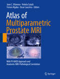
Atlas of Multiparametric Prostate MRI: With PI-RADS Approach and Anatomic-MRI-Pathological Correlation
by Joan C. Vilanova Violeta Catalá Ferran Algaba Oscar LauciricaThis atlas provides a comprehensive, state of the art review of the use of multiparametric MRI (mpMRI) for the imaging of prostate cancer, covering aspects from diagnosis and loco-regional staging through to the role of the technique after treatment and follow-up. The book contains a wealth of high-resolution images, many of them in color, and displays the anatomical-MRI-pathological correlation whenever appropriate. Readers will find a helpful overview on the current standardized method for reading and reporting on mpMRI, the Prostate Imaging Reporting and Data System (PI-RADS), version 2. Dedicated chapters focus on differential diagnosis and imaging pitfalls, and the inclusion of helpful diagrams and algorithms will further assist in image interpretation, enabling readers to ease and improve their use of mpMRI. Edited and written by very experienced radiologists, pathologists, and urologists; the Atlas of Multiparametric Prostate MRI will serve as a unique source of clinically relevant information and an aid to disease management for radiologists, urologists, pathologists, radiotherapists, and oncologists.
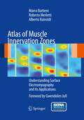
Atlas of Muscle Innervation Zones: Understanding Surface Electromyography And Its Applications
by Roberto Merletti Alberto Rainoldi Marco BarberoInvasive electromyography is a well-established diagnostic tool that has been used for decades by neurologists. Recently, new and alternative devices have increasingly become available that permit diagnosis without the use of needles. This developing area of science and the new tools have not, however, been sufficiently investigated in academic training. Consequently a gap exists between what science is making possible and the competence acquired during graduate studies. This handy volume has the aim of filling this gap by providing the information required by medical practitioners in rehabilitation, sports, and occupational health as well as by rehabilitation therapists, ergonomists, and sport coaches. The techniques that are presented and explained will help in monitoring and recording changes, evaluating the effectiveness of treatments and training, evaluating work stations, and preventing and documenting the evolution of occupational disorders of the neuromuscular system.
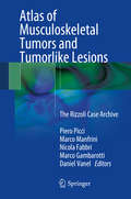
Atlas of Musculoskeletal Tumors and Tumorlike Lesions: The Rizzoli Case Archive
by Piero Picci Marco Manfrini Nicola Fabbri Marco Gambarotti Daniel VanelThis book reflects the experience of the Rizzoli Orthopedic Institute during more than 100 years of treatment of musculoskeletal tumor and tumorlike lesions. It presents a wide range of lesions from a multidisciplinary perspective, highlighting pertinent clinical, radiological, and histological correlations. Treatment is briefly reported for each entity. In addition, the more recent biomolecular findings of use for diagnosis, prognosis, and treatment are carefully analyzed. The Rizzoli case archive spans more than a century, the first treated case dating back to 28 September 1900, and contains the original material - clinical charts, imaging, paraffin blocks, and histological slides - of more than 40,000 cases, including about 29,000 bone lesions and 11,000 soft tissue lesions. This book reports the most relevant entities and reflects the improvements in knowledge of musculoskeletal tumors as presented during the yearly international course held at the Rizzoli Institute.
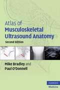
Atlas of Musculoskeletal Ultrasound Anatomy
by Mike Bradley Paul O'DonnellAtlas of Musculoskeletal Ultrasound Anatomy provides an essential grounding in normal ultrasound anatomy, enabling the reader to assess whether anatomy is disrupted through injury or disease. The book is structured systematically, with all commonly imaged areas illustrated by high quality ultrasound scans with accompanying concise descriptive text. Features of the second edition: · Over 100 individual anatomical descriptions · Numerous new images from the latest generation ultrasound machines · Improved surface anatomy diagrams indicating limb and probe optimal positions for each area of anatomy · Numerous radiographic anatomical diagrams showing ultrasound probe overlying the anatomical structure for improved visual understanding Atlas of Musculoskeletal Ultrasound Anatomy appeals to a wide range of practitioners who need to visualize the musculoskeletal system to diagnose injuries or locate blood vessels or nerves while undertaking clinical procedures. Radiologists, sonographers, anaesthetists, physiotherapists, rheumatologists, and orthopaedic surgeons will find this an invaluable practical reference.
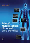
Atlas of Musculoskeletal Ultrasound of the Extremities
by Mohini RawatFeaturing nearly 700 illustrations, images, and photos, Atlas of Musculoskeletal Ultrasound of the Extremities by Dr. Mohini Rawat is a comprehensive visual guide to musculoskeletal ultrasound imaging for health care students and clinicians. Musculoskeletal ultrasound imaging is a new, rapidly growing field with applications across many health care disciplines. With its increased popularity comes a need for detailed training resources. The Atlas of Musculoskeletal Ultrasound of the Extremities presents information on scanning protocols for the joint regions and peripheral nerves of the upper and lower extremities in an easy-to-follow, highly visual format. Beginning with an overview of ultrasound physics, equipment, terminology, and technique, the book provides detailed instruction for musculoskeletal ultrasound of the shoulder, elbow, wrist, hip, knee, ankle and foot, concluding with a comprehensive chapter on peripheral nerves. Each chapter contains detailed images of scanning protocols, anatomy, sonoanatomy, patient positioning, and probe positioning for each joint region. Images are accompanied by explanatory text descriptions, along with clinical pearls under points to remember. Designed for students and clinicians in physical therapy, occupational therapy, athletic training, orthopedics, rheumatology, physiatry and podiatry, the Atlas of Musculoskeletal Ultrasound of the Extremities provides essential introductory training materials and serves as a helpful reference for busy clinical environments.
Atlas of Nail Disorders Across All Skin Colors: A Comprehensive Guide to Navigating Nails
by Shari R. LipnerThis atlas encompasses the spectrum of nail disorders and intends to provide an up-to-date and highly illustrated synopsis of both common and rare conditions. The diseases included are of infectious, inflammatory, neoplastic, and traumatic etiologies, and include both adult and pediatric conditions. There is an emphasis on images spanning a range of Fitzpatrick skin types, to demonstrate the similarities and differences in presentation of nail diseases in light, medium, and dark skin. Atlas of Nail Disorders Across All Skin Colors is not only an important tool for dermatologists in practice or in training but also for physicians in fields such as internal medicine, primary care, pediatrics, and surgery who often see patients with nail conditions as part of their day-to-day practice.

Atlas of Nail Signs and Disorders with Clinical and Onychoscopic Correlation
by Matilde Iorizzo Robert Baran Nilton Di Chiacchio Nilton Gioia Di Chiacchio Robertha Carvalho de Nakamura Michela StaraceThis atlas allows dermatologists and resident and qualified professionals in other disciplines to reach a quick diagnosis, with a wealth of clinical and dermatoscopic images for convenience.Key features: Provides a quick and straightforward guide to diagnosis for medical professionals. Allows residents and professionals in other disciplines easy access to the essential points. Presents the expertise of some of the most eminent international nail specialists.
Atlas of Neoplastic Pulmonary Disease: Pathology, Cytology, Endoscopy and Radiology
by Armin Ernst Philip T. Cagle Timothy Craig Allen Richard S. Irwin Shanda Blackmon Armando E. Fraire Megan K. Dishop Dina R. ModyUsing a multi-disciplinary approach to the diagnosis of pulmonary disease, this unique atlas will offer the busy practitioner a quick and reliable tool crossing over traditional boundaries and will be useful not only to pathologists and cytopathologists, but also to pulmonologists, internists, endoscopists and radiologists as well as students and residents in training. The atlas will contain a profusion of images (320 in color) accompanied by descriptive text and a restricted number of highly selected references.
Atlas of Neuro-ophthalmology
by Thomas C. SpoorThere are many great neuro-ophthalmology texts available ranging from huge encyclopedic tomes to small, detailed resources. Combining the best features of these books, Atlas of Neuro-ophthalmology offers a glimpse into a wide variety of rare and unusual neuro-ophthalmic disorders. Long recognized as a leading authority on the optic nerve, Tom Spoor
Atlas of Neuromuscular Diseases: A Practical Guideline
by James W. Russell Eva L. Feldman Wolfgang Grisold Wolfgang N. LöscherThis atlas presents a comprehensive outline of neuromuscular diseases, written by respected American and European authors. It discusses all aspects of neuromuscular disorders including cranial and spinal nerves, motor neuron diseases, nerve plexus, peripheral nerves, mono- and polyneuropathies, entrapment syndromes, neuromuscular junctions, and muscle disease. Each chapter is structured into the following sections: anatomy, symptoms, signs, pathogenesis, diagnosis and differential diagnosis, therapy and prognosis. The diagnostic tools in neuromuscular disease are explained and practical guidelines are offered on how to advance from symptoms to syndromes. The therapeutic options for each disease are also described. In this new edition, the structure of the chapters has been reorganized and chapters on principles of peripheral nerves, nerve pain, nerve surgery and rehabilitation have been added. The current trend of increased use of imaging techniques such as US and MRI in the diagnosis and follow-up of neuromuscular disorders is also reflected.
Atlas of Neuromuscular Diseases: A Practical Guideline
by James W. Russell Eva L. Feldman Wolfgang Grisold Wolfgang N. Löscher Stefan MengThis atlas offers a comprehensive overview of neuromuscular diseases. It discusses all aspects of neuromuscular disorders, including general tools, the cranial and spinal nerves, the nerve plexus, peripheral nerves, mono- and polyneuropathies, entrapment syndromes, the neuromuscular junction, motor neuron diseases, muscle disease, and autonomic involvement. Each chapter is structured into the following sections: anatomy, symptoms, signs, pathogenesis, diagnosis and differential diagnosis, therapy, and prognosis. The diagnostic tools used for neuromuscular disease are explained, and the therapeutic options for each disease are described. This updated third edition includes new chapters addressing a range of topics: from histology to molecular mechanisms, genetic aspects, the mechanisms of emerging new therapies, neuroimaging, neuromuscular disease, and new pathogenic mechanisms. The book aims to be a useful companion for neuromuscular disease. The homogenous structure, illustrations with figures, and representative images makes the atlas easy to read and helpful in understanding neuromuscular problems.
Atlas of Neurotologic and Lateral Skull Base Surgery
by John S. Oghalai Colin L. W. DriscollLong awaited, this fine surgical atlas covers all aspects of neuro-otology and lateral skull base procedures in comprehensively in detail. The lavishly illustrated step-by-step guide is written by two American experts to ensure continuity between topics. The text is highly structured with step-by-step explanation of each surgical procedure and TIPS and TECHNIQUES sections as well as a PEARLS section in each chapter. More than 200 superb artwork illustrations describe each surgical procedure with nearly 600 additional intraoperative pictures with CT and MRI images to teach specific case examples. This offers a complete educational experience for the skull base surgeon in training and a thorough reference for the experienced surgeon.
Atlas of Non-Desirable Outcomes in Cleft Lip and Palate Surgery: A Case-Based Guide to Preventing and Managing Complications
by Percy Rossell-PerryThis book encompasses diagnostic, treatment and prevention of bad results and complications associated with cleft lip and palate surgery. Illustrated by more than 200 images and based on a 25 years’ experience of the editor, the work includes the whole spectrum of complications based on case studies, as well as management and prevention protocols. Divided in nine didactic parts, the chapters present complications related to anesthesia, lip asymmetry, scar disorders, postoperative dehiscence, secondary nose deformities besides dentoeskeletal sequels. Atlas of Non-Desirable Outcomes in Cleft Lip and Palate Surgery will benefit plastic surgeons, pediatric surgeons, otolaryngologists, maxillofacial surgeons, head and neck surgeons, dentists and anesthesiologists.
Atlas of Non-Gynecologic Cytology (Atlas of Anatomic Pathology)
by Qing Kay Li Xin Jing Momin T. SiddiquiCytology is a unique subspecialty in anatomic pathology, and plays a critical role in the diagnosis of a variety of benign and malignant lesions by using minimally invasive procedures, such as fine needle aspiration, brushing, washing and lavage. Cytology not only provides an accurate diagnosis of the lesion based on the cytomorphological evaluation at cellular level, but also provides material for molecular characterization of a tumor for targeted therapy. In the era of personalized medicine, cytology has continued to grow and evolve as an important diagnostic tool. For example, some types of procedures are routinely used, diagnostic criteria have become more refined, and certain terminology has been changed based on current WHO classifications. Therefore, it is necessary to update our knowledge and terminology in cytology. In this volume, we have retained the quality and the clarity of the series, and like the other volumes, this volume aims to be concise and comprehensive yet clinically relevant to daily practice. Although this volume is written by multiple authors, all chapters follow a similar format: brief introduction of the specific organ/system (including types of specimens and techniques to obtain samples), description of normal findings, and a practice approach to diagnose benign and malignant lesions. In each chapter, the key cytomorphological feature and main differential diagnoses of the lesion are summarized in a concise table. Images in each chapter are instructive and clearly represent findings. Where necessary, we have also illustrated the important ancillary tests, such as flow cytometry, immunohistochemistry and molecular analysis, which are crucial for an accurate diagnosis and targeted therapy. The updated knowledge, including key cytomorphological features, current terminology and molecular diagnostic tests, is the highlight of this volume.
Atlas of Non-Invasive Imaging in Cardiac Anatomy
by Jagat Narula Francesco F. Faletra Siew Yen HoThis atlas provides a detailed visual resource of how sophisticated non-invasive imaging relates to the anatomy observed in a variety of cardiovascular pathologies. It includes investigation of a wide range of defects in numerous cardiac structures. Mitral valve commissures, atrioventricular septal junction and right ventricular outflow tract plus a wealth of other structures are covered, offering readers a comprehensive integrative experience to understand how anatomic subtleties are revealed by modern imaging modalities.Atlas of Non-Invasive Imaging in Cardiac Anatomy provides a detailed set of visual instructions that is of use to any cardiovascular professional needing to understand the orientation of a patient’s imaging. Therefore this is an essential guide for all trainee and practicing cardiologists, cardiac imagers, cardiac surgeons and interventionists.
Atlas of Normal Imaging Variations of the Brain, Skull, and Craniocervical Vasculature
by Alexander M. MckinneyThis atlas presents normal imaging variations of the brain, skull, and craniocervical vasculature. Magnetic resonance (MR) imaging and computed tomography (CT) have advanced dramatically in the past 10 years, particularly in regard to new techniques and 3D imaging. One of the major problems experienced by radiologists and clinicians is the interpretation of normal variants as compared with the abnormalities that the variants mimic. Through an extensive collection of images, this book offers a spectrum of appearances for each variant with accompanying 3D imaging for confirmation; explores common artifacts on MR and CT that simulate disease; discusses each variant in terms of the relevant anatomy; and presents comparison cases for the purpose of distinguishing normal findings from abnormalities. It includes both common variants as well as newly identified variants that are visualized by recently developed techniques such as diffusion-weighted imaging and multidetector/multislice CT. The book also highlights normal imaging variants in pediatric cases. Atlas of Normal Imaging Variations of the Brain, Skull, and Craniocervical Vasculature is a valuable resource for neuroradiologists, neurologists, neurosurgeons, and radiologists in interpreting the most common and identifiable variants and using the best methods to classify them expediently.
Atlas of Nuclear Cardiology (Atlas Of Ser.)
by Vasken Dilsizian Jagat NarulaThe aim of the 4th edition of the Atlas of Nuclear Cardiology is to provide physicians and students in cardiology, radiology, and nuclear medicine who want the latest information in the field of cardiovascular nuclear medicine up-to-date and comprehensive information on advances in instrumentation, radiotracers, protocols, and clinical studies. Unlike other books that are narrow in their scope of either technology and technique or clinical studies, the 4th edition of the Atlas will present diagnostic algorithms and schematic diagrams integrated with nuclear cardiology procedures generously interspersed with color illustrations to emphasize key concepts in cardiovascular physiology, pathology, and metabolism relevant for the clinical practice of cardiology. The atlas emphasizes today's most current information, meeting the requirements for those who will be using the book as a reference source for certifying or re-certifying in cardiology, nuclear cardiology, nuclear medicine or radiology. Hybrid PET/CT and SPECT/CT represent new technologies that were introduced recently in clinical medicine and are evolving rapidly with several improvements in instrumentation, imaging procedures as well as in clinical trials that support the expanded role of these technologies in clinical practice. As such, an updated 4th edition of the Atlas is critical in order for the clinicians remain current with the imaging field and maintain their skills. Imaging protocols with these technologies have to be updated and/or expanded in order to acquire high quality images at a reduced radiation burden to the patient while advancing the application of these techniques for more advanced disease detection. Accordingly, beyond significantly updating the chapters from the 3rd edition, 2 new chapters will be introduced in the 4th edition, which reflects the expanded clinical applications of the technologies in the past 3 years. The new chapters are as follows: "Hybrid SPECT/CT and PET/CT Imaging" and a dedicated chapter on "Radiation Safety and Exposure: Clinical Decision-Making and the Risk-Benefit Ratio". Chapter 7 from the 3rd edition will be deleted. The updated Atlas will serve as a reference source for all cardiologists, radiologists, and nuclear medicine physicians interested in the most up-to-date approaches to noninvasive diagnostic cardiovascular nuclear imaging techniques for the evaluation of patients with known or suspected coronary artery disease as well as non-coronary heart disease. It will also serve as a ready reference textbook for medical students and residents interested in the practice of cardiovascular medicine.
Atlas of Nuclear Cardiology (Atlas Of Ser.)
by Vasken Dilsizian Jagat NarulaThe fifth edition of this book presents clinical data, image acquisition, and interpretation of nuclear cardiology procedures through high quality illustrative image examples. It includes up-to-date and comprehensive coverage of advances in instrumentation, radiotracers, protocols, and clinical studies. New content includes indications in imaging cardiac sarcoidosis, amyloidosis, and device infections as well as recent advances in instrumentation (Hybrid PET/MR). It also provides fresh chapters on the history of nuclear cardiology imaging, radionuclide handling techniques and radiation safety, PET-based myocardial perfusion imaging, and vascular imaging. The entire field is presented in pictographic form that is visually pleasing and conforming to current trends of medical education. The fifth edition of the Atlas of Nuclear Cardiology is an essential reference for cardiologists, radiologists, and nuclear medicine physicians interested in the latest approaches to noninvasive diagnostic cardiovascular nuclear imaging techniques. It also serves as a ready reference textbook for medical students and residents as well as nuclear physicists, nuclear medicine technologists, and radiopharmacists.
Atlas of Nuclear Medicine in Musculoskeletal System: Case-Oriented Approach
by Seoung-Oh Yang So Won Oh Yun Young Choi Jin-Sook RyuNuclear medicine imaging in the musculoskeletal system with its ability to assess disease activities has contributed to accurate diagnosis and improved medical and surgical treatment. Several nuclear medicine textbooks and case studies in forms of atlases have been published so far, but there seems to be no in-depth nuclear medicine imaging atlas focused on diseases of the musculoskeletal system. Therefore, the authors have written about common cases as well as rare musculoskeletal disorders for which various imaging techniques of nuclear medicine (bone scan, SPECT, SPECT/CT, PET/CT, PET/MR, etc.) are useful based on their clinical experience in many different hospitals. This book intends to share the experiences of the authors with nuclear medicine and radiology residents and board specialists, and to help other clinicians who manage musculoskeletal disorders, such as orthopedic and rheumatology, through various cases of musculoskeletal disorders by providing algorithmic imaging utilization to support their patient care.
Atlas of Ocular Anatomy
by Mohammad Wakeel Ansari Ahmed NadeemThis book is a practical and concise atlas on ocular anatomy, with an emphasis on applied aspects and hints for easy retention strategies. The vast color illustrations and photographs consist of self-explanatory, precise, and meaningful representations of the points covered in the text. Covering chapters such as bony socket of the eye, extraocular muscles, eyelids, cornea and lens, and neurology of the eye, Atlas of Ocular Anatomy gives a summary of the important and relevant points for each topic, separating out the essential from the nonessential elements. Complete with representative schematic line diagrams and full color photographs, this atlas features the correlation between anatomic facts with their probable clinical presentations in disease.
Atlas of Ocular Optical Coherence Tomography
by Fedra HajizadehThis book provides a collection of optical coherence tomographic (OCT) images of various diseases of posterior and anterior segments. It covers the details and issues of diagnostic tests based on OCT findings which are crucial for ophthalmologists to understand in their clinical practice. Throughout the chapters all aspects of this non-invasive, popular imaging technique, known for ingenuity and accuracy, is clearly illustrated. Atlas of Ocular Optical Coherence Tomography has been categorized into eleven sections, discussing and illustrating distinct OCT features, as well as showing other image modalities such as fluorescein angiography, fundus autofluorescence, perimetry and laboratory examination. This book also covers choroidal pathologies and vitreous abnormalities. The last section has been allocated to anterior segment disease, including cornea, angle, iris and conjunctival abnormalities. Above all, the numerous images, and detailed descriptions of diseases, make this book an essential guide for general ophthalmologists and ophthalmology residences.
Atlas of Ocular Optical Coherence Tomography
by Fedra HajizadehThis book provides a collection of optical coherence tomographic (OCT) images of various diseases of posterior and anterior segments. It covers the details and issues of diagnostic tests based on OCT findings which are crucial for ophthalmologists to understand in their clinical practice. Throughout the chapters all aspects of this non-invasive, popular imaging technique, known for ingenuity and accuracy, is clearly illustrated. Atlas of Ocular Optical Coherence Tomography, 2nd Edition has been fully revised to include updates optic disc disease and advancements in OCT for the diagnosis and monitoring of glaucoma. In addition, many other recent developments in CSCR, ARMD and OCT-A are highlighted throughout the book with new image modalities featured throughout. This book is an essential guide for general ophthalmologists and ophthalmology residences seeking an easy to use resource with numerous images and detailed descriptions of diseases.
Atlas of Ocular Trauma (Ocular Trauma)
by Hua YanThis book aims to provide comprehensive pictures of ocular trauma illustrating signs, examinations and surgical procedures to clinical practitioners including the nurses, medical students, residents, fellows and ophthalmologists, and help them make the appropriate decision on the diagnosis and management of such patients. <p><p> The first chapter gives a general introduction of ocular trauma which helps clinical practitioners generate the basic ideas of classification of ocular trauma and understand general principles of examination and first-aid management of such patients. The following chapters cover all types of ocular trauma with the comprehensive pictures combined with brief case studies. For each disease, a brief introduction, explanation as well as management are offered to the readers. With the illustrative figures, making the right diagnose, offering the best advice or treatment to the patients, and understanding surgical procedures would be easily achieved. The highlight of this book is that the diagnosis and treatment of each disease are concentrated on the pictures and practitioners would understand a sign or even a disease in one visual sweep. Since ophthalmology is such an imaging-heavy specialty, and ocular trauma comes as an emergency condition at most of the time, making the right decision for ocular traumatic patients the first glance is necessary for daily clinical practice. Hopefully this book may help the audiences to be prepared for any challenge of ocular traumatic cases.
Atlas of Oculoplastic and Orbital Surgery
by Jonathan DuttonPublisher's Note: Products purchased from 3rd Party sellers are not guaranteed by the Publisher for quality, authenticity, or access to any online entitlements included with the product. Now with more than 1,100 detailed and accurate medical illustrations, this second edition of Atlas of Oculoplastic and Orbital Surgery offers detailed step-by-step instructions for 100 of the most common procedures—including 12 new ones—performed by eyelid, lacrimal, and orbital surgeons. In addition to technical steps, the book offers background material on pre-op prep and basic anatomy. You’ll also find critical tips to help you prepare effectively for all your operations and to minimize post-operative complications.
Atlas of Oculoplastic and Orbital Surgery
by Thomas C. SpoorSince its inception two generations ago, oculoplastic surgery has constantly evolved. What was once dogma may now be passe. Procedures that were once passe may be resurrected and utilized again. Providing simplified solutions to complex problems, Atlas of Oculoplastic and Orbital Surgery is a practical, problem-orientated guide to the management of