- Table View
- List View
Atlas of Postsurgical Neuroradiology: Imaging of the Brain, Spine, Head, and Neck
by Daniel Thomas Ginat Per-Lennart A. WestessonAs a result of the increasing number of surgical procedures on the brain, head, neck, and spine, postoperative changes are being encountered more frequently on neuroradiological examinations. However, these findings are often unfamiliar to neuroradiologists and neurosurgeons and can be difficult to interpret. This book, which contains numerous images and to-the-point case descriptions, is a comprehensive yet concise reference guide to postsurgical neuroradiology. It will enable the reader to identify the type of surgery performed and the hardware implanted and to differentiate expected sequelae from complications. Topics reviewed include trauma, tumors, vascular disorders, and infections of the head, neck, and spine; cerebrospinal fluid abnormalities; and degenerative diseases of the spine. This book will serve as a unique and convenient resource for both neuroradiologists and neurosurgeons.
Atlas of Practical Mohs Histopathology
by David J. Leffell Rossitza Z. Lazova Sumaira Z. AasiMohs surgery is microscopically controlled surgery used to treat common types of skin cancer and allows for the removal of a skin cancer with a very narrow surgical margin and a high cure rate. However, for those involved with the Mohs procedure, it is critical to understand the optimal preparation and interpretation of frozen sections.Complete with hundreds of high resolution figures, Atlas of Practical Mohs Histopathology is written by leading experts in the field and discusses everything from normal skin histology and rare tumors to pitfalls and incidental findings. Dermatologic surgeons, Mohs cutaneous surgeons, dermatopathologists and pathologists alike will find this book to be a comprehensive and indispensable reference. Complete with hundreds of high resolution figures, Atlas of Practical Mohs Histopathology is written by leading experts in the field and discusses everything from normal skin histology and rare tumors to pitfalls and incidental findings. Dermatologic surgeons, Mohs cutaneous surgeons, dermatopathologists and pathologists alike will find this book to be a comprehensive and indispensable reference.
Atlas of Preimplantation Genetic Diagnosis (Encyclopedia of Visual Medicine Series)
by Anver Kuliev Svetlana Rechitsky Oleg VerlinskyBased on one leading center's experience with over 100,000 cases, the new edition of this extensively illustrated atlas provides a detailed manual for procedures and techniques in preimplantation genetic diagnosis. New topics in this edition include de novo mutations, diseases with genetic predisposition, and HLA typing. The book provides insight f
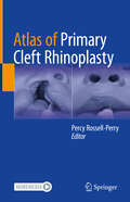
Atlas of Primary Cleft Rhinoplasty
by Percy Rossell-PerryCleft lip is a common congenital anomaly and results in an abnormal shape and position of the nasal septum and nasal tip cartilages that creates an uneven appearance of the nostril, nasal tip, and deviation of the nasal septum. These changes frequently cause breathing difficulties during childhood and remain a formidable challenge for any cleft surgeon. This unique book is about primary repair of nose deformities associated with cleft lip. Written by leading experts, it addresses anatomy, physiology, classification, pre- and postsurgical management, surgical techniques, and related complications. The work is based on the editor's extensive experience and supported by excellent outcomes and up-to-date scientific evidence. It is richly illustrated with pre- and postoperative photographs and well-designed figures. 3D technology designing presurgical orthopedics and postoperative nasal conformers is also presented. Atlas of Primary Cleft Rhinoplasty is an essential resource for plastic, pediatric, ENT, and maxillofacial surgeons. It is also useful for dentists and for members of the interdisciplinary cleft team.
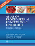
Atlas of Procedures in Gynecologic Oncology
by Douglas A. Levine Nadeem R. Abu-Rustum Richard R. BarakatA core text from the renowned Memorial Sloan-Kettering Cancer Center, this volume covers the latest developments in both open and minimally invasive surgery. Supplemented with full-color photographs, practical explanations, and video clips, the book provides a detailed overview of the major gynecologic oncology procedures. Topics include conization, surgical staging, vulvar surgery, radical hysterectomy, paracentesis, chest tube placement, and central venous access as well as sentinel node mapping and minimally invasive lymph node dissection. It also discusses intraoperative radiation therapy, inguinofemoral lymphadenectomy, and myocutaneous flap reconstruction.
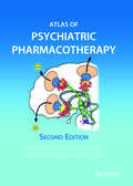
Atlas of Psychiatric Pharmacotherapy
by David J. Nutt Roni Shiloh Rafael Stryjer Abraham WeizmanThe second edition of this exceptional book provides a comprehensive understanding of the mechanisms of action involved in psychiatric pharmacotherapy. Using imaginative colourful double-page spreads, this exceptional book presents state-of-the-art information on all the basic principles of psychiatric pharmacotherapy, abused substances, drug inter
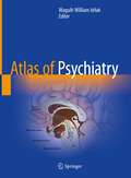
Atlas of Psychiatry
by Waguih William IsHakThis atlas is the first fully visual reference to cover psychiatry broadly, appealing to psychiatric as well as non-psychiatric clinicians and trainees who need an easy-to-use visual resource with holistic approach to patient care. Written by expert clinicians and educators, this text describes basic clinical and scholarly information across the field utilizing an easy-to-understand format. The rich figures and tables describe etiology, pathophysiology, phenomenology, and treatment even in areas that are difficult to illustrate, including substance-related disorders, neurodegenerative diseases, personality disorders, and others. The visual approach proves valuable to some of the most innovative techniques in psychiatry, including implications for neuroimaging. Comprehensive and unique, Atlas of Psychiatry is a landmark reference for all medical practitioners looking for an intricate yet accessible visual resource.
Atlas of Pulmonary Pathology: A Pattern Based Approach
by Yasmeen Mahmood Butt Henry D. TazelaarClosely mirroring the daily sign-out process, Atlas of Pulmonary Pathology: A Pattern Based Approach is a highly illustrated, efficient guide to accurate diagnosis. This practical reference uses a proven, pattern-based approach to clearly explain how to interpret challenging cases by highlighting red flags in the clinical chart and locating hidden clues in the slides. Useful as a daily “scope-side guide,” it features numerous clinical and educational features that help you find pertinent information, reach a correct diagnosis, and assemble a thorough and streamlined pathology report. More than 1,500 high-quality photomicrographs capture the subtle morphologic spectrum of both neoplastic and non-neoplastic lung biopsies. Each image is captioned with key diagnostic considerations and includes call-outs showing subtle features and diagnostic clues. Practical tools throughout the text include: Tables that emphasize salient clinicopathologic features, management implications, and therapeutic options Discussions of how and when to incorporate molecular tools Checklists for key elements of the diagnostic approach and sample notes for inclusion in pathology reports Relevant endoscopic images, photographs of select gross specimens, and medical figures Brief reviews of normal histology that provide contrast to succeeding patterns “Pearls and Pitfalls” and “Near Misses” sections with lessons from real life sign-out experience “Frequently Asked Questions” sections that discuss common diagnostic dilemmas “Sample Note” sections that offer a template of how to sign out cases from the simple to the complex Comprehensive quiz provides experience with high-yield, board-style teaching topics Enrich Your Ebook Reading Experience Read directly on your preferred device(s), such as computer, tablet, or smartphone. Easily convert to audiobook, powering your content with natural language text-to-speech.
Atlas of Radiology in Pediatric Kidney Disorders
by Sidharth Kumar Sethi Rupesh Raina Vivek Sharma Hui Kim YapThis book covers all forms of imaging in pediatric nephrology incorporating all radiologic modalities and imaging of pediatric kidney disorders. It covers from antenatal to post-natal imaging of kidney of a child, starting from normal to abnormal imaging and latest evidence based approach. Chapters cover all forms of abnormalities including cysts, neoplasms, infections, acute kidney injury and chronic kidney disease. The book includes chapter on basics of radiology in children, how to avoid radiation, and point of care ultrasound in pediatric intensive care among others. The editors and authors are leading experts in pediatric nephrology and pediatric radiology from across the world. The editors mentor pediatric nephrology sessions at multiple avenues across the world and therefore the book incorporates all the latest protocols. The book is relevant for radiologists, pediatric radiologists, pediatricians, pediatric nephrologists, adult nephrologists, fellows in nephrology and radiology. It also meets the curriculum requirements of fellowship training in nephrology and pediatric nephrology.
Atlas of Real Time 3D Transesophageal Echocardiography
by Hans-Joachim Nesser Natesa G. Pandian Itzhak Kronzon Stefano De Castro Francesco F. Faletra Siew Yen HoThis atlas provides a comprehensive description of normal anatomy of the internal structures of the heart (natives valves, interatrial septum, left atrial appendage, left atrium etc..) as seen by this revolutionary ultrasound technique. Normal TEE cardiac structures are described and compared with the corresponding anatomical specimens focusing on the fundamental as well as the details of the cardiac structures, providing a detailed understanding of the anatomy that has not previously been possible with either real-time transthoracic echocardiography (TTE) or reconstructed 3D TEE imaging technology. The atlas contains a large number of challenging cardiac pathology cases observed in clinical settings and based of the combined experience of five outstanding institutions in Europe and United States. Each case is accompanied by a brief presentation and discussion of the value of the imaging modality to effective diagnosis.
Atlas of Response to Immunotherapy
by Stefano Fanti Egesta LopciThis atlas is a concise but comprehensive guide to the diverse patterns of response to immunotherapy as observed on Positron Emission Tomography/Computed Tomography (PET/CT) and other conventional imaging modalities, including CT and Magnetic Resonance Imaging (MRI). The purpose for this publication is to fill the gap between the growing clinical relevance and utilization of immunotherapy in medical oncology, mainly based on checkpoint inhibitors, and the need for experienced imagers with reliable tools assessing response to treatment. A series of disease-oriented chapters will present the imaging findings during immunotherapy in the major oncological settings, with helpful comparison of functional (PET/CT) and morphological (CT/MRI) patterns of response in individual cases. To complete the atlas, a dedicated chapter will focus on major pitfalls and immune-related adverse events (irAEs) affecting image interpretation during the course of immunotherapy. The concluding chapter will lastly examine the available data and potential developments of immuno-PET, which is considered as the novel frontier of research in this oncological scenario. The atlas will be of high value for radiologists and nuclear medicine specialists at all levels of experience.
Atlas of Robotic Cardiac Surgery
by Jr. W. Randolph ChitwoodRobotic surgery is currently devoid of adequate didactic material necessary to facilitate daily application in cardiothoracic surgical practice. This book represents the definitive atlas that will lead both the practicing and new cardiothoracic surgeons in these methods. It will define the operative pathway of each procedure, from beginning to end, for surgeons who wish to be a complete robotic cardiac surgeon and include hints and procedural pitfalls derived from the experiences of chapter contributors. The book will be illustrated with high quality illustrations and color photographs from surgical operations where appropriate. Leading surgeons have contributed to the book and provided sample illustrations for their respective chapters. Anesthetic and cardiopulmonary support preparation for each operation will be included and selected references will be provided to emphasize evidence-based outcomes.
Atlas of Robotic Prostatectomy
by Peter Wiklund Jorn H. Witt Hubert JohnIn many centers of excellence in Urology, robotic prostatectomy has become the first choice for the surgical treatment of localized prostate cancer owing to benefits such as reduced pain and minimization of impotence and incontinence. This atlas, specifically designed for use by surgeons, provides a beautifully illustrated, step-by-step guide to all aspects of the procedure. The various techniques that can be employed to achieve excellent oncological and functional results are carefully depicted in appropriate detail; for example, nerve-sparing techniques, bladder neck reconstruction, and approaches aimed at the early restoration of continence are clearly described. Special situations, such as prior prostate surgery, a large prostate, and salvage prostatectomy, are also fully covered. The information contained in this atlas will be of great value in enabling surgeons to improve their results and to take full advantage of the benefits of robotic prostatectomy compared with open prostatectomy.
Atlas of Robotic Thoracic Surgery
by Kemp KernstineThis book represents the definitive robotic thoracic surgery atlas, containing didactic material necessary to facilitate effective practice in thoracic surgery and to provide learning tools in these methods both to practicing surgeons and to those in training. It defines the complete operative pathway for each procedure for surgeons who wish to be a complete robotic cardiothoracic surgeon and includes hints and procedural pitfalls derived from the experiences of chapter contributors. The Atlas of Robotic Thoracic Surgery is illustrated with high quality illustrations and color photographs from surgical operations and contains expert analysis from leading surgeons who provide the key visual features of their chosen topics. Anesthetic and cardiopulmonary support preparation for each operation are included and selected references are provided to emphasize evidence-based outcomes.This book has been designed to augment Atlas of Robotic Cardiac Surgery edited by Ranny Chitwood, both being developed from these same concepts of simplicity and practical instruction. It will therefore be an important resource for all involved in thoracic robotic surgery or interested in learning more about the techniques involved.
Atlas of Robotic Upper Gastrointestinal Surgery
by Omar Yusef Kudsi Peter P. GrimmingerDeep knowledge of anatomy and surgical technique will continue to remain the foundation of surgery despite advancement in surgical technology. Robotic surgery usage has increased drastically in the last decade. More than ever before, surgical community in great need for an updated atlas in the various upper GI surgical procedures and techniques available. This atlas demonstrates how to perform the most common Upper GI robotic procedure via a set of high-quality state-of-the-art annotated images showing step-by-step guidance providing pertinent and concise procedure descriptions spanning benign and malignant upper GI problems. Robotic upper GI procedures are considered technically demanding with attention to details thus are considered great teaching procedures especially with dual robotic consoles, simulation, and teleproctoring. Preoperative, intraoperative, and postoperative figures are integrated to highlight the importance of these step-by-step procedures, enhance skill and efficiency, and avoid surgical pitfalls. Detailed descriptive figures accompany step-by-step instructions and include specific anatomical annotations that describe the anatomy during upper GI procedures. Atlas of Robotic Upper Gastrointestinal Surgery will provide a comprehensive, insightful and state-of-art review of this field, and will serve as a valuable visual resource for surgeons, surgeons in training, and students with an interest in robotic upper GI surgery. All chapters are written by an international group of experts in their field, to provide a comprehensive atlas for specialists and trainees, this illustrated book will give a current and concise summary of all key topics and recent developments in upper GI surgery.
Atlas of Robotic Urologic Surgery (Current Clinical Urology Ser.)
by Li-Ming SuThe Atlas of Robotic Urologic Surgery provides a detailed, step-by-step guide to common robotic urologic procedures for the purpose of helping novice surgeons in their transition to robotic surgery and seasoned robotic surgeons to refine their surgical technique and expand their repertoire of robotic procedures. In addition, less commonly performed robotic procedures such as those for male infertility, pelvic organ prolapse, urinary tract reconstruction and pediatrics are included. Each chapter is written by the thought leaders in robotic urologic surgery with descriptive step-by-step text, complimented by figures and intraoperative photographs detailing the nuances of each procedure. Emphasis is placed on operative setup, instrument and equipment needs and surgical techniques for both the primary surgeon as well as the operative assistant. The use of ancillary equipment and robotic instrument and endoscope exchanges are highlighted throughout the procedural text by tables designed to aid surgeons and their teams in improving efficiency. This volume will provide unique insights into robotic urologic surgery and reduce the learning curve of accomplishing these increasingly popular procedures.
Atlas of Robotic Urologic Surgery (Current Clinical Urology)
by Li-Ming SuThe Atlas of Robotic Urologic Surgery provides a detailed, step-by-step guide to common robotic urologic procedures for the purpose of helping novice surgeons in their transition to robotic surgery and seasoned robotic surgeons to refine their surgical technique and expand their repertoire of robotic procedures. In addition, less commonly performed robotic procedures such as those for male infertility, pelvic organ prolapse, urinary tract reconstruction and pediatrics are included. Each chapter is written by the thought leaders in robotic urologic surgery with descriptive step-by-step text, complimented by figures and intraoperative photographs detailing the nuances of each procedure. Emphasis is placed on operative setup, instrument and equipment needs and surgical techniques for both the primary surgeon as well as the operative assistant. The use of ancillary equipment and robotic instrument and endoscope exchanges are highlighted throughout the procedural text by tables designed to aid surgeons and their teams in improving efficiency. This volume will provide unique insights into robotic urologic surgery and reduce the learning curve of accomplishing these increasingly popular procedures.
Atlas of Robotic, Conventional, and Single-Port Laparoscopy: A Practical Approach in Gynecology
by Tommaso Falcone Pedro F. EscobarMinimally invasive surgery has emerged as the standard treatment for many gynecologic diseases and conditions. In the past decade, numerous studies have demonstrated the superiority of laparoscopic approaches over standard open procedures in terms of improved quality of life for patients. Innovations in minimally invasive surgical technology—such as multichannel ports, articulating instruments, and flexible high-definition endoscopes—have made it possible for laparoscopic surgeons to perform increasingly complicated gynecologic surgeries through smaller incisions. As such, since the first edition of the atlas published in 2014, technologies and techniques once considered novel have become standard. This second edition, with five new chapters and content updated throughout to reflect the latest evolutions in the field, serves as a guide in robotic, conventional, and single-port laparoscopic surgery, presenting invaluable, up-to-date information about instrumentation, surgical technique, port systems, and the current research and development in robotics. Chapters address unique challenges associated with each technique, such as lack of haptic feedback or articulation and instrument crowding, and describe the advanced laparoscopic skills required to safely and efficiently perform procedures, such as how to move and control a flexible camera or use the robot. Specific topics include conventional laparoscopic myomectomy, adnexal surgery, total and supracervical hysterectomy, and excision of endometriosis excision, as well as related techniques in gynecologic oncology, urogynecology and pelvic reconstructive surgery, tubal surgery and ectopic pregnancy, isthmocele repair, and trachelectomy for early cervical cancer. For single-port laparoscopic techniques, chapters are presented on adnexal surgery, hysterectomy, and gynecologic oncology, while the section on robotic surgery offers guidance on instrumentation, platforms, and basic principles; robotic-assisted laparoscopic myomectomy, total hysterectomy for benign disease, endometriosis management, and total hysterectomy for cancer; as well as techniques for robotic adnexal surgery, urogynecology/pelvic reconstructive surgery, tubal surgery, and complication management, concluding with a review of new and emerging technologies. For students, residents, fellows, operating room personnel, and practicing gynecologic surgeons, the editors share experience amassed while developing novel surgical instrumentation and collaborating on presentations for numerous worldwide events. Internationally renowned experts contribute as well to this practical, illustrated resource on current minimally invasive techniques in gynecologic surgery.
Atlas of SPECT-CT
by Mohsen Farsad Stefano Fanti Luigi MansiThe field of nuclear medicine has evolved rapidly in recent years, and one very important aspect of this progress has been the introduction of hybrid imaging systems. PET-CT has already gained widespread acceptance in many clinical settings, especially within oncology, and now SPECT-CT promises to emulate its success. Useful applications of this new approach have been identified not only in oncology but also in endocrinology, cardiology, internal medicine, and other specialties. This atlas, which includes hundreds of high-quality images, is a user-friendly guide to the optimal use and interpretation of SPECT-CT. The full range of potential SPECT-CT applications in clinical routine is considered and assessed by acknowledged experts. The book is designed to serve as a reference text for both nuclear physicians and radiologists; it will also provide fundamental support for radiographers, technologists, and nuclear medicine and radiology residents.
Atlas of Salivary Gland Pathology
by Joaquín J. GarcíaSurgical pathologists play a central role in patient surveillance and treatment by surgeons, radiation oncologists, and medical oncologists. Although salivary gland tumors are uncommon overall, their histopathologic diversity and challenge command the attention of practicing surgical pathologists. Atlas of Salivary Gland Pathology focuses on the diagnostic approach to salivary gland neoplasia—one of the more challenging fields within surgical pathology—emphasizing the need to understand downstream implications with respect to patient surveillance and treatment. The presence of formidable histologic mimicry in salivary gland neoplasia is well-documented in the pathology literature and has also been observed in the consultation practice of the volume editor. This textbook is designed with the needs of practicing surgical pathologists and pathologists-in-training in mind, providing a comprehensive overview of both common and rare salivary gland neoplasms. The primary educational objectives for readers include the following: 1) distinguish benign from low-grade malignant salivary gland neoplasms, 2) effectively use histochemical, immunohistochemical, and cytogenetic studies in challenging cases, 3) understand which diagnoses merit additional surgery and/or adjuvant therapy (radiation, chemotherapy), and 4) enhance pathologist-to-clinician communication in the setting of salivary gland disease.
Atlas of Scar Treatment and Correction
by Igor SafonovThis atlas is a comprehensive guide to the treatment and correction of scars. It is divided into four sections covering the different types of scar: atrophic and stretch marks, keloid and hypertrophic, normotrophic, and mixed. For each scar type, the various invasive and minimally invasive procedures and their results are documented with the aid of numerous high-quality photographs. In the section on keloid and hypertrophic scars, treatment is presented according to scar localization. In addition, the influence of etiology on treatment is considered, with distinction between scars due to injuries, animal bites, inflammatory diseases (including acne and varicella), and burns. Care is taken to distinguish between approaches suitable for fresh scars (in the inflammation, proliferation, and maturation phases) and those appropriate for scars present for more than one year. Potential adverse effects and complications of treatment are also explored.
Atlas of Sciatica: Etiologies, Diagnosis, and Management
by Ali AkhaddarThis atlas is the first reference covering exclusively all aspects of sciatic pain. It is designed to serve as a brief and easy-to-comprehend review of the knowledge of spinal sciatica, with emphasis on classification, epidemiology, clinical presentations, neuroimaging, and treatment options. Sections on extraspinal sciatica and differential diagnosis of this multifaceted topic are also included. This atlas delivers more information in less space than traditional texts, allowing for a quick review of the essential facts of this clinical entity through plentiful images and tables. Pertinent imaging is combined with intraoperative photographs and hand-drawn illustrations to help readers visualize variable presentations and enhance their management. The comprehensive content of this richly-illustrated book covers different etiologies of sciatic pain seen in spinal, neurosurgical, neurologic, rheumatologic and emergency practices, divided into five thematic sections. After general considerations about sciatica and their differential diagnosis, the second section focuses on lumbosacral discogenic sciatica. The third section includes spinal non-discogenic sciatica. The fourth section focuses on extraspinal intrapelvic sciatica, and the fifth provides a description of the most important etiologies of extraspinal extrapelvic sciatica. Comprehensive and unique, Atlas of Sciatica is an excellent pictorial resource for neurosurgeons, spinal surgeons, neurologists, rheumatologists, and many other clinicians worldwide. It is a “one of a kind” book that stands head and shoulders above any other book on this subject (from the foreword of Professor Edward C. Benzel, MD, Founder of the World Spinal Column Society).
Atlas of Sectional Anatomy: Understanding the Anatomical Aspects of the Thorax, Abdomen and Pelvis
by Luciano Alves Favorito Natasha T. LogsdonSectional anatomy is a valuable resource for understanding and interpreting imaging exams, specially computed tomography (CT) and magnetic resonance imaging (MRI). Thus, health professionals should have a solid anatomical knowledge to properly evaluate such exams during clinical assessments of cardiac, thoracic, abdominal, proctologic, gynecological and urological diseases. The chapters in this book describe the thoracic anatomy, the abdominal wall, retroperitoneal space, and the male and female pelvis. Sectional images of cadaveric material illustrate the thoracic and the abdominal cavities, kidney, ureter, prostate, penis and other male and female organs. The images and descriptions build familiarity with the anatomical traits and can be applied in the fields of urology, gynecology, proctology, radiology and surgery. This work appeals to a wide range of readers, from health professionals to residents and students of different medical specialties.