- Table View
- List View
Atlas of Sectional Radiological Anatomy for PET/CT
by Mehmet T. KitapciThe horizons of sophisticated imaging have expanded with the use of combined positron emission tomography (PET) and computed tomography (CT). PET-CT has revolutionized medical imaging by adding anatomic localization to functional imaging, thus providing physicians with information that is vital for the accurate diagnosis and treatment of pathologies. Since the integration of PET and CT several years ago, PET/CT procedures are now routine at leading medical centers throughout the world. This has increased the importance of nuclear medicine physicians acquiring a broad knowledge in sectional anatomy for image interpretation. The Atlas of Sectional Radiological Anatomy for PET/CT is a user-friendly guide presenting high-resolution, full-color images of anatomical detail and focuses solely on normal FDG distribution throughout the head & neck, thorax, abdomen, and pelvis, the primary sites for cancer detection and treatment through PET/CT.
Atlas of Sellar and Parasellar Lesions: Clinical, Radiologic, and Pathologic Correlations
by Gabriel Zada M. Beatriz S. Lopes Srinivasan Mukundan Jr. Edward R. Laws Jr.This book presents, in a stepwise and interactive fashion, approximately 75 cases that reflect the wide spectrum of pathology encountered in this region. Each case description commences with a concise clinical scenario. High-quality radiologic, laboratory, and histopathologic images depicting the differentiating features of the lesion subtype in question are then presented, and key operative and clinical management pearls are briefly reviewed. The interdisciplinary nature of this easy-to-use color atlas and textbook reflects the fact that the management of patients with sellar and parasellar lesions is itself often interdisciplinary. The format is unique in that no similar interdisciplinary book is available on lesions of this region of the brain. Atlas of Sellar and Parasellar Lesions: Clinical, Imaging, and Pathologic Correlations is of great value for practitioners and trainees in a range of medical specialties, including radiology, neurology, endocriniology, pathology, oncology, radiation oncology, and neurosurgery.
Atlas of Sexually Transmitted Diseases: Clinical Aspects and Differential Diagnosis
by Mauro Romero Leal PassosThe Atlas of Sexually Transmitted Diseases and Differential Diagnosis is a complete course through the most important sexual diseases. Counting with about 1000 figures, this atlas focuses on the peculiar characteristics of each of these diseases and in the relevant aspects for a proper differential diagnosis. It is authored by MDs with a deep knowledge in the field, providing a very comprehensive casuistry. Throughout the book the reader will have access to hundreds of pictures discussing about the most important sexually transmitted diseases. Each chapter will approach a different disease, providing a broad sort of images, since a microscopic perspective until the most classical genital lesions and characteristics, considering also systemic manifestations. Every figure is presented with a descriptive and instructive legend, providing the reader with all possible related information. Additionally, every chapter begins with an explanatory and introductory text disclosing about key aspects of the disease, such as etiopathogenesis; clinical manifestation; clinical, laboratorial and differential diagnosis; treatment and prevention.
Atlas of Single-Incision Laparoscopic Operations in General Surgery
by Shuodong Wu Ying Fan Yu TianThis atlas presents an extensive collection of single-incision laparoscopic operations in general surgery performed from 2008 to 2013 in Shengjing Hospital of China Medical University. 2000 operations are reported in it. All the figures in it were collected using real time recordings of the operations which will be particularly interesting to the readers. In each specific surgery, authors provide its key surgical steps, complications and management and contraindication. This atlas is intended for clinical surgeons who can evolve these operations soon after reading about them. Patients will also benefit from this book because of the combination of minimal invasiveness and cosmetic effect of single-incision laparoscopic surgery.
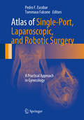
Atlas of Single-Port, Laparoscopic, and Robotic Surgery: A Practical Approach in Gynecology
by Tommaso Falcone Pedro F. EscobarMinimally invasive surgery has become the standard treatment for many diseases and conditions. In the last decade, numerous studies have demonstrated that laparoscopic approaches have improved patients' quality of life if compared with standard open procedures. Atlas of Single-Port, Laparoscopic, and Robotic Surgery serves as a guide in single-port, standard laparoscopy, and robotic surgery and shows how novel techniques, such as single-port laparoscopy and robotics, have recently evolved. The atlas illustrates the unique challenges that the new single-port surgery modality presents, including instruments crowding and articulation, and the advanced laparoscopic skills required to perform these procedures, such as the ability to move and control a flexible camera. It also illustrates how to efficiently and safely utilize the robot to perform most gynecologic procedures. This exceptional resource provides students, residents, fellows, operating room personnel, and practicing gynecologic surgeons with invaluable information about instrumentation, surgical technique, port systems, and the current research and development in robotics.
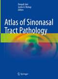
Atlas of Sinonasal Tract Pathology
by Deepali Jain Justin A. BishopThis book reviews normal anatomy and immunohistochemical profile of sinonasal tract (SNT) and covers histomorphology of infections some of which are unique to Asia, tumor-like lesions, and rapidly evolving spectrum of benign and malignant tumors of SNT. It describes all the entities with the help of over 200 full color photomicrographs with their detailed descriptions. The book includes relevant clinical and radiology photographs and anatomical drawings complemented by short bulleted legends describing the picture and providing relevant information. In addition to surgical pathology, it includes a chapter on the cytology of SNT lesions, which is described rarely in available books and literature. It serves as a concise and informative resource for easy understanding. The book is written by leading experts in pathology around the world from USA, Europe and Asia. It highlights the expanded role of pathologists in patient care of SNT lesions. This book is suitable for clinicians, practicing pathologists, faculty, pathology residents and students. This will help the post-graduate students in pathology to understand this area in detail and to be able to answer questions on SNT resection/biopsy cases asked in their board exams of residency.
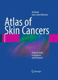
Atlas of Skin Cancers: Practical Guide to Diagnosis and Treatment
by Ali Hendi Juan Carlos MartinezThe incidence of skin cancer has risen rapidly in recent decades, and patients often present initially to practitioners in many different specialties. Because skin cancer can vary in clinical appearance, even dermatologists may experience difficulty in reaching a clinical diagnosis. For primary care physicians and physician extenders (physician assistants, nurses, and nurse practitioners), who have had very little or no formal training in dermatology, the task can be still more daunting. In this atlas, the authors set out to provide a practical resource that will help improve the 'visual vocabulary' of physicians and physician extenders, helping them identify lesions that should be biopsied. Hundreds of high-quality color images are included to assist the reader in the task of recognition and identification. All of the common cutaneous malignancies are illustrated, with a number of examples of each entity and of common mimickers. In addition, biopsy techniques and treatment options are presented in step-by step detail with the use of high resolution clinical images, and potential complications of treatment are discussed. This atlas is ideal for all providers who wish to sharpen their clinical acumen and gain confidence in identifying skin cancers.
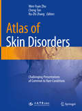
Atlas of Skin Disorders: Challenging Presentations of Common to Rare Conditions
by Wen-Yuan Zhu Cheng Tan Ru-Zhi ZhangThis book was written to assist the dermatologists and practitioners in the management of rare and challenging skin disorders. It is the most valuable collection of such skin disorders from more than 274 outstanding contributors over 4 decades in China. In this book, a comprehensive coverage of about 387 conditions are illustrated by 1215 superb images, and each is introduced with an initial summary of clinical characters. This atlas incorporates a wide range of skin disorders from the mildest and common conditions to the most severe conditions. The objective of this book is to provide readers with a clinical reference, which can be easily approachable and possesses the necessary expertise to sharpen a dermatologist’s diagnostic and clinical acumen.
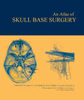
Atlas of Skull Base Surgery
by John K. Niparko Bert W. O’Malley S. James ZinreichOperative procedures directly on the base of the brain, inner ear, and cranial nerves are inherently delicate undertakings and are further complicated by the difficulty of achieving easy access to this confined space. Featuring extensive diagrams, illustrations, and photographs, this book comprehensively covers all of the principal surgical approaches to the base of the skull. Written by pioneers working at one of the world's leading centers for advanced neurosurgery, it clearly describes the steps by which each of the key anatomical structures at the skull base and inner ear may be accessed in order to perform advanced surgical interventions.
Atlas of Sleep Medicine
by Lois E. Krahn Timothy I. Morgenthaler Michael H. SilberWritten by experienced contributors from the renowned Mayo Clinic, the Atlas of Sleep and Sleep Medicine covers the history, humanities, and comparative biological aspects of sleep. This highly illustrated resource includes photographs, reproductions, graphics, segments of sleep studies, and clinical algorithms to aid the clinician in the correct d
Atlas of Sleep Medicine
by Robert J. Thomas Sudhansu Chokroverty Sushanth BhatThis authoritative and updated Atlas provides a comprehensive span of topics across all of sleep medicine, including old to futuristic approaches. It captures the significant changes and advances in the field and a wealth of new visual information available since the last edition. Edited and contributed by leaders in the art and science of sleep medicine, the Atlas highlights how the field of sleep medicine is truly a mix of several medical specialties. The field continues to rapidly evolve with research leading to some future directions. This Atlas remains a standard reference for Sleep Physicians, including Sleep Fellows and other trainees in Sleep Medicine, Sleep Technologists, and Sleep researchers.
Atlas of Small Animal CT and MRI
by Allison Zwingenberger Erik WisnerAtlas of Small Animal CT & MRI is a highly illustrated diagnostic imaging guide to common clinical disorders of dogs and cats. Contains over 3,000 high quality CT, MRI and related diagnostic images Offers a unique approach emphasizing comparative imaging and pathologic correlation Focuses on important imaging features relevant to imaging diagnosis of disease in dogs and cats Written by internationally renowned experts in the field
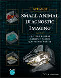
Atlas of Small Animal Diagnostic Imaging
by Clifford R. Berry Nathan C. Nelson Matthew D. WinterATLAS OF Small Animal Diagnostic Imaging Comprehensive and up-to-date resource on the interpretation of diagnostic images in small animals using survey radiographs and other modalities Atlas of Small Animal Diagnostic Imaging provides a comprehensive, multimodality atlas of small animal diagnostic imaging, with high-quality images depicting radiography, scintigraphy, ultrasonography, computed tomography, and magnetic resonance imaging. Taking a traditional body systems approach, the book offers an image-intensive resource to survey radiographs with some other imaging modalities being used to emphasize interpretation of survey radiographs. The Atlas offers clinically relevant information for small animal practitioners and students. Each body structure is thoroughly covered and well-illustrated, with discussion of the strengths and weaknesses of each modality in various scenarios. Edited by three experienced radiographers, The Atlas of Small Animal Diagnostic Imaging contains information on: Basics of diagnostic imaging, physics of diagnostic imaging, CT and MRI physics, US physics, and nuclear medicine physics Musculoskeletal normal anatomic variants, developmental orthopedic disease, joint disease, fracture and fracture healing, aggressive bone disease, and head and spine imaging Thorax anatomy, variants, and interpretation paradigm, extrathoracic structures, pleural space, pulmonary parenchyma, and mediastinum Abdomen anatomy, variants, and interpretation paradigm, extra-abdominal and body wall, peritoneal and retroperitoneal, liver and biliary, and spleen With its expansive coverage of the subject and hundreds of high-quality images to aid in efficient and seamless reader comprehension, Atlas of Small Animal Diagnostic Imaging is an invaluable and must-have resource for small animal practitioners, veterinary students, veterinary radiologists, and specialists in a number of areas.
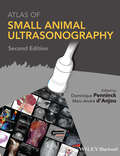
Atlas of Small Animal Ultrasonography
by Dominique Penninck Marc-André D'AnjouAtlas of Small Animal Ultrasonography provides a highly visual guide to the use of diagnostic ultrasound in small animal practice. This up-to-date atlas of the most commonly performed ultrasound examinations will be a valuable reference to well-established users of ultrasound as well as those just being introduced to the technology.Each chapter provides the reader with valuable information on the use of ultrasound diagnostics in small animal veterinary medicine. Taking a systems-based approach, Atlas of Small Animal Ultrasonography is divided into chapters on ultrasound procedures and techniques for examination of all major body parts and systems. Each chapter provides numerous illustrations and ultrasound images to aid the reader in diagnostic interpretation.Atlas of Small Animal Ultrasonography is an in-depth reference that provides thorough coverage of ultrasound techniques and interpretation. This volume will serve as an indispensable guide to the use of this important clinical modality.
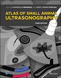
Atlas of Small Animal Ultrasonography
by Dominique Penninck Marc‐André D’AnjouComprehensive reference covering ultrasound techniques and findings in small animal practice with more than 2500 high-quality sonograms and illustrations Atlas of Small Animal Ultrasonography, Third Edition is a comprehensive reference for ultrasound techniques and findings in small animal practice. Offering more than 2500 high-quality sonograms and illustrations of normal structures and disorders, the book takes a systems-based approach to ultrasound examinations in small animals. With complete coverage of small animal ultrasonography, this reference guide is an essential resource for veterinary sonographers of all skill levels. In addition to updates reflecting current diagnostic imaging practice, the Third Edition adds two new chapters, on Point of Care Ultrasonography (POCUS) and on vascular diseases of the abdomen. Also, pertinent ultrasound-assisted interventional procedures were added in several chapters. The Third Edition of Atlas of Small Animal Ultrasonography features: More than 2500 figures of normal and abnormal ultrasound features of the thorax, abdomen, neck, eye/orbit and musculoskeletal systemComplementary imaging modalities when clinically pertinent to the clinical situation Additional surgical or histopathological specimens to best highlight the main features and complete case presentationsAccess to a companion website offering more than 150 annotated video loops of real-time ultrasound evaluations, illustrating the appearance of normal structures and common disorders Atlas of Small Animal Ultrasonography, Third Edition remains an essential teaching and reference tool for novice and advanced veterinary sonographers alike.
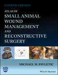
Atlas of Small Animal Wound Management and Reconstructive Surgery
by Michael M. PavleticA one-stop reference for the surgical treatment of wounds in small animal patients Wound management and reconstructive surgery are among the most challenging and innovative subspecialties of veterinary surgery for the management of traumatic injuries and neoplastic conditions commonly encountered in small animals. Atlas of Small Animal Wound Management and Reconstructive Surgery, Fourth Edition presents detailed procedures for surgical reconstruction and essential information on the principles of wound healing and wound management for dogs and cats. Coverage encompasses the pathophysiology and management of the wide variety of wounds encountered in small animal practice and the most current reconstructive techniques for closing the most challenging defects. This updated edition is presented with additional full color images and now includes color in each illustration, to enhance the reader’s understanding of each subject and successful execution of the surgical techniques covered. It imparts new and updated information on a wide variety of topics, including skin and muscle flap techniques, skin fold disorders, facial and nasal reconstructive surgery, foot pad surgery, urogenital reconstructive surgery, and includes a new chapter on reconstructive surgery of the pinna. Provides a key reference for general practitioners, surgeons, surgical residents, veterinary students, and small animal technicians in a single source Discusses current and new wound management and reconstructive surgical techniques for all body regions, including the face, ear, mouth, eye, nose, trunk, external genitalia, and foot Contains 35brand-new plate illustrations, updated and enhanced color plate illustrations and drawings, and additional clinical photographs throughout Features a new chapter dedicated to the surgical management of pinnal injuries and disorders, including new reconstructive surgical options developed by the author Includes information boxes to emphasize important points and the author’s personal observations, drawing on more than forty years of surgical experience Atlas of Small Animal Wound Management and Reconstructive Surgery, Fourth Edition is a valuable one-stop reference for veterinary surgeons, residents, and small animal practitioners.
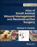
Atlas of Small Animal Wound Management and Reconstructive Surgery
by Michael M. PavleticFully updated new edition of the state-of-the-art, image-driven reference on surgical reconstruction and wound management in dogs and cats Taking a visual approach to the topic with detailed line drawings and high-quality clinical photographs to demonstrate the techniques described, Atlas of Small Animal Wound Management and Reconstructive Surgery provides detailed step-by-step procedures for surgical reconstruction and key information on wound management in dogs and cats. The Atlas covers all body regions, including the face, ears, mouth, eyelids, nose, trunk, external genitalia, and feet. The Fifth Edition has been thoroughly revised to include new techniques, updated information, and references. Over 30 new technique plates have been added and the atlas now includes over 200 colored plates and illustrations. There is a dramatic increase in photographic case series supporting the illustrations. Written by an accomplished veterinary surgeon based on the author’s years of research and clinical experience, Atlas of Small Animal Wound Management and Reconstructive Surgery includes detailed information on: Anatomy of the skin, principles of wound healing and wound management, topical wound care products, management of common complications, and bandages, dressings, external splints, and protective devicesWound drainage techniques, negative pressure wound therapy, and management of specific wounds including bites, thermal, frostbite, gunshot/arrow wounds, pressure ulcers, hygromas, snake/spider bites, and porcupine quillsTension relieving techniques, skin stretching techniques, tissue expanders, local and distant skin flaps, axial pattern skin flaps, skin grafting techniques, and surgical dermatomeFacial, nasal, eyelid, and oral reconstructive surgery, including cleft lip and cleft palate repair, muscle flaps and myocutaneous flaps, pinnal reconstruction, and aural hematoma managementFoot pad reconstruction, reconstructive surgery of the prepuce and vulva, techniques for fecal incontinence and atresia ani, use of the omentum, and a supplemental reconstructive surgery case series The Fifth Edition of Atlas of Small Animal Wound Management and Reconstructive Surgery is a practical, complete, and state-of-the art reference for veterinary surgeons, surgical oncologists, residents, interns, and small animal practitioners to help them effectively manage wounds and reconstruct the variety of soft tissue defects encountered in dogs and cats.
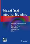
Atlas of Small Intestinal Disorders: Integrated Case Presentation
by Hsiu-Po Wang Cheng-Tang Chiu Yang-Yuan ChenThis atlas composes of original contributions of characteristic cases of small intestinal diseases. The typical small intestinal diseases related cases are discussed in this book. Each case in the atlas is introduced with relevant clinical history and exemplified with educatory endoscopy images.With abundant illustrations, this atlas provides valuable information learned from the diagnoses and treatments of the patients with small intestinal diseases. It also introduces the advances of the latest medical technologies in small intestinal medicine, such as capsule endoscopy, double-balloon enteroscopy, and single-balloon enteroscopy. Small intestinal medicine is an emerging, fascinating and important discipline in gastroenterology. This Atlas can serve as a useful and educational guide to young doctors and a refreshing reference book to educators in this field, and hence promoting better clinical care for their patients.
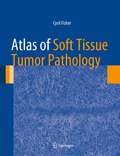
Atlas of Soft Tissue Tumor Pathology (Atlas of Anatomic Pathology)
by Cyril FisherSoft tissue tumors are a large and heterogeneous group of tumors and pseudotumors with a spectrum of behavior from benign to frankly malignant. This Atlas of Soft Tissue Tumor Pathology provides an overview of reactive, pseudoneoplastic, benign and intermediate neoplasms, sarcomas and related conditions arising in subcutaneous and deep soft tissues. Emphasis is placed on microscopic appearances with correlation with gross diagnostic findings where relevant. In addition, the immunohistochemical and molecular genetic features of the major soft tissue tumor subtypes are presented. This compendium of soft tissue tumors illustrates the vast majority of diseases you are likely to encounter in surgical pathology.
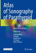
Atlas of Sonography of Parathyroid
by Alka Ashmita SinghalThis comprehensive atlas covers various common and uncommon cases of parathyroid disorders encountered in the day to day radiological practice. It serves as an illustrative database with numerous state of the art images of the diverse imaging features of parathyroid nodules and their correlation with various other imaging modalities. The book includes videos as well. It covers the typical and atypical ultrasound features of parathyroid nodules, their color Doppler findings and Sestamibi correlation. Size of the nodules detected range from small sub-centimeter nodules measuring between 6-15 mm, where often the sestamibi scan is negative, and diagnostic ultrasound scan is quite vital in management of these cases. The larger nodules range in size to be even larger than the thyroid gland and completely compressing and displacing the thyroid gland, which often leads to a missed diagnosis on initial imaging.Chapters cover tips and tricks in scanning along with various pitfalls. Additionally, it covers parathyroid nodules diagnosed on ultrasound in Sestamibi negative cases, localization of additional parathyroid nodules and parathyroid carcinoma. It covers correlation with surgical findings and includes histopathology images where relevant. The book serves as a teaching tool for radiologists, ENT surgeons, head and neck surgeons, general surgeons, endocrinologists, nephrologists, gastroenterologists, medical oncologists and radiation oncologists involved in the management of parathyroid disorders.
Atlas of Spleen Pathology (Atlas of Anatomic Pathology)
by Dennis P. O'MalleyThe spleen is an organ which has diverse functions including immunologic and hematologic. The Atlas of Spleen Pathology describes and selectively illustrates the normal and pathologic conditions that afflict the spleen. This extraordinary collection of high quality digital images will materially aid in continuing efforts to recognize, understand, and accurately interpret the gross and light microscopic findings in spleens.
Atlas of Strobolaryngoscopy: Laryngeal Disorders
by Wen XuSpecializing in viewing of vocal fold vibration, strobolaryngoscopy is a valuable tool for laryngologists and speech-language pathologists in diagnosis of pharyngolaryngeal diseases. This book presents 300 high-quality images and 18 videos from selected representative cases, which help practitioners to grasp the key diagnostic points of srobolaryngoscopy quickly. By watching the videos, readers can observe the vibratory characteristics of vocal folds in details.The book is presented in two parts: The first part is the overview of the strobolaryngoscopy, and the second part focuses on the strobolaryngoscopic signs of common pharngolaryngeal diseases. With the illustrative figures and videos, this book is a practicable reference to laryngologists and speech pathologists.
Atlas of Surgical Anatomy
by Alain C. MasqueletThe author of a number of acclaimed, best-selling surgical atlases has collaborated again with an award-winning artist to produce another invaluable surgical resource. This highly regarded team provide a master-class in the demonstration of surgically relevant anatomy. Masquelet has attained world-renown in particular for his innovative flaps for r
Atlas of Surgical Approaches to Paranasal Sinuses and the Skull Base
by Ziv Gil Dan M. FlissThis richly illustrated atlas, compiled by authors with extensive experience in the field, offers a step-by-step guide to the surgical treatment of tumors, and congenital diseases of the skull base and nasal sinuses. Particular attention is devoted to the various techniques employed for extirpation of tumors and reconstruction of the skull base and Paranasal Sinuses. In order to facilitate understanding of the different approaches, clear surgical illustrations are presented alongside the high-quality intraoperative photographs. Whenever appropriate, technical tips are provided and briefly discussed. This atlas will appeal to a broad audience of residents, fellows, and consultants in different fields of medicine, including surgeons (head and neck, neurosurgery, otolaryngology, plastic and reconstructive surgery, ophthalmology, maxillofacial surgery) and oncologists.
Atlas of Surgical Approaches to Soft Tissue and Oncologic Diseases in the Dog and Cat
by Marije RisseladaThis book offers practical guidance to making approaches for surgery to treat soft tissue and oncologic conditions in canine and feline patients. Every approach is outlined with step-by-step descriptions on how to handle the incision and covers indications and patient positioning. Detailed, high-quality medical illustrations are also included for each, and topics are logically laid out with images on the left and text on the right. Atlas of Surgical Approaches to Soft Tissue and Oncologic Diseases in the Dog and Cat starts with a chapter on oromaxillofacial approaches, followed by chapters covering the cervical area and ear, forelimb, hindlimb, thorax, and abdomen. The book finishes with complete coverage of the approaches to the perineal area and pelvic canal and digits and tail, making it an excellent guide for surgeons to plan and execute their approach to soft tissue and oncologic diseases. Describes the complete approach to surgical incisions for soft tissue and oncologic disease, with alternative positions or approaches where appropriate Provides a high-quality medical line drawing depicting each approach Offers practical guidance for surgeons to direct their approach during surgery Covers indications, patient positioning, and step-by-step summaries of each approach Follows a logical two-page layout with text on one side and illustrations on the other Atlas of Surgical Approaches to Soft Tissue and Oncologic Diseases in the Dog and Cat is an essential reference for any veterinary surgeon or clinician treating soft tissue and oncologic diseases surgically.