- Table View
- List View
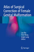
Atlas of Surgical Correction of Female Genital Malformation
by Lan Zhu Felix Wong Jinghe LangThis book discusses a rare form of female genital tract abnormality. It starts with an introduction to modern pre-surgical diagnostic tools, including hysterosalpingography, 3D ultrasound and MRI. From external to internal genitalia, this book introduces all commonly known malformations, as well as a variety of rare congenital malformations of the female genital tract. For each malformation, it includes indications, timing, surgical illustration, contraindication, anaesthesia, surgical procedure, postoperative treatment and key points. In order to facilitate understanding, extensive illustrations and surgical images are also provided. This text offers a comprehensive guide for clinicians, helping them to accurately diagnose and treat these patients, who may present with different clinical manifestations. The authors are well-known gynecologists actively involved in the diagnosis and treatment of difficult diseases.
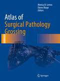
Atlas of Surgical Pathology Grossing (Atlas of Anatomic Pathology)
by Monica B. Lemos Ekene OkoyeThis book is a unique surgical pathology grossing atlas, comprised of a collection of photos of various anatomic specimens frequently encountered in routine and frozen surgical pathology practice, including various organ systems. The photos in this atlas have been collected over many years of practicing surgical pathology in one of the largest medical centers in the world, and include emphasis on important anatomic landmarks and explanations on how to properly orient, section and sample anatomic specimens. The use of actual gross images allows readers to more readily apply the grossing tips to actual specimens that they encounter at the grossing bench. Each chapter is arranged by organ system and includes essential tips for grossing each specimen and sample dictations with all the essential elements that must be addressed for proper assessment of each organ specimen.Written by expert pathologists, Atlas of Surgical Pathology Grossing is an excellent resource for pathologists, medical and pathology assistant students, residents, both in surgery and pathology, and pathology assistants.
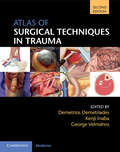
Atlas of Surgical Techniques in Trauma
by Demetrios Demetriades George C. Velmahos Kenji InabaAs surgical specialization becomes more focused, there is a growing lack of expertise amongst surgeons in life-preserving management of severely injured patients. This comprehensively updated second edition provides an in-depth, visual guide to both commonly and uncommonly performed trauma procedures. It includes over 900 high-quality color photographs and illustrations of step-by-step procedures on fresh, perfused and ventilated cadavers. Practical surgical anatomy, procedural sequencing, and common technical pitfalls are all clearly outlined. A number of new techniques have been introduced since the first edition, from REBOA (resuscitative endovascular balloon occlusion of the aortic), to ribplating for flail chest and skin grafting. Informed by the editors' experience in some of the busiest trauma centres in the world, the text has been updated throughout and includes additional photographs. This Atlas is an essential resource for trainee and operating trauma surgeons, and general surgeons distant from academic centres, as well as emergency medicine and critical care personnel.
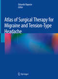
Atlas of Surgical Therapy for Migraine and Tension-Type Headache
by Edoardo RaposioThe approaches to the complex field of chronic headache are rapidly evolving; accordingly, this timely and well-illustrated volume offers an in-depth description of all currently available surgical options for the treatment of migraine and headache (MH). With more than 300 high-quality figures, and written by an international panel of experts, this first edition of the Atlas of Surgical Therapy for Migraine and Tension-Type Headache, provides detailed, step-by-step instructions on how to perform state-of-the-art MH surgical techniques, while reviewing relevant anatomical issues and their implications for treatment. In light of the interdisciplinary nature of migraine treatment, this book will prove an invaluable resource for MH practitioners from the resident plastic surgeon to the neurosurgeon and neurologist, and health care professionals across various fields of clinical medicine.
Atlas of Surgical Treatment of Inflammatory Bowel Disease
by Tomas M. HeimannThis atlas provides high-quality images of surgical inflammatory bowel diseases (IBD). The treatment of IBD continues to be developed and updated surgical imaging of IBD is critical to the care of patients with IBD. To better aid readers, chapters are organized by breaking down different diseases, including: Crohn's disease with Jejunoileitis, Crohn’s disease with Ileocolitis, Crohn’s disease with granulomatous colitis, Recurrent ileitis, Ulcerative Colitis, Indeterminate Colitis, Colorectal Cancer in Ulcerative Colitis, and Intestinal Cancer in Crohn’s disease. Emphasizing core surgical images of IBD, this atlas will be of interest to all physicians who care for patients with IBD, including gastroenterologists, surgeons, internists and even ancillary personnel such as nurses and nutritionists.
Atlas of Swept Source OCT and OCT Angiography
by Youxin Chen Xianzhao PengThe aim of this book is to explain this new technology and its advantages over previous imaging devices and to illustrate how it may be used in to define eye diseases, and in their treatment and facilitate treatment options. This atlas introduces the technical principles of SS-OCT imaging, OCTA stratification algorithm, and blood flow quantization. Through a collection of images and annotation of the microstructures of both the normal fundus and diseased eyes (for example, vitreous related disease, retinal diseased and choroidal disease) captured on SS-OCT to deepen the understanding of this new technology, as well as the interpretation of images. This atlas serves as a practical guide and is written for retinal specialist and clinicians with an interest in retinal diseases.
Atlas of Swept Source Optical Coherence Tomography
by Zofia Michalewska Jerzy NawrockiThis atlas presents an overview of Swept Source Optical Coherence Tomography (OCT) and its implications on diagnostics of vitreous, retina and choroid. As the sensitivity of OCT imaging devices has increased, updated technologies have become available for engineers, scientists and medical specialists to adopt, and recent developments have led to the creation of a new generation of devices. The aim of this resource is to explain this new technology and its advantages over previous imaging devices and to illustrate how it may be used in to define eye diseases, aid in their treatment and facilitate treatment options.

Atlas of Temporomandibular Joint Surgery
by Peter D. Quinn Eric J. GranquistThis second edition of the Atlas of Temporomandibular Joint Surgery is a major revision of Dr. Quinn's classic work, taking into account new procedures, equipment, and evidence-based findings from the latest research in TMJ treatment. Assuming that readers are familiar with non-surgical therapies to correct temporomandibular pain and disorders, Drs. Quinn and Granquist focus on the surgical remedies for disorders that are beyond conservative treatment. This concise, how-to surgical atlas guides both the novice and experienced surgeon through the intra-articular and extra-articular procedures that have proven efficacious in the treatment of advanced craniomandibular dysfunction. Chapters take readers through decision making for TMJ surgery, diagnostic imaging methods, surgical approaches, surgery for internal derangements, trauma, osseous surgical procedures, total joint replacement, and pathologies.
Atlas of Thoracoscopic Lobectomy with Bronchoplasty
by Jian Li Zhiqiang LongThis book introduces techniques and methods of the lymphadenectomy and the angioplasty under thoracoscope. It also presents the latest 2 cm uniportal lobectomy, which represents the highest level of thoracoscopic lung surgery technique. Compared to traditional thoracoscopic surgery, the survival and quality of postoperative life of patients undergoing lung cancer surgery have been greatly improved. This book provides a brand-new vision for thoracic surgeons to understand and learn the latest and most advanced development status and direction of modern minimally invasive techniques in thoracic surgery. The video screenshots fully reflect the real process of the operation and unique surgical methods. Thoracic surgeons will benefit from this book and improve the level of minimally invasive techniques in thoracic surgery.
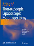
Atlas of Thoracoscopic-lapacoscopic Esophagectomy
by Yi Zhang Tiecheng Pan Xiang WeiThis Atlas provides an easy-to-follow operational guide to laparoscopic techniques. It features a wealth of photos to illustrate esophageal carcinoma surgery.Through step-by-step anatomical photographs, it clearly depicts the Ivor-Lewis operative and Ivor-Lewis-Mckeown operative techniques. Using a consistent format, it addresses the clinical anatomy, pre-operative considerations, operative steps, post-operative care, and pearls and pitfalls to make it easy-to-read.The authors emphasize the similarities of the principles and steps between open and laparoscopic surgery, which significantly simplifies the transition from one practice to the other. This Atlas also includes a description of anesthesia techniques, a guide to the use of staplers in laparoscopic surgery, and a comparison of the energy sources available for laparoscopic surgery, while also outlining future developments, e.g. the increasing prevalence of robotic surgery for these procedures. The Atlas offers an essential guide for practitioners and trainees, laparoscopic and thoracoscopic surgeons, and experienced esophageal surgeons who are preparing to change to minimal invasive techniques for the management of esophageal carcinoma. It will also benefit all surgeons who are seeking clear photos detailing how to perform these esophageal carcinoma operations.
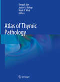
Atlas of Thymic Pathology
by Mark R. Wick Deepali Jain Justin A. BishopThis book reviews normal thymic structure and covers the histomorphology of benign and malignant tumors and rare lesions of the thymus, with detailed descriptions of over 300 full color photomicrographs accompanied by relevant clinical and radiology photographs and anatomical drawings. It includes chapters on the surgical pathology and cytology of thymic lesions (which is rarely described in the available literature), and all grades and stages of the most common thymic lesions. Written and edited by leading experts on pathology, the book highlights the expanded role of pathologists in patient care for thymic lesions. It will help students to understand thymic cytology and surgical pathology in detail and enable them to answer questions on thymic surgical resections and cytology cases in their pathology post-graduation examination. In addition, the book offers a comprehensive, richly illustrated guide to thymic pathology for clinicians, practicing pathologists, post-graduate residents and students alike.
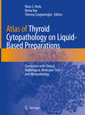
Atlas of Thyroid Cytopathology on Liquid-Based Preparations: Correlation with Clinical, Radiological, Molecular Tests and Histopathology
by Rana S. Hoda Rema Rao Theresa ScognamiglioThis illustrated volume serves as a handy guide to diagnostic fine needle aspiration (FNA) cytology of thyroid on liquid-based preparations (LBP). It is intended to be a ready resource to accurately diagnose thyroid lesions on LBP using key cytomorphologic features. Key cytologic differential diagnosis, gross, and histopathological correlations accompany the cytological findings. The Atlas of Thyroid Cytopathology on Liquid-Based Preparations is lavishly illustrated with color images of various thyroid diseases that should familiarize pathologists with the differences between conventional smears and LBP, and between the two commonly used LBPs. Authored by leaders in the field, this atlas provides clear, concise, and practical guidance pertaining to cytomorphology and the implications of thyroid FNA diagnoses for patient care in this era of precision medicine.
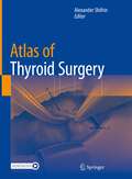
Atlas of Thyroid Surgery
by Alexander ShifrinThis surgical atlas illustrates the various successful thyroid surgery techniques of world-renowned surgeons. Some approaches presented include total thyroidectomy, right thyroid lobectomy, left thyroid lobectomy, minimally invasive approach to thyroidectomy, video-assisted approach to thyroidectomy, and endoscopic approaches, such as the transaxillary approach, bilateral axillo-breast approach (BABA), and trans-oral thyroid surgery (TOEVA). The Atlas is intended for a broader audience than publications for other procedures since thyroidectomy is among the most common neck procedures. The Atlas of Thyroid Surgery will help all surgeons caring for patients with thyroid disease minimize complications and perform successful thyroidectomy.
Atlas of Thyroid Ultrasonography
by Milan Halenka Zdeněk FryšákCombining high-quality ultrasound scans with clear and concise explanatory text, this atlas includes side-by-side depictions of various conditions of the thyroid both with and without indicative marking. Each ultrasound finding is displayed twice, six figures per page: The left-hand image is a native figure without marks; the right-hand image depicts marked findings. In this way, readers have the opportunity to see the native picture to assess it by themselves and then correct their opinion, if necessary. Five sections comprise this atlas, including the normal thyroid, diffuse thyroid lesions, both benign and malicious lesions (including various carcinomas), and rare findings. Including nearly 1500 ultrasound scans and covering the range of thyroid conditions, Atlas of Thyroid Ultrasonography will be a key reference for endocrinologists, radiologists, and primary care physicians, residents and fellows treating patients with thyroid problems.
Atlas of Thyroid and Neuroendocrine Tumor Markers
by Luca GiovanellaThis book highlights the increase in thyroid tumors and NET and demonstrates the growing importance of circulating markers in diagnosis as well as treatment and follow-up. Dramatic technical improvements have heightened the clinical impact of well-established, conventional biochemical markers. In addition, more recent genetic and molecular approaches have provided innovative molecular markers. In this context, effective communication between clinicians and laboratory physicians/scientists is essential in allowing all those involved to fully profit from these exciting advances. In this comprehensive, up-to-date book, authors from different laboratory and clinical areas link laboratory and clinical topics. Analytical problems such as interferences, false-negative and false-positive results are discussed in depth, and flow-charts offer insights into identifying and avoiding them. Illustrated clinical cases detail the clinical role and limitations of different tumor markers. Lastly, it explores health technology assessment and economic issues. This is a valuable resource for endocrinologists, oncologists, nuclear medicine physicians, scientists and technologists who want to keep abreast of the latest developments.
Atlas of Time Lapse Embryology
by Alison Campbell Simon FishelUnlike conventional single daily observations, time lapse technology provides hundreds of images, which allows pinpointing of key events in the embryo's in vitro development as well as the detection of brief but significant critical changes. This information is beneficial in selecting the most viable embryos from a cohort and increases the chances
Atlas of Topographical and Pathotopographical Anatomy of the Head and Neck
by Z. M. SeagalWritten by an experienced and well-respected physician and professor, this new volume, building on the previous volume, Ultrasonic Topographical and Pathotopographical Anatomy, also available from Wiley-Scrivener, presents the ultrasonic topographical and pathotopographical anatomy of the head and neck, offering further detail into these important areas for use by medical professionals. This atlas of topographic and pathotopographic human anatomy is a fundamental and practically important book designed for doctors of all specializations and students of medical schools. Here you can find almost everything that is connected with the topographic and pathotopographic human anatomy, including original graphs of logical structures of topographic anatomy and development of congenital abnormalities, topography of different areas in layers, pathotopography, computer and magnetic resonance imaging (MRI) of topographic and pathotopographic anatomy. Also you can find here new theoretical and practical sections of topographic anatomy developed by the author himself which are published for the first time. They are practically important for mastering the technique of operative interventions and denying possibility of iatrogenic complications during operations. This important new volume will be valuable to physicians, junior physicians, medical residents, lecturers in medicine, and medical students alike, either as a textbook or as a reference. It is a must-have for any physician’s library.
Atlas of Toxicological Pathology
by Chirukandath Gopinath Vasanthi MowatThis atlas contains more than 700 illustrations that the authors have collected over the years as well as references and information pertaining to recently developed drug classes, including biologics. It is a useful bench reference for practicing pathologists and may also be used as a reference text by other experts from related fields. The atlas is organised into different chapters based on systemic pathology. Each chapter has illustrations with legends, and the atlas includes some rare examples of unique lesions found during toxicity studies over many years.
Atlas of Transnasal Endoscopic Orbital Surgeries
by Nishi GuptaThis book incorporates a wide range of cases. With around 1000 images this atlas takes readers through case studies covering the clinical presentation, radiological findings, surgical techniques, and outcome. The pertinent anatomical information required to execute these orbital procedures via the trans-nasal route successfully is covered in a separate section. Schematic diagrams are presented to explain surgical procedures. This book is helpful to otorhinolaryngologists, ophthalmic and oculoplastic surgeons, and neurosurgeons who encounter such cases and need to perform orbital surgeries alone or in conjunction with the team, depending on the extent of the disease.
Atlas of Transvaginal Endoscopy
by Ivo Brosens Rudi Campo Stephan Gordts Hugo Christian VerhoevenTransvaginal endoscopy of the female genital tract is a powerful new technique for the evaluation and management of pelvic disease. This highly illustrated book details the possibilities and advantages of these techniques, including transvaginal laparoscopy, salpingoscopy, patency testing, and vaginocervicohysteroscopy for the exploration of the ut
Atlas of Trauma: Operative Techniques, Complications and Management
by Paula Ferrada Ricardo FerradaThis atlas is the collaborative work of surgeons from Latin America and North America and describes techniques that can aid in the treatment of trauma patients. Trauma surgeons need to perform procedures efficiently and expeditiously for patients that are crashing. The surgeries and exposures are narrated by experts in the field, and include pitfalls and complications, as well as ways to avoid and treat them. Detailed illustrations add clarity to each procedure and help surgeons improve their technique when treating trauma patients. Atlas of Trauma provides a visual aid to the trauma surgeon and acts as a great asset for the training of these specialists.
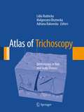
Atlas of Trichoscopy: Dermoscopy in Hair and Scalp Disease
by Adriana Rakowska Lidia Rudnicka Malgorzata OlszewskaThe aim of this atlas is to provide detailed and comprehensive, easy-to-use information, sufficient to perform trichoscopy in clinical practice. From basics to advanced knowledge, everything in one book. In this sense it is rather an "illustrated textbook" than solely an atlas. It includes evidence based information, acknowledged algorithms, which help easy diagnosis and "take home messages", which aid memorizing specific features of diverse diseases. The atlas consists of two major parts. In the first part the authors describe structures and patterns seen in trichoscopy. The second part consists of detailed description of characteristic trichoscopy features of diverse diseases of hair and scalp. Consecutive chapters illustrate genetic hair disorders, acquired hair loss and scalp diseases.
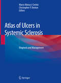
Atlas of Ulcers in Systemic Sclerosis: Diagnosis And Management
by Christopher P. Denton Marco Matucci-CerinicThis Atlas examines skin ulcers in patients affected by systemic sclerosis. Although they are not life threatening, digital ulcers cause enormous pain, impact function and quality of life, and can lead to serious local complications such as gangrene, osteomyelitis, and permanent tissue loss. Knowledge and understanding of these ulcers is pivotal in everyday practice in order to arrive at an accurate early diagnosis, a correct analysis of ulcer characteristics, and an appropriate direct and systematic treatment. To that end, this book describes all the major characteristics of skin ulcers in systematic sclerosis, including classification, evaluation, and categorization. Richly illustrated for practical use, Atlas of Ulcers in Systemic Sclerosis: Diagnosis and Management is an essential resource for physicians and related professionals, residents, and graduate students in rheumatology, dermatology, and angiology.
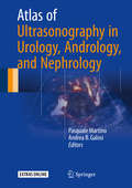
Atlas of Ultrasonography in Urology, Andrology, and Nephrology
by Pasquale Martino Andrea B. GalosiThis book provides the latest recommendations for ultrasound examination of the entire urogenital system, particularly in the male. The coverage encompasses the role of ultrasound in imaging of disorders of the kidneys, urinary tract, prostate, seminal vesicles, bladder, testes, and penis, including male infertility disorders. In addition, detailed consideration is given to intraoperative and interventional ultrasound and recently developed ultrasound techniques. Each chapter defines the purpose of and indications for ultrasound, identifies its benefits and limitations, specifies the technological standards for devices, outlines performance of the investigation, establishes the expected accuracy for differential diagnosis, and indicates the reporting method. Most of the recommendations are based on review of the literature, on previous recommendations, and on the opinions of the experts of the Imaging Working Group of the Italian Society of Urology (SIU) and the Italian Society of Ultrasound in Urology, Andrology, and Nephrology (SIEUN). The book will be of value for all physicians involved in the first-line evaluation of diseases of the renal/urinary system and male genital disorders.
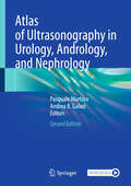
Atlas of Ultrasonography in Urology, Andrology, and Nephrology
by Pasquale Martino Andrea B. GalosiThis second edition provides updated recommendations for ultrasound examination of the whole urogenital system. Most of the chapters is updated, with new images and video clips; others are completely rewritten according to recent developments and guidelines. New chapters are added, mainly about in contrast-enhanced ultrasound, fusion transperineal prostate biopsy, focal ablation in prostate cancer, microultrasound and multiparametric US, bladder outlet obstruction, and computerized analysis of ultrasound through artificial neural networks. Coverage includes the role of ultrasound in imaging disorders of the kidneys, urinary tract of the prostate, seminal vesicles, bladder, testicles, and penis, including male infertility disorders. Detailed consideration is given to intraoperative and interventional ultrasound and recently developed ultrasound techniques. Each chapter defines the purpose and indications for ultrasound; identifies its benefits and limitations; specifies technology standards for devices; outlines performance of investigation; establishes the expected accuracy of the differential diagnosis; and indicates the reporting method. Most recommendations are based on literature review; precedent recommendations; and the opinions of the recognized experts, of the Section of Urological Imaging (ESUI), of the European Society of Urology (EAU), of the Italian Society of Integrated Diagnostics in Urology, Andrology, and Nephrology (SIEUN), of the Italian Society of Urology (SIU) and Nephrology (SIN). This book can be of support both to those taking their first steps in the field of ultrasound, and to subject expert and ultrasound experts, who want to clarify some aspects in the field of urinary tract and male genitalia.