- Table View
- List View
Atlas of Ultrasound Guided Musculoskeletal Injections (Musculoskeletal Medicine)
by Joseph E. Herrera David A. Spinner Jonathan S. KirschnerThe use of ultrasound guidance to perform diagnostic and therapeutic injections is growing at a rapid rate, as is the evidence to support its use. Even with the increased popularity of ultrasound, there remains a lack of formal training or a standard reference book. Atlas of Ultrasound Guided Musculoskeletal Injections fills this void in the literature and will be useful to physiatrists, orthopedists, rheumatologists, pain medicine and sports medicine specialists alike. Broken down by anatomic structure and heavily illustrated, this book is both comprehensive and instructive. The Editors and their contributors break down the basics (both the fundamentals of ultrasound to needle visibility and the role of injections) and explore ultrasound-guided injection for structures in the shoulder, elbow, wrist and hand, hip and groin, knee, ankle and foot, and spine. Using a clear, heavily illustrated format, this book describes the relevant clinical scenarios and indications for injection, the evidence to support ultrasound use, relevant local anatomy, injection methods, and pearls and safety considerations. It will be a valuable reference for trainees and experienced clinicians alike, for experienced sonographers or those just starting out.
Atlas of Ultrasound in Obstetrics and Gynecology: A Multimedia Reference
by Peter M. Doubilet Carol B. Benson Beryl R. BenacerrafPacked with more sonographic images than ever before, the third edition of the Atlas of Ultrasound in Obstetrics and Gynecology helps you better understand and interpret sonographic imagery, and improve diagnostic accuracy of ultrasound images. This comprehensive visual tutorial is split into two distinct sections: obstetrical ultrasound and gynecological ultrasound. Each section covers normal and abnormal anatomy, pathology, and interventional procedures.
Atlas of Ultrasound-Guided Central Venous Catheter Placement: Neonatal and Pediatric Approach
by Fernando Montes-TapiaCentral venous catheter (CVC) placement is the most frequently performed invasive procedure in pediatric and adult patients. The indications for a CVC include hemodynamic monitoring, administration of parenteral solutions, antibiotics, and parenteral nutrition, among others. The internal jugular, subclavian, and femoral veins are the most frequently used sites. Traditionally, vascular access is performed using anatomical guides; however, its effectiveness depends on the operator's expertise. The success of the anatomical guide procedure decreases as the patient's age and weight decrease or the patient has had previous catheterizations or abnormal anatomy. In addition, the risk of associated complications such as cervical hematomas, pneumothorax, cardiac tamponade, or deaths are reported when using this technique. Ultrasound-guided Central Venous Catheter Placement is recognized by the National Institute for Clinical Excellence (2002) and recommends ultrasound guidance as the preferred method for elective CVC insertion in adults and children. However, it has not yet become the standard due to a lack of training or adequate equipment (ultrasound). This innovative atlas intends to cover all the technical details of 11 types of vascular access in the pediatric age. It is richly illustrated with over 200 images. Atlas of Ultrasound-guided Central Venous Catheter Placement is a valuable book for medical professionals who perform vascular access in pediatric patients and a guide for anyone who wants to place an ultrasound-guided central venous catheter. It is a useful resource for pediatric surgeons, surgeons, anesthesiologists, pediatric emergency care, pediatric intensive care, neonatologists, pediatricians, and nurses.
Atlas of Ultrasound-Guided Procedures in Interventional Pain Management
by Samer N. NarouzeThis book is the first and definitive reference in the growing field of ultrasonography in pain medicine. Each chapter details all you need to know to perform a specific block. Comparative anatomy and sonoanatomy of the various soft tissues are featured, and tips and tricks for correct placement of the ultrasound probe and admimistration of the injection are described in detail. All the major peripheral nerve blocks are discussed as well as the various injections of the spine, pelvis, and musculoskeletal system.
Atlas of Uniportal Video Assisted Thoracic Surgery
by Diego Gonzalez-Rivas Calvin Sze Hang Ng Gaetano Rocco Thomas A. D’AmicoThis introduces the history, development and current status of uniportal VATS by pioneers and authorities of this technique. The highly illustrated content in the chapters enhances readers to rapidly understand the techniques of uniportal VAT. The use of video clips adds value to the learning experience and applicability of the techniques. The contents will be of great interest to thoracic surgeons who are already practicing video-assisted thoracic surgery, as well as those who are starting training. It will also serve as authoritative reference text for doctors, students and allied health professionals who would like to learn more about the new technique of uniportal VATS.
Atlas of Upper Extremity Trauma: A Clinical Perspective
by W. Andrew EglsederDemonstrating current management techniques for traumatic fractures and dislocations of the upper extremity, this atlas utilizes a practical, how-to structure, discussing philosophy, approach, patient positioning, prepping, draping, and surgical techniques for each type of injury. Generously illustrated with intraoperative photos, the chapters of this atlas are arranged by anatomic location, from the clavicle and shoulder down to the fingers, with each chapter briefly describing the thought processes involved in choosing surgical interventions and applied anatomy approaches, fixation selections, and techniques. Actual case examples, cadaver photos and plentiful radiographs illustrate the text for a strongly visual presentation, and a list of "Eglsederisms" - pithy bits of advice for residents and veteran surgeons alike - set the stage for a highly demonstrative resource for orthopedic and trauma surgeons, residents and fellows.
Atlas of Upper Gastrointestinal and Hepato-Pancreato-Biliary Surgery
by Michael G. Sarr Pierre-Alain Clavien Yuman Fong Masaru MiyazakiThe first edition of this book was judged by many to be the best available atlas of upper gastrointestinal and hepato-pancreato-biliary surgery. The second edition builds upon this success by covering all of the significant intervening developments, including both new procedures and adaptations of established techniques. The atlas opens with an introductory section on the basic principles of operative surgery. All of the important surgical techniques are then presented in a series of sections devoted to the esophagus, stomach, and duodenum, the liver, the biliary system, portal hypertension, the pancreas, and the spleen. General, oncologic, and transplantation surgical procedures are described step by step with the aid of more than 900 superb illustrations drawn by a team of artists with extensive experience. The contributors were selected on the basis of their extensive experience in the procedures discussed and most are established educators.
Atlas of Urogynecological Endoscopy
by Peter L. DwyerThe study of gynecology and pelvic floor disorders is a rapidly developing area for gynecologists all around the world; and, moreover, an essential aspect to the diagnosis and treatment of pelvic floor disorders is a need for expertise in cystourethroscopy to accurately diagnose conditions of the lower urinary tract that frequently present to the g
Atlas of Uterine Pathology (Atlas of Anatomic Pathology)
by Oluwole Fadare Andres A. RomaThis Atlas of Uterine Pathology is comprehensive overview of the major pathologic processes that may be encountered in the uterine corpus and cervix. Each section is lavishly illustrated and covers normal histology as well as neoplastic and non-neoplastic diseases. Emphasis is placed on presenting the full morphologic and immunophenotypic spectrum of entities, including classical and variant pathology, in a manner that maximizes the utility of the information in routine diagnostic practice.
Atlas of Uveitis: Diagnosis and Treatment
by Peizeng YangThis Atlas provides cutting-edge information on uveitis, which represents a major achievement in clinical studies on uveitis. It includes more than three thousand imaging photos of uveitis patients, showing the disease’s complete profile and the spectrum of variations commonly encountered. Numerous therapeutic regimens are also presented, each of which is designed for a specific form of uveitis. The Atlas also incorporates the latest advances in uveitis studies, making it a unique and valuable resource for a broad readership, including ophthalmologists, postgraduate students, medical students and doctors in ophthalmology.
Atlas of Vaginal Natural Orifice Transluminal Endoscopic Surgery (vNOTES)
by Omar F Dueñas Garcia Jan F. Baekelandt Grover MayVaginal natural orifice transluminal endoscopic surgery (vNOTES) is a relatively modern approach. Different disciplines have used natural orifices to insert endoscopes as a way to provide a minimally invasive approach to multiple procedures. The transvaginal approach is probably one of the least invasive access points into the abdomen. In a similar way laparoscopic surgery changed the history of minimally invasive surgery in the 1980-1990s, vNOTES is reshaping the definition of minimally invasive gynecologic surgery. The vNOTES approach provides surgeons with an alternative access point to the abdomen with quicker recovery times, faster surgery, better cosmetic outcomes and better anesthesia requirements with less cardiopulmonary compromise during the surgery. This is the first surgical atlas of vNOTES surgery in the world that describes different transvaginal laparoscopic procedures for benign and malignant gynecology. Opening chapters present the history of the approach, relevant pelvic anatomy, perioperative considerations, candidate identification and operating room set-up. Stepwise chapters on procedures follow, including hysterectomy, gender affirmation, adnexal surgery, myomectomy and more. Closing chapters discuss the teaching of vNOTES, gynecological oncology, robotic surgery and more. The book is authored and edited by leading experts in the world including multiple photographs and steps to provide surgeons guidance into vNOTES.
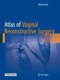
Atlas of Vaginal Reconstructive Surgery
by Shlomo RazThis volume provides a comprehensive, step by step description of the surgical techniques that the author has used in more than 13,000 vaginal reconstructive procedures. Chapters cover surgery for incontinence, prolapse, urethral diverticula, vaginal fistula, reconstruction, surgery for urethral obstruction and vaginal cysts and masses, and lastly, complications of vaginal surgery. Each surgical chapter covers a succinct summary of diagnosis, indications for surgery, and the surgical procedure itself. Sequential operative photographs, drawings, and videos guide are also included to guide the reader through the procedural steps. The Atlas of Vaginal Reconstructive Surgery will serve as a valuable resource for urologists, gynecologists and other specialists with an interest in vaginal reconstructive surgery.
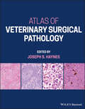
Atlas of Veterinary Surgical Pathology
by Joseph S. HaynesATLAS OF VETERINARY SURGICAL PATHOLOGY An indispensable next-to-the-microscope diagnostic resource for veterinary pathologists Atlas of Veterinary Surgical Pathology delivers a comprehensive exploration of the lesions and diseases most commonly encountered by veterinary practitioners in small animals and horses. The book includes coverage of diseases of the skin, eye, and musculoskeletal systems, and male and female reproductive tracts. It also offers descriptions of relevant microscopic features and color microphotographs of the included lesions. More than 500 images depict lesions. With fully detailed discussions of degenerative, inflammatory, and neoplastic lesions, the book is an authoritative guide to quickly and accurately identifying common and uncommon lesions in small animals and horses. It also offers: A thorough introduction to the techniques relevant to surgical pathology, including specimen preparation and the interpretation of biopsy and the reporting of results Comprehensive explorations of skin and adjacent soft tissue, including developmental and degenerative diseases, inflammatory diseases, and neoplasms Practical discussions of reproductive tract lesions Complete treatments of lesions of the musculoskeletal system, and eye and periocular tissues Perfect for veterinary pathologists and residents, Veterinary Surgical Pathology is a practical handbook to the lesions and diseases encountered by veterinary professionals in small animal and equine surgical pathology.
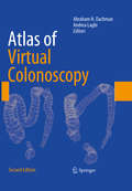
Atlas of Virtual Colonoscopy: Comprehensive Atlas And Fundamentals
by Abraham H. Dachman Andrea LaghiAtlas of Virtual Colonoscopy thoroughly revises and updates Abraham Dachman's bestselling first edition. Joined in this edition by co-editor Andrea Laghi, Dr. Dachman has expanded the focus of the text to cover fundamental topics of this rapidly evolving technology, including the history of virtual colonoscopy, a review of clinical trial data from throughout the world, and a presentation of clinical background information. Also included are chapters covering patient preparation and tagging, performing and reporting virtual colonoscopy, viewing methods, MR colonography, and computer aided detection. The second part of the text presents an atlas of high-resolution images with detailed explanations of teaching points, covering normal anatomy; sessile, pedunculated, diminutive and flat lesions; masses; stool and diverticula; and common pitfalls. Atlas of Virtual Colonoscopy is a valuable resource for all radiologists and gastroenterologists interested in learning the fundamentals of this exciting technique.
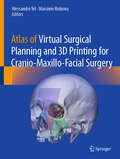
Atlas of Virtual Surgical Planning and 3D Printing for Cranio-Maxillo-Facial Surgery
by Alessandro Tel Massimo RobionyThis book is the first comprehensive atlas dedicated to virtual surgical planning and 3D printing in cranio-maxillo-facial surgery. As the field rapidly evolves, this atlas serves as an essential resource, offering a unified learning platform with detailed examples of virtual surgical planning across various anatomical regions. Each clinical case is meticulously categorized, guiding readers through the intricacies of radiological acquisition protocols, computational design methods, and surgical planning strategies, culminating in 3D printing applications and surgical outcomes. Key concepts explored include point-of-care 3D printing, engineering principles, and the integration of artificial intelligence in surgical planning. Esteemed authors and leading opinion leaders delve into these topics, providing insights into the regulatory aspects crucial for point-of-care laboratories. These labs are increasingly vital in hospitals worldwide, showcasing the potential for advanced case studies using cutting-edge medical software. This atlas is indispensable for a diverse audience, including students, postdoctoral fellows, cranio-maxillo-facial surgeons, neurosurgeons, ENT surgeons, plastic surgeons, bioengineers, clinical engineers, and industry representatives. It not only equips medical professionals with the skills necessary for modern surgical planning but also offers guidance to companies involved in designing and manufacturing medical devices.

Atlas of Vitrified Blastocysts in Human Assisted Reproduction
by Thomas Ebner Pierre Vanderzwalmen Barbara WirleitnerThe method of vitrification of oocytes and embryos is fundamental for the outcome of IVF. This atlas presents data on both closed system and open vitrification techniques, and the consequences of each method for survival rates, aiding the comparison of vitrification methods. Structured on a patient-by-patient basis, the atlas describes 100 clinically documented case studies that follow the evolution of cryopreserved blastocysts between warming and blastocyst transfer. It relates fresh to post-warming blastocyst morphology and to response to controlled ovarian hyperstimulation. For each case, pronuclear morphology and synchrony, as well as embryo morphology, are reported and described. Data on indications for treatment, stimulation type and duration, are accompanied by over 400 high-quality images of vitrified blastocysts. Covering the state-of-the-art techniques, this atlas is an essential aid in selecting the vitrification method for clinical embryologists and physicians in reproductive medicine.
Atlas of Vulvovaginal Disease in Darker Skin Types
by Nina Madnani Kaleem Khan Nisha ChaturvediThere is a significant increase in cross-migration of women from various ethnicities, cultures, and with varying skin color. With publications focused mainly on Fitz Skin types 1–4, accurate vulvar disease diagnosis in darker skin types may be compromised, resulting in suboptimal therapeutic outcomes. Diseases look quite different as the color disguises much of the erythema, which is prominently seen in the Caucasian population. This book is focused on female genital dermatoses and fills in those gaps for healthcare workers looking to accurately diagnose and manage such patients. Key features: Provides inclusive and consolidated contents on disorders of female genital skin and mucosa. Features more than 300 high-quality illustrations and impressive treatment algorithms, making it a ready reckoner for dermatologists, gynecologists, and general physicians. Facilitates diagnosis and further management of vulvar disease in the darker skin types, thus optimizing patient care. Features collective contributions from experts across the country. Simplifies the patient consult by providing ready answers to frequently asked questions.
Atlas of Whole Body Contouring: A Practical Guide
by Richard J. Zienowicz Ercan KaracaogluThis atlas presents current and proven strategies and techniques for body contouring in plastic and reconstructive surgery. In the majority of plastic surgeries, and likewise in body contouring surgeries, surgeons don't have the luxury of long learning curves. The book's simple, descriptive, and didactic structure addresses this issue. The topics have been carefully selected to avoid redundancies and, where there is more than one surgical option to choose from, expert contributors present the technique that has been broadly accepted as ‘the procedure of choice’. The easily accessible information will guide surgeons in their daily practice, helping them minimize complications and achieve consistent results. The book also presents a number of complicated cases, supported by descriptive figures and high-quality illustrations, offering a unique resource for plastic surgeons around the globe.
Atlas of Wide-Field Retinal Angiography and Imaging
by Igor Kozak J. Fernando ArevaloWritten by experts in the field of ophthalmology, this new text assists in understanding new imaging technology and the clinical information it can provide. It allows the reader to review numerous images of various pathologies and would be of interest to retina specialists and ophthalmologists. Retinal imaging has undergone dramatic changes in the last decade, characterized by constantly improving image resolution, most notably, as it applies to macular diseases, thanks to ever evolving optical coherence tomography. However, imaging retina outside of the vascular arcades has come a long way with advent of new cameras and lenses. The most profound change has been the introduction of wide-angle angiography, which has demonstrated pathologies previously not seen or suspected and gives rise to new theories of pathophysiology of some retinal diseases. Understanding this new imaging technology and the clinical information it can provide requires a basis of knowledge and experience in associating the findings with other clinical signs in the eye and the rest of the body. Atlas of Wide-Field Retinal Angiography and Imaging helps with this experience by allowing the reader to review numerous images of various pathologies and analyzes angiographic findings in the retinal periphery. Furthermore, as this technology increasingly fills ophthalmologists' offices around the world, this book will prove to be an invaluable resource, written by experts in the field for retina specialists and ophthalmologists.

Atlas of Wisdom Teeth Surgery
by Dapeng LuThis atlas as a guidebook with twenty chapters covers the latest surgical techniques and clinical cases based on procedure-based approach. Each chapter consists of two to five sections providing detailed descriptions of the basic techniques of evaluation, diagnosis and treatment to allow immediate clinical application. Impacted wisdom teeth are divided into 5 major categories in three-dimensional structure detected by CBCT on the position and growth status of impacted teeth, which helps the classification of 6 degree of difficulty determination for the treatment and the time and surgery procedure needed for each degree. About 1800 colour clinical photos and line drawings with case reports provided by dental expert surgeons and educators in this atlas offer detailed, procedure-based instructions, illustrations and approach that demonstrate how to plan for and perform wisdom teeth surgical procedures safely and efficiently for dentists and students to manage the patients with wisdom teeth disorders.
Atlas of Women's Dermatology: From Infancy to Maturity
by Jennifer L. Parish Lawrence Charles Parish MD Sara Brenner Marcia Ramos e SilvaProving again that a picture is worth a thousand words, Atlas of Women's Dermatology: From Infancy to Maturity is an encyclopedia in pictorial format. The book illustrates diseases and conditions that demonstrate the very different morphology between the sexes. It includes clinical entities that help complete a section or because the lack of gender
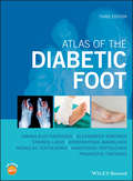
Atlas of the Diabetic Foot
by Konstantinos Makrilakis Stavros Liatis Ioanna Eleftheriadou Alexandros Kokkinos Nicholas Tentolouris Anastasios Tentolouris Panagiotis TsapogasThe revised and updated third edition of the essential guide to the diagnosis and treatment of the diabetic foot The revised third edition of the Atlas of the Diabetic Foot is an updated guide to the information needed for the prevention and treatment of diabetic foot problems with the aim of reducing amputations. In addition to offering the theoretical knowledge, the book is filled with more than 500 color photos from real-life cases. The cases explore a wide-variety of foot issues and the text includes information about differential diagnosis and treatment. The authors—noted experts in the field—describe the epidemiology, pathophysiology and classification of diabetic foot ulcers. In addition, the book highlights the diagnosis of the main risk factors for the diabetic foot, namely diabetic neuropathy, peripheral arterial disease and the anatomic deformities of the lower extremities. The updated third edition, include 5 videos that clearly demonstrate the methods of examination for diabetic neuropathy and peripheral arterial disease. The text incldues methods of callus removal and debridement as well as offloading. In addition, it contains information on how a total contact cast is constructed. This essential resource: Contains a full colour presentation of diabetic foot cases Explains the prevention and treatment of diabetic foot problems in a revised and updated edition Includes a larger format that allows for a better quality images Offers a companion website with high-resolution digital files of photographs of the case studies presented Presents a multidisciplinary approach appropriate for a wide audience foot and diabetic professionals Written for diabetes specialists, endocrinologists and diabetic nurse specialists, podiatrists and podiatry nurse specialists, Atlas of the Diabetic Foot offers the information needed to help with the prevention, diagnosis and management of the diabetic foot.
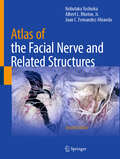
Atlas of the Facial Nerve and Related Structures
by Nobutaka Yoshioka Albert L. Rhoton, Jr. Juan C. Fernandez-MirandaThe second edition of the esteemed atlas provides an in-depth exploration of craniofacial anatomy, enhanced by images of real cadavers that vividly depict the details of human anatomy. Updated content includes a detailed examination of the intratemporal facial nerve, the extraparotid peripheral branches, and their innervation of the mimetic muscles. Delving into the nuances of the intracranial region, skull anatomy, and the complexities of the upper and lower facial regions, this edition captures every detail with stunning clarity. Organized by region for comprehensive learning, each dissection reveals an intricate network of nerves and structures with accuracy. Supported by precise clinical explanations, the book offers profound insights into neurosurgical-related pathologies and beyond. From mimetic muscle myectomy and neurectomy to nerve transfer for facial paralysis, readers will find the knowledge within these pages indispensable for performing precise and safe procedures on the face. Beneficial across various medical specialties including plastic surgery, otolaryngology, neurosurgery, and craniofacial surgery, this atlas is essential for physicians seeking a comprehensive understanding of craniofacial anatomy. Physiotherapists and practitioners of chemodenervation therapy will also discover invaluable insights within this volume.
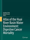
Atlas of the Huai River Basin Water Environment: Digestive Cancer Mortality
by Gonghuan Yang Dafang ZhuangThis atlas shows the relationship between water pollution and cancer in the Huai River Basin in China over the last 30 years. Drawing on a five-year study conducted by the China Centre for Disease Control (CDC), Professor Gonghuan Yang & Dafang Zhuang present a spatial and longitudinal analysis of regular pollution monitoring and disease surveillance data. A review of variation in trends in the causes of death in the Huai River Basin over the past 30 years shows that precisely those areas which were the most seriously polluted for the longest time were the areas with the highest increase in digestive cancer deaths - several times that of the national average increase for the respective cancers. Spatial analysis shows a high level of correspondence between the seriously polluted areas and areas with high mortality from cancer, the most important finding in the atlas. Dr. Gonghuan Yang is a Professor at the Institute of Basic Medical Sciences, Chinese Academy of Medical Sciences. She serves as Director of the Center of NCD & BRFS of the Institute of Basic Medical Sciences, Chinese Academy of Medical Science, Beijing, China. She is an expert on Public Health and an epidemiologist focusing on chronic non-communicable diseases. Dr. Dafang Zhuang is a Professor at the Institute of Geographical Sciences and Natural Resources Research, Chinese Academy of Sciences, Beijing, China. He is an expert of geographical information system and its application in correlating public health with environmental changes.