- Table View
- List View
Treating Urothelial Bladder Cancer
by Paolo Gontero Francesco SoriaThis book aims to bring together the current research and discussions surrounding bladder cancer management. New technologies and therapeutic agents have become increasingly effective at treating urothelial bladder cancer and have created a range of opportunities to change the ways in which the disease is treated. Chapters cover the changes in adjuvant treatment, confocal laser endomicroscopy, and the roles of oncologists, pathologists, and radiotherapists in managing bladder cancer. Treating Urothelial Bladder Cancer is particularly relevant to urologists, oncologists, and radiotherapists, but is also applicable to practitioners, geriatricians, and public health managers due to its multidisciplinary approach to bladder cancer management.
Treating Violence: An Emergency Room Doctor Takes On a Deadly American Epidemic
by Rob GoreThe inspiring story of a Black doctor who was deeply affected by the violence that plagued his Brooklyn childhood and later dedicated himself to addressing trauma and violence as public health issuesRob Gore first encountered violence when he was beaten and robbed as a 10-year old; it was treated as an inevitable fact of life, but after another brush with violence as a teen, he began to reject that prevalent attitude. As he matured and became a doctor, he grew in his determination to find treatments for what he saw not as an unavoidable fact for most people living in vulnerable, underserved neighborhoods especially, but as a public health issue that could be addressed by early intervention and solid support, beginning in the medical community. He also became deeply involved in efforts to diversify the entire field of medicine, starting with the &“front lines&” in the Emergency Department.Seeing his brother Angel and close friend Willis fall prey to the epidemic of violence with profound—and in Willis&’s case—deadly consequences, Rob began seriously researching the issue and went on to found an organization which is one of the models for successful approaches to reducing violence and protecting victims, who are disproportionately BIPOC, living in impoverished neighborhoods, or members of the LGBTQ+ community. Here he provides not only statistics, but stories of what he witnessed in NYC neighborhoods, in Atlanta, Chicago, Buffalo and even in medical work in Haiti and Kenya. His work with the Kings Against Violence Initiate (KAVI) and allied organizations is a blueprint for treating violence not as a police matter, but as a public health crisis, which can and should be addressed and substantially reduced. The people he introduces us to in these pages are not merely victims, but often advocates, paving the way for eliminating the epidemic of violence in our country.
Treating the Complete Denture Patient
by Carl F. Driscoll William Glen GoldenThis book presents step-by-step procedures for all techniques, materials, and methods associated with the use of complete dentures in dental practice. Written for dental students, dental general practitioners, and laboratory technicians, the book provides a practical approach to the complete denture patient. More than 800 photographs illustrate the text, making it easy to follow and apply in the practice or laboratory. Treating the Complete Denture Patient covers all topics related to complete dentures, from the initial appointment and impressions to insertion and troubleshooting. Chapters discuss the diagnostic appointment, covering the analyzing of and treatment planning for edentulous patients; the making of preliminary impressions for the beginning stages of treatment; custom trays for final impression appointments; and much more. Presents easy-to-use, clinically relevant information on all topics related to complete dentures Covers all the steps associated with providing complete dentures, from the initial appointment and impressions to inserting and troubleshooting problems Features hundreds of high-quality color photographs to depict the concepts discussed Includes access to a companion website offering video clips Treating the Complete Denture Patient is an essential resource for dental general practitioners, dental students, and laboratory technicians.
Treating the Dental Patient with a Developmental Disorder
by Karen A. Raposa Steven P. PerlmanTreating the Dental Patient with a Developmental Disorder provides a basic understanding of patients with developmental and intellectual disorders and offers help in communicating with and treating these patients. The book opens with an overview of the major types of developmental disabilities-autism spectrum disorders, Down Syndrome, attention deficit, cerebral palsy-and others such as spina bifida and learning difficulties. Following chapters also discuss how to gather personal information, medical histories, dental experiences, and oral habits; determine family dynamics; and understand how to communicate with patients and model desired patient behavior. The authors also cover aspects of the dental exam and hygiene appointment, and restorative treatment, both in the office and hospital setting. A review of follow-up care and the long-term impact on the practice, the patient, and the families when caring for these patients is covered. Treating the Dental Patient with a Developmental Disorder is a must-have book for practicing and student pediatric dentists, general dentists, and dental hygienists whose patients include families with developmentally or intellectually disabled members.
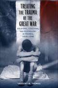
Treating the Trauma of the Great War: Soldiers, Civilians, and Psychiatry in France, 1914-1940 (Southern Literary Studies)
by Gregory M. ThomasFrom the outset of World War I, French doctors faced an apparent epidemic of puzzling neurological and psychiatric illnesses among soldiers. As they attempted to understand the causes of these illnesses, doctors organized specialized centers near the front, where they submitted soldiers to swift, humiliating treatments and then returned them to duty. At home, they interned the scores of civilians who succumbed to the war's strains in decrepit asylums or left them to fend for themselves. In Treating the Trauma of the Great War, Gregory M. Thomas explores the psychological effects of the war on French citizens, showing how doctors' understanding of mental illness produced deep, tangible effects in the lives of the men and women who suffered.Doctors vigorously debated the war's role in the genesis of the neuropsychiatric disturbances observed in soldiers and civilians, but most psychiatrists ultimately concluded that mental illnesses appeared primarily in individuals predisposed to disease. Consequently, doctors granted their patients few favors when making decisions about diagnostic labels, treatment regimes, and pension allocations, leaving many to endure illnesses without adequate care or sufficient financial support. In their quest to understand the psychological impact of war, Thomas argues, doctors focused more on demonstrating the capabilities of their medical specialties and serving a state at war than on treating patients. Those aims significantly affected doctors' scientific conclusions, their medical and legal decisions, and their treatment practices. When the war ended, psychiatric reformers used the trauma of war to their advantage, promoting the perception of France as a traumatized nation in need of new psychiatric institutions that could accommodate a large and growing pool of psychologically wounded citizens.Thomas draws on the vast medical literature produced during and after the war, including veterans' journals, parliamentary debates, newspaper articles, and medical administrative reports, infusing his narrative with a vivid human element. Though psychiatrists ultimately failed to raise the status of their specialty, Thomas reveals how the war helped precipitate lasting changes in psychiatric practice.

Treatise on Controlled Drug Delivery: Fundamentals-optimization-applications
by Agis KydonieusAn introductory but detailed treatise which includes some 1,000 references and solved examples and end-of-chapter problems, making it useful to both students and practitioners. The pharmokinetics, pharmacodynamics, and biological and biopharmaceutical parameters pertinent to each route of administra
Treatment Adherence in Dermatology (Updates in Clinical Dermatology)
by Steven R. Feldman Abigail Cline Adrian Pona Sree S. KolliPatient adherence to a given treatment plan directly correlates to the quality of disease outcome. In looking at the how and why behind patient adherence and non-adherence to a treatment regimen, understanding realistic expectations of a patient can provide a beneficial guide towards providing optimal healthcare.Treatment Adherence in Dermatology offers insight and strategies for understanding and promoting treatment adherence with a focus on dermatological conditions, specifically psoriasis, atopic dermatitis, and acne. Reasons for non-adherence are investigated particularly in populations such as children and patients with multiple co-morbidities. The proposed text provides patient and physician-centered strategies alongside technological advancements to promote adherence. Written for the practicing dermatologist, this title will find audience with primary care physicians, students, residents, and other practicing doctors alike.
Treatment Dilemmas for Vulnerable Patients in Oral Health: Clinical and Ethical Issues
by Alexander MerselThis book equips the reader with a sound understanding of the treatment of neglected and vulnerable patients in the dental office. It offers a comprehensive multidisciplinary approach to atraumatic carious treatment, minimal intervention dentistry, TMJ affections and nutritional consequences. Maintaining proper oral health includes managing of oral hygiene, healthy diet and seeking for a treatment when needed. Untreated dental disease can have a significant adverse impact on the health, wellbeing and quality of life. This book helps practitioners to understand and address the needs of dental neglected adults and children, and guide them through prevention procedures, diagnostics and treatment planning. Important techniques like physiologic impression for removable denture, single crown impression, digital planning and prosthodontic bridges are explained, and socio-demographic and economic changes in Oral Health are discussed. The comprehensive coverage of the topic and the evidence based references make this book a must have for dental practitioners.
Treatment Kind and Fair: Letters to a Young Doctor
by Perri Klass'Treatment Kind and Fair' offers a glimpse inside the doctor's mind for aspiring physicians, medical buffs, and anyone who's ever been a patient.
Treatment Manual for Anorexia Nervosa, Second Edition
by Daniel Le Grange James LockThis indispensable manual presents the leading empirically supported treatment approach for adolescents with anorexia nervosa (AN). What sets family-based treatment apart is the central role played by parents and siblings throughout therapy. The book gives practitioners a clear framework for mobilizing parents to promote their child's weight restoration and healthy eating; improving parent & child relationships; and getting adolescent development back on track. Each phase of therapy is described in session-by-session detail. In-depth case illustrations show how to engage clients while flexibly implementing the validated treatment procedures. New to This Edition Reflects the latest knowledge on AN and its treatment, including additional research supporting the approach. More user friendly clarifies key concepts and techniques. Chapter on emerging directions in training and treatment dissemination. Many new clinical strategies.
Treatment Methods for Early and Advanced Prostate Cancer
by Roger S Kirby Alan W Partin J Kellogg Parsons Mark R FeneleyProstate cancer is treated in a number of different ways, depending on a host of different factors, ranging from the severity of the cancer, the health of the patient, their age, and their own personal choice of treatment. Whether the choice is open or laparoscopic surgery, laser treatment or cryoablation, ultimately, the options open to
Treatment Options Before and After Edentulism: Implant Overdenture
by Yasemin Kulak ÖzkanThis richly illustrated book presents different implant supported overdenture application strategies to help clinicians face a widening range of possibilities in edentulous patients. Edentulism is generally correlated with a functional and esthetic challenge for the patient and brings psychological problems, possibly affecting routine activities. Throughout the book, much attention is devoted to preventive prosthodontics emphasizing the importance of any procedure that can delay or eliminate future prosthodontic problems. Overdenture is an important part of the preventive treatment modality. The introductory chapter of this book explains the theoretical and technical background as well as clinical considerations for overdenture. Different attachment systems for overdentures are then described in detail covering stud attachments, bar attachments, magnetic and telescopic attachments. The final part of the volume focuses on the clinical evaluation parameters of an overdenture treatment, supporting tooth preparation and post treatment protocol. This book is a must have for all clinicians who treat complete denture patients. Two companion volumes covering treatment options before and after edentulism, including tooth supported overdentures and implant supported fixed restorations, are available separately.
Treatment Options Before and After Edentulism: Tooth Supported Overdentures
by Yasemin ÖzkanThis richly illustrated book presents different treatment protocols in preventive prosthodontics to help clinicians face a widening range of possibilities in partially edentulous patients. The introductory chapter explains the theoretical and technical background as well as clinical considerations for overdenture. Different attachment systems for overdentures are then described in detail covering stud attachments, bar attachments, magnetic and telescopic attachments. The final part of the volume focuses on the clinical evaluation parameters of an overdenture treatment, supporting tooth preparation and post treatment protocol. Throughout the book, much attention is devoted to preventive prosthodontics and the importance of any procedure that can delay or eliminate future prosthodontic problems. Two companion volumes covering treatment options after edentulism, including implant overdenture and implant supported fixed restorations, are available separately.
Treatment Planning In Implant Dentistry (BDJ Clinician’s Guides)
by Saj Jivraj Winston CheeThis book outlines a comprehensive interdisciplinary practice philosophy designed for developing the foundation of optimal diagnosis and treatment planning in implant dentistry. Each chapter will have a comprehensive literature review followed by diagnosis, treatment planning and comprehensive patient case presentations. The goal of modern implant dentistry is no longer represented solely by successful osseointegration. In order to claim success, the definitive restorations must be surrounded by a soft and hard tissue environment in harmony with the existing dentition. This book focuses on analogue and digital workflows from single tooth to full mouth. It discusses the approach of interdisciplinary therapy which involves the combination of knowledge, skills, and experience of all the disciplines of dentistry. Information on the rationale for dental implants and surgical guidelines is provided as well as on the biomechanics of occlusion and the medical evaluation of the implant patient. Subsequent chapters cover treatment planning in the aesthetic zone, impression techniques and scanning, the role of orthodontics in implant dentistry, and screw and cemented restorations. Implant maintenance and failures and complications are discussed in the final part of this book, which is a valuable resource for a wide range of practitioners.
Treatment Planning in Psychotherapy
by Sheila R. Woody Jerusha Detweiler-BedellThis user-friendly book helps clinicians of any theoretical orientation meet the challenges of evidence-based practice. Presented are tools and strategies for setting clear goals in therapy and tracking progress over the course of treatment, independent of the specific interventions used. A wealth of case examples illustrate how systematic treatment planning can enhance the accountability and efficiency of clinical work and make reporting tasks easier without taking up too much time. Special features include flowcharts to guide decision making, sample assessment tools, sources for a variety of additional measures, and instructions for graphing client progress. Ideal for busy professionals, the book is also an invaluable text for graduate-level courses and clinical practica.
Treatment Program Evaluation: Public Health Perspectives on Mental Health and Substance Use Disorders
by Allyson KelleyThis invaluable text provides a rigorous guide to the assessment and evaluation of treatment programs through a multi-disciplinary, holistic model of care. It highlights issues of race, social justice, and health equity, and offers real-world guidance to effect community healing and transformation. Written by a researcher and experienced evaluator, the book begins by outlining the theories and research which frame our understanding of substance misuse, and upon which treatment programs are based. It then examines the principles which should underpin any evaluation, before detailing the practical various steps required to conduct an evaluation, from data collection to outcome measurement. The book shows, too, through detailed and effective evaluation, policy changes can be made and treatment programs improved. Including practical examples of evaluation and assessment throughout, and also assessing the numerous social systems which can support recovery, the book builds to a four-step public health model for establishing sustainable treatment programs. In an era where substance misuse has reached epidemic proportions in the United States and beyond, this book will be essential reading for anyone involved in public health policy and practice in this important area.
Treatment Resistance in Psychiatry: Risk Factors, Biology, And Management
by Yong-Ku KimThis book reviews all the important aspects of treatment-resistant psychiatric disorders, covering issues such as definitions, clinical aspects, neurobiological correlates, treatment options, and predictors of treatment response. The book is divided into three sections, the first of which examines the most recent thinking on treatment resistance in psychiatry, including definition and epidemiology, paradigm shift in the study of the subjects, individual susceptibility and resilience, abnormal structural or functional connectivity, and insights from animal models. The second section then discusses treatment resistance in each of the major psychiatric disorders, with particular focus on the responsible clinical and biological factors and the available management strategies. Finally, more detailed information is presented on diverse pharmacological and non-pharmacological therapeutic interventions. The book, written by leading experts from across the world, will be of value to all who seek a better understanding of the clinical-neurobiological underpinnings and the development of management for treatment resistance in psychiatric disorders.
Treatment Resource Manual For Speech-Language Pathology
by Froma Roth Colleen WorthingtonTREATMENT RESOURCE MANUAL FOR SPEECH-LANGUAGE PATHOLOGY, Fifth Edition, is an ideal text for students entering a clinical practicum or preparing for certification and licensure, as well as practicing professionals who need a thorough guide to reliable intervention materials. This detailed, evidence-based text includes complete coverage of common disorder characteristics, treatment approaches, information reporting techniques, and patient profiles across a wide range of child and adult client populations. In addition, the text contains a wealth of practical resources, including information on intervention strategies, goals, and techniques, as well as forms available in digital format for easy duplication and customization to specific experiences. The Fifth Edition features extensive new and updated material to reflect recent developments in the field and current evidence-based best practices. Two new chapters are dedicated to communication deficits in patients with autism or traumatic brain injury, both rapidly growing client populations. Additional updates include discussion of the impact of Common Core Standards on school-based interventions and the reaction-to-intervention (RTI) instructional model, new documentation trends such as self-reporting, expanded coverage of "telepractice," and new information on group therapy strategies. Widely respected this is a must-have resource for Speech-Language Pathology students and professionals.
Treatment Strategy for Unexplained Infertility and Recurrent Miscarriage
by Satoru Takeda Keiji Kuroda Jan J. Brosens Siobhan QuenbyThis book offers a highly informative guide to treating unexplained infertility and recurrent miscarriage (RM). In particular, it provides detailed treatment strategies for infertility or RM derived from uterine circumstance such as chronic endometritis and perturbation of endometrial decidualization, as well as maternal immunological rejection of an embryo as semi-allograft.Unexplained infertility refers to those types that cannot be detected by the general screening test. The causes are sometimes detected in the course of treatment with assisted reproductive technology including IVF. However, some unexplained infertility is intractable even after intracytoplasmic sperm injection or repeated implantation of morphologically suitable embryos. Patients with unexplained RM also have a high likelihood of undetectable risk factors of miscarriage. As a result, gynecologists often repeatedly provide these couples with general treatments for infertility and miscarriage or even discontinue treatment because they cannot detect the reason, which places serious financial, physical and mental burdens on the couples affected. This book offers gynecologists essential insights into the pathological condition of unexplained infertility and RM, equipping them to identify it, explain it to patients, and consider further examinations and more aggressive fertility treatments.
Treatment and Care of the Geriatric Veterinary Patient
by Mary Gardner Dani McvetyTreatment and Care of the Geriatric Veterinary Patient offers veterinarians a complete guide to treating and managing geriatric canine and feline patients. Offers practical guidance on managing all aspects of veterinary care in geriatric pets Takes a holistic approach to managing the geriatric patient, from common diseases and quality of life to hospice, euthanasia, client communications, and business management Focuses on dogs and cats, with a chapter covering common exotic animals Provides clinically oriented advice for ensuring quality of life for older pets Includes access to a companion website with videos, client education handouts, and images
Treatment and Management of Bladder Cancer
by Mark P Schoenberg Cora N Sternberg Seth P LernerBladder cancer is an international public health problem affecting millions of people in both developed and underdeveloped nations. This major urologic malignancy was one of the first cancers to be linked to occupational and environmental exposure to carcinogens. Bladder cancer is the 5th most common cancer of American men and the 8th most common c
Treatment and Management of Cancer in the Elderly
by Hyman B. Muss Carrie P. Hunter Karen A. JohnsonBuilding upon the strengths of the popular reference, Cancer in the Elderly, this guide outlines novel approaches in the identification and management of cancer in geriatric populations by world-renowned experts on the topic. Presenting new trends and strategies in surgery, radiation therapy, and chemotherapy, this source presents a multidisciplina
Treatment and Prevention of Malaria
by Henry M. Staines Sanjeev KrishnaMalaria has defeated previous efforts at eradication and remains a massive global public health problem despite being readily preventable and treatable. It is a devastating disease that also extracts huge economic costs from the poorest countries in endemic regions. Starting with an overview of the disease and its current political, financial and technical context, this Milestones in Drug Therapy volume describes the history, chemistry, mechanisms of action and resistance, preclinical and clinical use, pharmacokinetics and safety and tolerability of the current range of antimalarial drugs. There is particular emphasis on artemisinins and related peroxides, as these drugs have now become the frontline treatment for malaria. Next generation antimalarials, molecular markers for detecting resistance, the importance of diagnostics and disease prevention are also covered in detail.
Treatment for Body-Focused Repetitive Behaviors: An Integrative Psychodynamic Approach (Routledge Focus on Mental Health)
by Stacy K. NakellTreatment for Body-Focused Repetitive Behaviors is the first book to establish the theory and practice of a psychodynamic approach to treating body-focused repetitive behavior disorders (BFRBDs), such as hair pulling, skin picking, and cheek, lip and cuticle biting. Chapters set out a new framework for understanding and treating BFRBDs, one grounded in attachment theory and neurobiological research.
Treatment for Posttraumatic Stress Disorder in Military and Veteran Populations: Final Assessment
by Committee on the Assessment of Ongoing Efforts in the Treatment of Posttraumatic Stress DisorderPosttraumatic stress disorder (PTSD) is one of the signature injuries of the U. S. conflicts in Afghanistan and Iraq, but it affects veterans of all eras. It is estimated that 7-20% of service members and veterans who served in Operation Enduring Freedom and Operation Iraqi Freedom may have the disorder. PTSD is characterized by a combination of mental health symptoms - re-experiencing of a traumatic event, avoidance of trauma-associated stimuli, adverse alterations in thoughts and mood, and hyperarousal - that last at least 1 month and impair functioning. PTSD can be lifelong and pervade all aspects of a service member's or veteran's life, including mental and physical health, family and social relationships, and employment. It is often concurrent with other health problems, such as depression, traumatic brain injury, chronic pain, substance abuse disorder, and intimate partner violence. The Department of Defense (DoD) and the Department of Veterans Affairs (VA) provide a spectrum of programs and services to screen for, diagnose, treat for, and rehabilitate service members and veterans who have or are at risk for PTSD. The 2010 National Defense Authorization Act asked the Institute of Medicine to assess those PTSD programs and services in two phases. The Phase 1 study, "Treatment for Posttraumatic Stress Disorder in Military and Veteran Populations: Initial Assessment," focused on data gathering. "Treatment for Posttraumatic Stress Disorder in Military and Veteran Populations Final Assessment" is the report of the second phase of the study. This report analyzes the data received in Phase 1 specifically to determine the rates of success for each program or method. "Treatment for Posttraumatic Stress Disorder in Military and Veteran Populations Final Assessment" considers what a successful PTSD management system is and whether and how such a system is being implemented by DoD and VA. This includes an assessment of what care is given and to whom, how effectiveness is measured, what types of mental health care providers are available, what influences whether a service member or veteran seeks care, and what are the costs associated with that care. This report focuses on the opportunities and challenges that DoD and VA face in developing, implementing, and evaluating services and programs in the context of achieving a high-performing system to care for service members and veterans who have PTSD. The report also identifies where gaps or new emphases might be addressed to improve prevention of, screening for, diagnosis of, and treatment and rehabilitation for the disorder. The findings and recommendations of "Treatment for Posttraumatic Stress Disorder in Military and Veteran Populations: Final Assessment" will encourage DoD and VA to increase their efforts in moving toward a high-performing, comprehensive, integrated PTSD management strategy that addresses the needs of current and future service members, veterans, and their families.