- Table View
- List View
MPC-Based Reference Governors: Theory and Case Studies (Advances in Industrial Control)
by Martin Klaučo Michal KvasnicaThis monograph focuses on the design of optimal reference governors using model predictive control (MPC) strategies. These MPC-based governors serve as a supervisory control layer that generates optimal trajectories for lower-level controllers such that the safety of the system is enforced while optimizing the overall performance of the closed-loop system.The first part of the monograph introduces the concept of optimization-based reference governors, provides an overview of the fundamentals of convex optimization and MPC, and discusses a rigorous design procedure for MPC-based reference governors. The design procedure depends on the type of lower-level controller involved and four practical cases are covered:PID lower-level controllers;linear quadratic regulators;relay-based controllers; andcases where the lower-level controllers are themselves model predictive controllers.For each case the authors provide a thorough theoretical derivation of the corresponding reference governor, followed by illustrative examples.The second part of the book is devoted to practical aspects of MPC-based reference governor schemes. Experimental and simulation case studies from four applications are discussed in depth:control of a power generation unit;temperature control in buildings;stabilization of objects in a magnetic field; andvehicle convoy control.Each chapter includes precise mathematical formulations of the corresponding MPC-based governor, reformulation of the control problem into an optimization problem, and a detailed presentation and comparison of results.The case studies and practical considerations of constraints will help control engineers working in various industries in the use of MPC at the supervisory level. The detailed mathematical treatments will attract the attention of academic researchers interested in the applications of MPC.
MR Angiography of the Body
by Mirco Cosottini Davide Caramella Emanuele NeriMagnetic resonance angiography (MRA) continues to undergo exciting technological advances that are rapidly being translated into clinical practice. It also has evident advantages over other imaging modalities, including CT angiography and ultrasonography. With the aid of numerous high-quality illustrations, this book reviews the current role of MRA of the body. It is divided into three sections. The first section is devoted to issues relating to image acquisition technique and sequences, which are explored in depth. The second and principal section addresses the clinical applications of MRA in various parts of the body, including the neck vessels, the spine, the thoracic aorta and pulmonary vessels, the heart and coronary arteries, the abdominal aorta and renal arteries, and peripheral vessels. The final section considers the role of MRA in patients undergoing liver or pancreas and kidney transplantation. This book will be an invaluable aid to all radiologists who work with MRA.
MR Enterography
by Silvio Mazziotti Alfredo BlandinoIn recent years, there has been huge interest in developing new methods that offer improved accuracy for the detection of small bowel pathology, and in particular for the assessment of inflammatory bowel diseases (IBD). Cross-sectional imaging, such as CT and MR, has advantages over traditional barium fluoroscopic techniques in terms of direct visualization of the bowel wall and improved visualization of extraluminal findings and complications. This means a complete change in the diagnostic approach to the patient with IBD: from analysis of the bowel surface to direct evaluation of parietal alterations and assessment of peri and extraintestinal involvement. The ideal imaging test is reproducible, well tolerated by patients and, above all, free of ionizing radiation. MR enterography, currently performed only in a few reference centers, meets these criteria and offers accurate diagnosis, particularly in respect of the wide spectrum of intra and extraintestinal complications of IBD.This book provides a thorough overview of the indications, techniques, diagnostic advantages and limitations of MR enterography. Particular attention is paid to patient preparation in relation to the particular study type and to the potential advantages of the most up-to-date MR studies in specific cases, e.g., allergy or renal failure. A separate chapter is devoted to MR of perianal region for the detection and staging of perianal fistula, a common complication in patients with Crohn’s disease. Numerous high-quality illustrations are included and help to ensure that the book will be a valuable source of information for every radiologist involved in abdominal MR imaging.
MR Enterography: Crohn’s Disease and Beyond
by Silvio Mazziotti Alfredo Blandino Giuseppe CiceroThis second edition is primarily dedicated to MR-Enterography, an imaging modality whose reliability for intestinal assessment in patients suffering from inflammatory bowel diseases has gained increasing consensus.The performance of this technique has been significantly enlarged over the last years, due to its undoubted advantages in terms of exhaustive evaluation of the abdominal cavity and safeness due to the lack of radiation exposure. The first part is focused on MR-Enterography standard protocol. Nevertheless, additional sequences, technical novelties and common pitfalls are reported and discussed. In the second section, the typical intestinal and extra-intestinal findings related to Crohn’s disease are described in detail. In this edition, more emphasis has been put on practical topics, useful in the daily clinical practice: anatomical changes after surgical procedures, structured reporting and emerging clinical applications over inflammatory bowel diseases.Another part is dedicated to Magnetic Resonance Imaging of the anal canal, including technical aspects and clinical implications.The text is largely complemented by diagnostic images. Nonetheless, at the end of the book 30 illustrated cases show different clinical scenarios evaluated with MR-Enterography and Magnetic Resonance Imaging of the anal canal.
MR Imaging of the Fetus
by R. RajeswaranThis book presents the anatomy and MRI features of the normal fetus and describes the anomalies of each system in a systematic way. The normal fetal brain at different gestational ages is also extensively illustrated. It features a treasure of MR images illustrating several clinical conditions. Sonographic images, line diagrams and post-natal images are supplemented for easy learning. It also addresses the differential diagnoses and prognostic indicators of the various fetal anomalies.This book will help the consultants and postgraduates of radiology, obstetrics, fetal medicine and pediatrics in understanding various fetal anomalies and in patient counseling.
MR Spectroscopy of Pediatric Brain Disorders
by Ashok Panigrahy Stefan BlümlMagnetic resonance spectroscopy (MRS) is a modality available on most clinical MR scanners and readily integrated with standard MR imaging (MRI). For the brain in particular, MRS has been a powerful research tool providing additional clinically relevant information for several disease families such as brain tumors, metabolic disorders, and systemic diseases. The most widely-available MRS method, proton (1H; hydrogen) spectroscopy, is FDA approved for general use in the US and can be ordered by clinicians for patient studies if indicated. There are several books available that describe applications of MRS in adults. However, to the best of our knowledge there is currently no book available that focuses exclusively on applications in pediatrics. MR spectroscopy in the pediatric population is different from adults for two main reasons. Particularly in the newborn phase the brain undergoes biochemical maturation with dramatic changes of the "normal" biochemical fingerprint. Secondly, brain diseases in the pediatric population are different from adult disorders. For example, brain tumors, which are mostly gliomas in the adults, often originate from different cell types and are also more diverse even within the same type and grade of tumor. This diversity of diseases and its implications for MR spectroscopy has not been addressed sufficiently in the literature, we believe. The target audience for "MR Spectroscopy of Pediatric Brain Disorders" are thus both clinicians and researchers involved with pediatric brain disorders. This includes radiologists, neurologists, neurooncologists, neurosurgeons, and more broadly the neuroscience and neurobiology community. This book will provide the necessary background information to understand the basics of MR spectroscopy. This will be followed by a detailed discussion of the normal biochemical maturation which will highlight the metabolic differences between the pediatric and adult brain. Thereafter, in SECTION I individual chapters will address various pediatric brain disease families. Of particular importance for pediatrics are case studies. For that reason, SECTION II will contain a large number of case studies. This will be particularly important for clinicians who may want to see examples of MRS for various conditions. A standardized format will be used for case reports that allow the reader to quickly understand the history of each case presented and the significance of the findings. The case reports will also include information from other imaging modalities to point out any added value of MRS in addition to conventional studies and clinical information. This section is necessary because the format of providing more complete information about individual patients is not practical for the chapters in SECTION I.
MRI Handbook
by Muhammed Elmaoğlu Azim ÇelikMRI Handbook presents a concise review of the physical principles underlying magnetic resonance imaging (MRI), explaining MR physics, patient positioning, and protocols in an easy-to-read format. The first five chapters of the book introduce the reader to the basics of MR imaging, including the relaxation concept, MR pulse sequences, and MR imaging parameters and options. The second part of the book (chapters 6-11) uses extensive illustrations, images, and protocol tables to explain tips and tricks to achieve optimal MR image quality while ensuring patient safety. Individual chapters are devoted to each major anatomic region, including the central nervous, musculoskeletal, and cardiovascular systems. By using annotated MR images and examples of patient positions used during scanning correlated with sample protocols and parameters, MRI Handbook is a practical resource for imaging professionals to use in the course of their daily practice as well as for students to learn the basic concepts of MR imaging.
MRI Pulse Sequences: Physics, Methods and Clinical Applications
by Suraj D. SeraiThis book explains MRI pulse sequences in a simple, easy-to-understand way. As MRI use grows rapidly due to its detailed imaging and faster technology, it's important for radiology trainees to learn core pulse sequences early. The authors clearly describe the physics behind commonly used clinical MRI sequences, like spin-echo, gradient-echo, and MR angiography, etc., while simplifying complex concepts and including clinical examples. The book also addresses challenges in MRI education and standardization, offering a comprehensive guide for radiologists, residents, physicists, researchers, and students.
MRI from A to Z
by Gary LineyThis second edition of Gary Liney's MRI from A-Z, much expanded from the first edition, is both a reflection of and an apt companion for the dramatic growth of the field of MRI. The MRI-trainee to the most seasoned practitioner in MRI will find this A-Z of the field, with 1,300 entries and 100 illustrations, an indispensable reference tool. Providing the reader with concise, clear and eloquent definitions of MRI terminology, this book is both highly practical and a pleasure to read.
MRI in Epilepsy
by Horst UrbachMRI can play an important role in identifying and localizing epileptogenic foci. This book aims to provide the clinical and imaging information required in order to decide whether an MRI scan is appropriate and whether it is likely to be sufficient to detect a lesion. The first part of the book presents background information on epilepsy patients and explains how to perform an MRI examination. Detailed attention is paid to functional MRI and post-processing, and the examination of subcategories of patients is also discussed. The second part of the book then documents the MRI findings obtained in the full range of epileptogenic lesions with the aid of high-quality images. Throughout, emphasis is placed on guiding the reader in the correct interpretation of the imaging findings. Both radiologists and referring physicians will find this book to be an indispensable guide to the optimal use of MRI in epilepsy.
MRI of Cardiovascular Malformations
by Bruno KastlerMRI is a non-invasive and non-ionizing imaging modality that is perfectly suited for the diagnosis and follow-up of both pediatric and adult congenital heart disease. It provides a large field of view and has the unique ability to depict complex cardiac and vascular anatomy and to measure cardiac function and flow within one examination. MRI is the ideal complement to echocardiography whenever the information provided by the latter is limited. This book has been conceived as a self-teaching manual that will assist qualified radiologists, cardiologists, and pediatricians, as well as those in training. It is richly illustrated with numerous images and drawings that cover all usual and most unusual anomalies. The principal author, Professor Bruno Kastler, is head of radiology at Besançon University Hospital, France and is board certified in both radiology and cardiology.
MRI of Degenerative Disease of the Spine: A Case-Based Atlas
by Paola D'Aprile Alfredo TarantinoThis is the second edition of an acclaimed, richly illustrated and comprehensive case-based atlas focusing on MRI of degenerative changes in the osteoarticular structures of the spine. Spinal degenerative disease is highly prevalent in the general population and its incidence increases with age. At the same time, degenerative spinal conditions are one of the most common causes of pain.The book presents a comprehensive overview of the MR findings observed in degenerative disease of spinal joints, ligaments and paravertebral muscles, and offers guidance on selecting the appropriate imaging protocol, which is critical in detecting the potentially very subtle changes. The MR study protocols presented include T2-weighted sequences with fat saturation and contrast-enhanced T1-weighted sequences with fat saturation, since these sequences permit better visualization of inflammatory changes of both anterior and posterior elements of the spine.This richly illustrated second edition highlights the inflammatory component of the degenerative pathology of the spine, which in most cases is responsible for the painful symptomatology. It also discusses in detail the use of contrast medium in MRI of spinal degenerative disease. The “case-based” structure of the atlas allows easy but effective consultation by radiologists, neuroradiologists, rheumatologists, orthopedists and physiatrists, as well as students.
MRI of Gynaecological Diseases: Illustrations and Cases
by Guofu ZhangThis book systematically covers MRI imaging findings of a spectrum of gynaecological diseases, including ovarian cysts, uterine fibroids, cervical cancer, and endometrial cancer, etc. All chapters are arranged in the same format: simply clinical history is firstly presented in order to help readers mimic the real clinical scenario, and then untypical features in many common diseases and typical features in many uncommon diseases are interpreted. In the end of each chapter, tips in differential diagnosis were summarized by analysing a class of cases with similar imaging performance. All cases in the book have the detailed pathological results, and the pathological specimens are also shown in some cases. It will be an ideal reference for physicians involved in the diagnosis and treatment of gynaecological diseases.
MRI of Rectal Cancer
by Mathias Langer Arnd-Oliver SchäferToday magnetic resonance imaging (MRI) of rectal cancer is one of the most interesting topics in radiology. Over the last decade technical refinements have strengthened the role of MRI as a first-line diagnostic strategy for accurate staging of rectal cancer. MRI is perfectly suited to predict tumor-free circumferential resection margins. Additionally, high accuracies have been reported for the T and N stages, which is comparable to transrectal ultrasound (TRUS). Novel approaches to moving table MRI serve as a platform for thoracic, abdominal and pelvic staging of rectal cancer patients within one examination. The broad spectrum of high-quality illustrations and the contribution of clinical experts provide substantial insight into the sophisticated field of rectal cancer management.
MRI of Short and Ultrashort-T_2 Tissues: Making the Invisible Visible
by Graeme M. Bydder Jiang DuThis book comprehensively covers ultrashort echo time (UTE), zero echo time (ZTE), and other magnetic resonance imaging (MRI) acquisition techniques for imaging of short and ultrashort-T2 tissues. MRI uses a large magnet and radio waves to generate images of tissues in the body. The MRI signal is characterized by two time constants, spin-lattice relaxation time (T1) which describes how fast the longitudinal magnetization recovers to its initial value after tipping to the transverse plane, and spin-spin relaxation time (T2) which describes how fast the transverse magnetization decays. Conventional MRI techniques have been developed to image and quantify tissues with relatively long T2s. However, the body also contains many tissues and tissue components such as cortical bone, menisci, ligaments, tendons, the osteochondral junction, calcified tissues, lung parenchyma, iron containing tissues, and myelin, which have short or ultrashort-T2s. These tissues are “invisible” with conventional MRI, and their MR and tissue properties are not measurable. UTE and ZTE type sequences resolve these challenges and make these tissues visible and quantifiable.This book first introduces the basic physics of conventional MRI as well as UTE and ZTE type MRI, including radiofrequency excitation, data acquisition, and image reconstruction. A series of contrast mechanisms are then introduced and these provide high resolution, high contrast imaging of short and ultrashort-T2 tissues. A series of quantitative UTE imaging techniques are described for measurement of MR tissue properties (proton density, T1, T2, T2*, T1p,magnetization transfer, susceptibility, perfusion and diffusion). Finally, clinical applications in the musculoskeletal, neurological, pulmonary and cardiovascular systems are described.This is an ideal guide for physicists and radiologists interested in learning more about the use of UTE and ZTE type techniques for MRI of short and ultrashort-T2 tissues.
MRI of the Gastrointestinal Tract
by Jaap StokerMRI has become an important tool in the management of patients with diseases of the gastrointestinal tract, such as rectal cancer and inflammatory bowel diseases. This book, written by distinguished experts in the field, discusses in detail the technical, practical, and clinical aspects of MRI of the gastrointestinal tract. The chapters on technique encompass the most recent developments and address such topics as contrast media, high field strength MRI, and perfusion MRI. Subsequently, individual chapters are devoted to the clinical applications of MRI in the different parts of the gastrointestinal tract. Both established applications and new frontiers are considered, with the aid of numerous high-quality illustrations. By combining chapters dedicated to technical aspects and clinically oriented chapters, this book will prove very instructive for the novice while simultaneously offering experienced practitioners further insights into the value of MRI of the gastrointestinal tract.
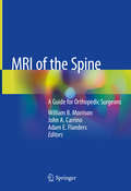
MRI of the Spine: A Guide for Orthopedic Surgeons
by William B. Morrison John A. Carrino Adam E. FlandersUtilizing plentiful radiological images to illustrate each topic, this text is a comprehensive and descriptive review of magnetic resonance imaging (MRI) interpretation for the spine, emphasizing standardized nomenclature and grading schemes. The book begins with current MR imaging protocols, including indication, sequencing and advanced imaging techniques, and a review of the relevant anatomy of the spine and its anomalies. Subsequent chapters encompass topics of trauma, degenerative disease, infection, inflammatory disease, as well as neoplastic and metabolic disease. Spinal cord and dural lesions will also be presented, with additional chapters dedicated to MRI evaluation of the post-operative patient. The format is reader-friendly, utilizing an efficient presentation of the essential principles and important findings on MR images of the spine, with a wealth of high-quality figures, graphics and tables for differential diagnosis as well as tips and tricks from experts in the field. Presenting the most up-to-date protocols and suggested interpretations, MRI of the Spine will be a solid reference for orthopedic surgeons, sports medicine specialists, neurosurgeons, radiologists and all clinicians and support staff caring for the spine.
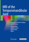
MRI of the Temporomandibular Joint: Correlation Between Imaging and Pathology
by Tiziana Robba Carlotta Tanteri Giulia TanteriThis book is the outcome of a fruitful, long-standing cooperation between expert radiologists and clinicians, and explains the most relevant features and technical requirements that are needed to optimally conduct and assess MR examinations for temporomandibular joint (TMJ) pathologies. TMJ conditions are increasingly gaining attention, as the underlying diseases involved can vary considerably and be difficult to diagnose. Similarly, several imaging sub-specialties (e.g. dental radiology, neuroradiology, and musculoskeletal radiology) now find themselves dealing with the temporomandibular joints. The authors provide essential information on TMJ anatomy, dynamics, function and dysfunction. Correlations between clinical aspects and MRI findings are discussed and guidance for the correct interpretation of results is offered. Special findings that are helpful for differential diagnosis (arthritis, osteochondroma, synovial chondromatosis) are also examined. Given its extensive and varied coverage, the book offers a valuable asset for radiologists, dentists, gnathologists, maxillofacial surgeons, orthodontists and other professionals seeking a thorough overview of the subject
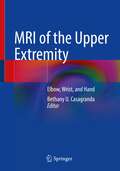
MRI of the Upper Extremity: Elbow, Wrist, and Hand
by Bethany U. CasagrandaThis book systematically discusses the anatomy and pathology of three specific regions of the upper extremity: the elbow, wrist, and hand. Divided into three sections, by body part, chapters cover anatomy and pathology. The anatomy chapters give a comprehensive view of each body part and normal variants found there. Although the primary modality emphasized will be MRI, illustrations and other modalities, including plain radiograph and CT, will be used to comprehensively discuss the anatomy of each region. Liberally illustrated, the pathology chapters then cover both traumatic and non-traumatic causes for imaging and detail how to perform and interpret each MRI. Specific examples include: osseous trauma, soft tissue trauma, and tumor imaging. Chapters are written with the deliberate intention to be of value to all levels of radiology training while remaining a reliable resource for attending radiologists.
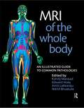
MRI of the Whole Body: An Illustrated Guide for Common Pathologies
by Edward Hoey Nikhil Bhuskute Amit Lakkaraju Kshitij MankadThe optimal use of magnetic resonance imaging poses a constant challenge as the technology is continually and rapidly advancing. This leaves the MR practitioner, beginner or experienced, in constant need of up-to-date, easily read and well illustrated material presenting the clinical constellation of pathologies as seen by an MRI scanner in such an
MRI: Essentials for Innovative Technologies
by Giuseppe PlacidiMRI: Essentials for Innovative Technologies describes novel methods to improve magnetic resonance imaging (MRI) beyond its current limitations. It proposes smart encoding methods and acquisition sequences to deal with frequency displacement due to residual static magnetic field inhomogeneity, motion, and undersampling. Requiring few or no hardware
MRI: Physics, Image Reconstruction, and Analysis (Devices, Circuits, and Systems)
by Krzysztof Iniewski Angshul Majumdar Rabab Kreidieh WardThe field of magnetic resonance imaging (MRI) has developed rapidly over the past decade, benefiting greatly from the newly developed framework of compressed sensing and its ability to drastically reduce MRI scan times. MRI: Physics, Image Reconstruction, and Analysis presents the latest research in MRI technology, emphasizing compressed sensing-based image reconstruction techniques. The book begins with a succinct introduction to the principles of MRI and then: Discusses the technology and applications of T1rho MRI Details the recovery of highly sampled functional MRIs Explains sparsity-based techniques for quantitative MRIs Describes multi-coil parallel MRI reconstruction techniques Examines off-line techniques in dynamic MRI reconstruction Explores advances in brain connectivity analysis using diffusion and functional MRIs Featuring chapters authored by field experts, MRI: Physics, Image Reconstruction, and Analysis delivers an authoritative and cutting-edge treatment of MRI reconstruction techniques. The book provides engineers, physicists, and graduate students with a comprehensive look at the state of the art of MRI.
MULTIDISCIPLINARY APPROACHES FOR SUSTAINABLE DEVELOPMENT: International Conference on MULTIDISCIPLINARY APPROACHES FOR SUSTAINABLE DEVELOPMENT IN SCIENCE & TECHNOLOGY
by Monica Sharma Shilpi Birla Anshuman Tripathi Jagrati Sahariya Mamta SoniIn a world where the pace of technological advancement continues to accelerate, the imperative to ensure sustainable development has never been more pressing to address the same, the 1st International Conference on Multidisciplinary Approaches for Sustainable Development in Science & Technology (MASDST - 2024), took place at Manipal University Jaipur, Rajasthan, India, from 28th to 29th March 2024. Embracing the spirit of innovation and collaboration, this conference marks a significant milestone in the pursuit of sustainable solutions for our global challenges.
MWW-Type Titanosilicate: Synthesis, Structural Modification and Catalytic Applications to Green Oxidations
by Hao Xu Mingyuan He Yueming Liu Le Xu Peng WuThis book provides a comprehensive review of a new generation of selective oxidation titanosilicate catalysts with the MWW topology (Ti-MWW) based on the research achievements of the past 12 years. It gives an overview of the synthesis, structure modification and catalytic properties of Ti-MWW. Ti-MWW can readily be prepared by means of direct hydrothermal synthesis with crystallization-supporting agents, using dual-structure-directing agents and a dry-gel conversion technique. It also can be post-synthesized through unique reversible structure transformation and liquid-phase isomorphous substitution. The structural conversion of Ti-MWW into the materials usable for processing large molecules is summarized. Taking advantage of the structure diversity of the lamellar precursor of Ti-MWW, it can be fully or partially delaminated, and undergo interlayer silylation to obtain a novel structure with larger porosity. In the selective oxidation (alkene epoxidation and ketone/aldehyde ammoximation) with hydrogen peroxide or organic peroxide as an oxidant, the unique catalytic properties of Ti-MWW are described in comparison to conventional titanosilicates such as TS-1 and Ti-Beta.
MXene Nanocomposites: Design, Fabrication, and Shielding Applications
by Poushali Das Sayan Ganguly Andreas RosenkranzMXenes offer single step processing, excellent electrical conductivity, easy heat dissipation behavior, and capacitor-like properties and are used in photodetectors, lithium-ion batteries, solar cells, photocatalysis, electrochemiluminescence sensors, and supercapacitors. Because of their superior electrical and thermal conductivities, these composites are an ideal choice in electromagnetic interference (EMI) shielding. MXene Nanocomposites: Design, Fabrication, and Shielding Applications presents a comprehensive overview of these emerging materials, including their underlying chemistry, fabrication strategies, and cutting-edge applications in EMI shielding. • Covers modern fabrication technologies, processing, properties, nanostructure formation, and mechanisms of reinforcement. • Discuss biocompatibility, suitability, and toxic effects. • Details innovations, applications, opportunities, and future directions in EMI shielding applications. This book is aimed at researchers and advanced students in materials science and engineering and is unique in its detailed coverage of MXene-based polymer composites for EMI shielding.
