- Table View
- List View
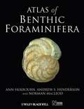
Atlas of Benthic Foraminifera
by Norman Macleod Ann Holbourn Andrew S. HendersonAn up-to-date atlas of an important fossil and living group, with the Natural History Museum. Deep-sea benthic foraminifera have played a central role in biostratigraphic, paleoecological, and paleoceanographical research for over a century. These single–celled marine protists are important because of their geographic ubiquity, distinction morphologies and rapid evolutionary rates, their abundance and diversity deep–sea sediments, and because of their utility as indicators of environmental conditions both at and below the sediment–water interface. In addition, stable isotopic data obtained from deep–sea benthic foraminiferal tests provide paleoceanographers with environmental information that is proving to be of major significance in studies of global climatic change. This work collects together, for the first time, new morphological descriptions, taxonomic placements, stratigraphic occurrence data, geographical distribution summaries, and palaeoecological information, along with state-of-the-art colour photomicrographs (most taken in reflected light, just as you would see them using light microscopy), of 300 common deep-sea benthic foraminifera species spanning the interval from Jurassic - Recent. This volume is intended as a reference and research resource for post-graduate students in micropalaeontology, geological professionals (stratigraphers, paleontologists, paleoecologists, palaeoceanographers), taxonomists, and evolutionary (paleo)biologists.
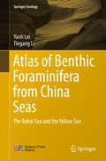
Atlas of Benthic Foraminifera from China Seas: The Bohai Sea and the Yellow Sea (Springer Geology)
by Yanli Lei Tiegang LiThis atlas gives a comprehensive account on the benthic foraminiferal fauna in the China Seas, especially on the Bohai and the Yellow Seas. Details of about 183 species, subjected to 5 orders, 52 families and 92 genera are included. For each species there is a brief description of the morphological characteristics, synonymised names, measurements and geographical distribution worldwide, as well as a top-level elegant plate illustrated the fossil and live specimens. It could be used as a reference book for researchers working at marine biology, marine geology, micropaleontology, paleoceanography, paleobiology and related fields.
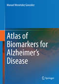
Atlas of Biomarkers for Alzheimer's Disease
by Manuel Menéndez GonzálezA lot of research on biomarkers for Alzheimer is being done in the last few decades. The aim of these studies is to find some method to ease the diagnosis of Alzheimers as early as possible. Such methods are a range of blood or CSF tests on one hand and several types of neuroimaging scans on the other. Many of the images coming both from laboratory and neuroimaging are very visual and illustrative. These images, accompanied by a short description, can perfectly explain the main results and usefulness of every biomarker. The objective of this book would be to summarize the most important studies made in this field. Few publications have systematically compiled results on this topic and only one as an atlas. Readers would be interested in this publication because it allows reviewing the current status of research by handily visualizing the results.
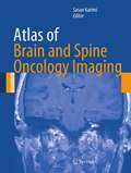
Atlas of Brain and Spine Oncology Imaging (Atlas of Oncology Imaging)
by Sasan KarimiAtlas of Brain and Spine Oncology Imaging presents a comprehensive visual review of pathologic disease variations of cancers of the brain and spine through extensive radiologic images. The focus of the book is on algorithmic strategies for identifying neoplastic pathologies commonly found in brain and spinal tumors through a visual representation of the variety of appearances that each neoplasm takes, within both benign and malignant manifestations. With contributions from radiologists on staff at a National Cancer Institute-designated comprehensive cancer center, who draw from an extensive collection of diagnostic images across all imaging modalities, this book will be valuable to practicing radiologists, radiation oncologists, surgeons and other practitioners involved in the diagnosis and treatment of brain and spinal neoplasms in all patient populations.
Atlas of Breast Implant Ultrasound
by Jae Hong KimThis atlas is the first book on the use of high-resolution ultrasound to assess breast implants and identify the various potential breast implant-related complications, which are frequently asymptomatic. The aim is to provide radiologists, breast surgeons, plastic surgeons, and other medical staff with a comprehensive guide of high clinical value during not only the diagnostic but also the treatment process. To this end, a wealth of ultrasound images and videos are presented, along with surgical photos and videos and pathological findings. The coverage includes the role of ultrasound in the management of breast implant-associated anaplastic large cell carcinoma, with explanation of its value in distinguishing the type of implant shell, which is highly relevant in this disease. A concluding chapter presents a large series of instructive cases. The author has extensive experience in breast surgery and has been collecting implant-related data using high-resolution ultrasound, including data on the diagnosis of side effects, for more than a decade.
Atlas of CT Angiography: Normal and Pathologic Findings
by Gratian Dragoslav Miclaus Horia PlesThe second edition of this atlas presents a wealth of normal and pathologic findings observed on CT angiography with 3D reconstruction in diverse clinical applications, including the imaging of cerebral, carotid, thoracic, coronary, abdominal, and peripheral vessels. The superb illustrations display the excellent anatomic detail obtained with CT angiography and depict the precise location of affected structures and lesion severity. Careful comparisons between normal imaging features and pathologic appearances will assist the reader in image interpretation and treatment planning, and the described cases include some very rare pathologies. In addition, the technical principles of the modality are clearly explained and guidance provided on imaging protocols. This edition is the outcome of 18 years of work by a renowned radiological team whose research focuses specifically on vascular pathology of the whole body and the role of CT angiography in its assessment. Numerous new images are presented and three additional chapters address cerebral arteriovenous malformations, congenital cardiac malformations in children and CT venography. The atlas will be invaluable for radiologists, neurologists, neurosurgeons, cardiologists, cardiovascular surgeons, and medical students..
Atlas of Cell Organelles Fluorescence
by Elli Kohen Rene Santus Joseph G. Hirschberg Nuri OzkutukContaining over 150 original photomicrographs accompanied by protocol information, Atlas of Cell Organelles Fluorescence delineates organelles structures, interaction, and organization into complexes. It provides a collection that shows living cells under physiopathological conditions and in the context of treatment with carcinogens, xenobi
Atlas of Chest Imaging in COVID-19 Patients
by Jinxin Liu Xiaoping Tang Chunliang LeiThis book presents X-ray and CT findings of patients with 2019 Novel Coronavirus (2019-nCoV) pneumonia in early, progressive, critical, and recover stage. It starts with a general review of CT features of new coronavirus pneumonia. In the following chapters, imaging manifestations in different groups and stages are presented, especially CT findings of asymptomatic COVID-19 patients and those first nucleic tests for the novel coronavirus are negative, but turn positive in one day or two. In addition, imaging and pathological analysis of COVID-19 death are summarized in the last chapter. The book provides a valuable reference source for radiologists and doctors working in the area of coronavirus pneumonia.
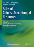
Atlas of Chinese Macrofungal Resources: Volume 1: Overview, Macrofungal Ascomycetes, Jelly Fungi and Coral Fungi
by Yu Li Taihui Li Zhuliang Yang Tolgor Bau Yucheng DaiThis book is part of the 4-volumes collection of Atlas of Chinese Macrofungal Resources . This atlas documented 1819 species (or varieties) in 509 genera of macrofungi known from China, which are, according to their morphological characteristics, practically divided into 10 groups, including 196 larger ascomycetes, 21 jelly fungi, 47 coral fungi, 637 polyporoid, hydnaceous and lephoroid fungi, 11 cantharelloid fungi, 653 agarics, 130 boletes, 75 gasteroid fungi, 16 larger pathogenic fungi on crops, and 33 larger myxomycetes.All species are evidenced with voucher sand photographs. About 370 of the listed species (occupying 1/5 of the totals pieces) have their type localities in China, among which over 260 species (accounting for 1/7 of the species) were firstly discovered and published by the present authors. Some of the species are endemic to China and East Asia. Introduction to all species are accompanied with color photographs showing their macro-morphology and (or) habitat. The macroscopic and microscopic diagnostic characters ecological habits, economic importance (edibility, medicinal availability or toxicity) and geographical distribution in China of all species are described in brief and easy-to-understand style. In the guide, the characteristics and using method of the book, related mycological vocabulary, common taxonomic techniques and positions of the fungal genera in modern taxonomic system are briefly introduced.The knowledge of this book should be interesting to mycologists, mycology fans and mushroom lovers, as well as researchers, teachers and students studying on edible fungi, plant pathology, healthcare and biomedicine sciences, bioresources and biodiversity, ecology and other related disciplines. It is an ideal reference for those who are interested in the Chinese macrofungi and larger slime molds. In this first volume, it covers Macrofungal Ascomycetes, Jelly Fungi and Coral Fungi.
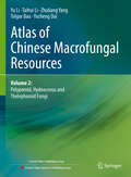
Atlas of Chinese Macrofungal Resources: Volume 2: Polyporoid, Hydnaceous and Thelephoroid Fungi
by Yu Li Taihui Li Zhuliang Yang Tolgor Bau Yucheng DaiThis book is part of the 4-volumes collection of Atlas of Chinese Macrofungal Resources. This atlas documented 1819 species (or varieties) in 509 genera of macrofungi known from China, which are, according to their morphological characteristics, practically divided into 10 groups, including 196 larger ascomycetes, 21 jelly fungi, 47 coral fungi, 637 polyporoid, hydnaceous and lephoroid fungi, 11 cantharelloid fungi, 653 agarics, 130 boletes, 75 gasteroid fungi, 16 larger pathogenic fungi on crops, and 33 larger myxomycetes. All species are evidenced with vouchers and photographs. About 370 of the listed species (occupying 1/5 of the total species) have their type localities in China, among which over 260 species (accounting for 1/7 of the species) were firstly discovered and published by the present authors. Some of the species are endemic to China and East Asia. Descriptions of all species are accompanied with color photographs showing their macro-morphology and (or) habitat. The macroscopic and microscopic diagnostic characters, ecological habits, economic importance (edibility, medicinal availability or toxicity) and geographical distribution in China of all species are described in brief and easy-to-understand style. In the guide, the characteristics and using method of the book, related mycological vocabulary, common taxonomic techniques and positions of the fungal genera in modern taxonomic system are briefly introduced. The knowledge of this book should be interesting to mycologists, mycology fans and mushroom lovers, as well as researchers, teachers and students studying on edible fungi, plant pathology, healthcare and biomedicine sciences, bioresources and biodiversity, ecology and other related disciplines. It is an ideal reference for those who are interested in the Chinese macrofungi and larger slime molds. In this second volume, it covers Polyporoid, Hydnaceous and Thelephoroid Fungi.
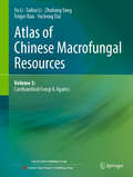
Atlas of Chinese Macrofungal Resources: Volume 3: Cantharelloid Fungi & Agarics
by Yu Li Taihui Li Zhuliang Yang Tolgor Bau Yucheng DaiThis book is part of the 4-volumes collection of Atlas of Chinese Macrofungal Resources. This atlas documented 1819 species (or varieties) in 509 genera of macrofungi known from China, which are, according to their morphological characteristics, practically divided into 10 groups, including 196 larger ascomycetes, 21 jelly fungi, 47 coral fungi, 637 polyporoid, hydnaceous and lephoroid fungi, 11 cantharelloid fungi, 653 agarics, 130 boletes, 75 gasteroid fungi, 16 larger pathogenic fungi on crops, and 33 larger myxomycetes. All species are evidenced with vouchers and photographs. About 370 of the listed species (occupying 1/5 of the total species) have their type localities in China, among which over 260 species (accounting for 1/7 of the species) were firstly discovered and published by the present authors. Some of the species are endemic to China and East Asia. Descriptions of all species are accompanied with color photographs showing their macro-morphology and (or) habitat. The macroscopic and microscopic diagnostic characters, ecological habits, economic importance (edibility, medicinal availability or toxicity) and geographical distribution in China of all species are described in brief and easy-to-understand style. In the guide, the characteristics and using method of the book, related mycological vocabulary, common taxonomic techniques and positions of the fungal genera in modern taxonomic system are briefly introduced. The knowledge of this book should be interesting to mycologists, mycology fans and mushroom lovers, as well as researchers, teachers and students studying on edible fungi, plant pathology, healthcare and biomedicine sciences, bioresources and biodiversity, ecology and other related disciplines. It is an ideal reference for those who are interested in the Chinese macrofungi and larger slime molds. In this third volume, it covers Cantharelloid fungi & Agarics.
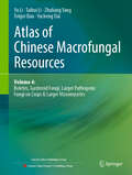
Atlas of Chinese Macrofungal Resources: Volume 4: Boletes, Gasteroid Fungi, Larger Pathogenic Fungi on Crops & Larger Myxomycetes
by Yu Li Taihui Li Zhuliang Yang Tolgor Bau Yucheng DaiThis book is part of the 4-volumes collection of Atlas of Chinese Macrofungal Resources. This atlas documented 1819 species (or varieties) in 509 genera of macrofungi known from China, which are, according to their morphological characteristics, practically divided into 10 groups, including 196 larger ascomycetes, 21 jelly fungi, 47 coral fungi, 637 polyporoid, hydnaceous and lephoroid fungi, 11 cantharelloid fungi, 653 agarics, 130 boletes, 75 gasteroid fungi, 16 larger pathogenic fungi on crops, and 33 larger myxomycetes. All species are evidenced with vouchers and photographs. About 370 of the listed species (occupying 1/5 of the total species) have their type localities in China, among which over 260 species (accounting for 1/7 of the species) were firstly discovered and published by the present authors. Some of the species are endemic to China and East Asia. Descriptions of all species are accompanied with color photographs showing their macro-morphology and (or) habitat. The macroscopic and microscopic diagnostic characters, ecological habits, economic importance (edibility, medicinal availability or toxicity) and geographical distribution in China of all species are described in brief and easy-to-understand style. In the guide, the characteristics and using method of the book, related mycological vocabulary, common taxonomic techniques and positions of the fungal genera in modern taxonomic system are briefly introduced. The knowledge of this book should be interesting to mycologists, mycology fans and mushroom lovers, as well as researchers, teachers and students studying on edible fungi, plant pathology, healthcare and biomedicine sciences, bioresources and biodiversity, ecology and other related disciplines. It is an ideal reference for those who are interested in the Chinese macrofungi and larger slime molds. In this fourth volume, it covers Boletes, Gasteroid fungi, Larger pathogenic fungi on crops & Larger myxomycetes.
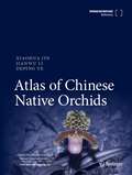
Atlas of Chinese Native Orchids
by Xiaohua Jin Jianwu Li Deping YeThis book updates taxonomy information of orchids in China. China is one of the countries with the richest biodiversity. In China, all five subfamilies of Orchidaceae are represented, about 1600 orchid species in 198 genera. All orchids are rare and endangered plants. They are among the flagships for biological conservation, listed in CITES appendix I or II. This book provided an updated classification system of Orchidaceae with newly recorded and published species in China and new combinations. 1026 species in 157 genera of native species in China are included, about half of which are newly recorded or published species in China in the last two decades. Indexes to genera and species are included. For each species, one to four photos, most of which were taken by the authors, are utilized to illustrate habitats, morphological characters, and phenology. Furthermore, the geographic distribution of them also demonstrated in a map. This book can be used as a reference for researchers working on Orchidaceae, as well as practitioners in the horticulture community.
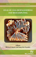
Atlas of Cilia Bioengineering and Biocomputing
by Richard Mayne Jaap Den ToonderCilia are microscopic finger-like cell-surface organelles possessed by a great many eukaryotic organisms, including humans, whose purposes include generating local fluid movements via rhythmic whip-like beating and environmental sensing. Despite intense research efforts since their discovery by van Leeuwenhoek in the 1670's, several key questions regarding ciliary functions, experimental manipulation and in silico imitation remain unanswered. Major justifications for cilia research lie in their involvement in various forms of human disease (ciliopathies) and their ability to instantiate decentralised, asynchronous sensorial-actuation of adjacent matter through modulation of beating characteristics. Further elucidation of these characteristics, which is a problem requiring the combined expertise of mathematicians, computer scientists, engineers and life scientists, will lead to novel biomedical therapies, creation of `smart' actuating surfaces for microfluidics/lab-on-chip applications and a greater understanding of fluid mechanics in real-world scenarios. This lavishly-illustrated anthology presents recent advances in the fields of ciliary investigation, manipulation, emulation, mimesis and modelling from key researchers in their fields: its goal is to explain the state-of-the-art in cilia bioengineering and bio-computation in a uniquely creative, accessible manner, towards encouraging further transdisciplinary work in the field as well as educating a broad spectrum of scientists and lay people. The volume is split into three distinct but interwoven themes:Biology: Biological preliminaries for the study of cilia; the state-of-the-art in genetic engineering of ciliated cells for biomedical purposes; reprogramming of cilia dynamics in live cells.Engineering: Creation of macro cilia robots for object sorting applications; pneumatic cilia for the optimization of fluid motion; electrostatic, magnetic and MEMS cilia for microfluidic mixing; reviews in artificial cilia fabrication, actuation and flow induction methods.Numerical and computational modelling. Analyses of thin film cilia for `lab on chip' microfluidic mixing applications; modelling of gel-based artificial cilia towards simulating dynamic behaviors of responsive cilia layers in complex fluids across a wide range of potential applications.
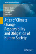
Atlas of Climate Change: Responsibility and Obligation of Human Society (Springer Atmospheric Sciences)
by Fumin Ren Jianbin Huang Wenjie Dong Yan GuoThis atlas and reference resource assembles the latest research findings on the responsibility and obligation of human society for historical climate change. It clearly and quantitatively estimates to what extent the developed and developing world are responsible for historical climate change with regard to anthropogenic carbon and sulfur emissions as well as global carbon trade, and so provides a potential tool to address the controversial issue of carbon emission reduction in international climate negotiations. Since the quantitative attribution of historical climate change is calculated based on CMIP5 models, the fidelity of these models in representing the observed climate change is also evaluated. In addition to evaluation, future climate change based on CMIP5 models is also shown both on global and regional scales (especially for China and its surrounding areas ) in terms of surface air temperature, precipitation, sea surface temperature, atmospheric circulations and Arctic Sea ice. The atlas also makes various comparisons among different multi-model ensemble methods in order to obtain the most reliable estimation.
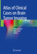
Atlas of Clinical Cases on Brain Tumor Imaging
by Yelda Özsunar Utku ŞenolThis book presents and analyzes clinical cases of brain tumors and follows the classification provided by the WHO in 2016. After introductory chapters reviewing the international literature on the topic, the advances made in all imaging modalities (especially Magnetic Resonance and Computed Tomography) are examined.All radiological findings are supplemented with a wealth of images and brief explanations. The clinical information is given as part of the case discussion, as are the characteristics and differential diagnosis of the tumors. Radiologic-pathologic correlations round out the description of each clinical case.Intended as a quick and illustrative reference guide for radiology residents and medical students, this atlas represents the most up-to-date, practice-oriented reference book in the field of Brain Tumor Imaging.
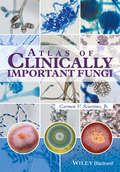
Atlas of Clinically Important Fungi
by Carmen V. Sciortino Jr.Although there are many texts that provide quality information for the identification of fungi, researchers and technologists rarely have time to read the text. Most are rushed for time and seek morphological information that helps guide them to the identification of fungi. The Atlas of Clinically Important Fungi provides readers with an alphabetical list of fungi as well as listing the division of fungi by both sporulation and morphology. The characteristic traits for a particular fungus are displayed through a series of images, with the fungi appearing as they did in the author's lab on the day(s) that testing was performed. For this reason, numerous (6-20) color photographs are included so that technologists will have sufficient reference photos for identifying the various morphologies of a single organism. Organism photographs begin with the macroscopic colony views followed by the microscopic views. Also included for some microorganisms, are clinical pathology photographs demonstrating how the organism appears in human tissues.A collection of literature citations are also provided to enable further reading. This user-friendly fungi atlas provides a resource for those seeking information in the field of medical mycology, specifically with regards to identifying an organism using the parameters of culture morphology.
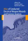
Atlas of Conducted Electrical Weapon Wounds and Forensic Analysis
by Jeffrey D. Ho Mark W. Kroll Donald M. DawesAtlas of Conducted Electrical Weapon Wounds and Forensic Analysis provides a comprehensive publication on the subject of Conducted Electrical Weapon (CEW) wounds and signature markings created by this class of weapon. This volume will serve as a very useful resource for all professions tasked with assisting persons that have allegedly been subjected to a CEW exposure. The volume provides an introduction to basic CEW technology and the types of CEWs currently available. It also serves as a comprehensive pictorial atlas of signature markings that CEW exposures make in the immediate and more remote post-exposure periods. Also, it discusses the ability of forensic specialty examinations of the CEW itself to aid in the determination of whether the alleged CEW exposure is consistent with the objective evidence and the subjective statements. Finally, this text addresses the important and growing area of factitious CEW markings that will be useful for consideration by investigators and litigators. Atlas of Conducted Electrical Weapon Wounds and Forensic Analysis provides an objective atlas of evidence for reference that will benefit those professionals who often must make diagnostic, treatment or legal judgments on these cases including Emergency and Primary-Care Physicians, Medical Examiners, Forensic Pathologists, Coroners, Law Enforcement Investigators, and Attorneys.
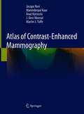
Atlas of Contrast-Enhanced Mammography
by Jacopo Nori Maninderpal Kaur Anat Kornecki J. Devi Meenal Martin J. YaffeThis superbly illustrated atlas serves as a basic introduction to contrast-enhanced mammography (CEM), a breakthrough functional breast imaging modality, which is rapidly growing. This book is an essential guide for the latest developments with correlative findings, practical interpretation tips, physics, and information on how contrast mammography differs from conventional 2D and 3D Full Field of View digital mammography (FFDM). It includes: · over 1000 high-quality 2D, 3D and recombined contrast mammography images representing the spectrum of breast imaging · findings obtained in the full range of benign, pre-malignant and malignant conditions, including artefacts and postoperative changes, presented with high-quality illustrations from case examples · image interpretation tips using mammographic and DCE-MRI descriptors of the BI-RADS lexicon to effectively read and interpret this advanced imaging modality · practical tips to interpret this new modality and how it is used as an adjunct to 2D mammography · details on how integration of contrast-enhanced mammography drastically changes lesion work-up and overall workflow in the department · "imaging pearls" boxes offering interpretation tips for expert clinical guidance · a case on the recently introduced CEM guided biopsy procedure The book’s target audience consists of diagnostic radiologists, residents, fellows, technologists and clinicians involved in the care of breast cancer patients, including surgeons and oncologists. The goal is to provide a concise introduction to CEM and to lead to enhanced interpretation and better patient staging prior to surgery.
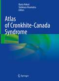
Atlas of Cronkhite-Canada Syndrome
by Tadakazu Hisamatsu Ryota HokariThis manual treats both conceptual and practical aspects of gastrointestinal polyposis syndrome, which is characterized by dermatologic manifestations. The book offers both detailed and comprehensive analysis of the endoscopic findings achieved from 88 cases, which led to novel concepts and classification of the dermatologic manifestations according to distribution pattern, the thickness of mucosa, and intervening inflamed mucosa between polyps. The book also offers comparisons of the endoscopic features to pathological features. Abundant illustrations show the observations of the malignant lesions using special lights, such as NBI and BLI, to provide a comprehensive view of the Cronkhite-Canada syndrome (CCS) polyp.Atlas of Cronkhite-Canada Syndrome is essential guidance for gastroenterologists and pathologists who pursue the new diagnostic approach and treatment for CCS. It also attracts clinicians interested in the latest updates on this field and those who encountered the CCS patients with refractory to corticosteroid treatment. The risk of colorectal cancer in patients with CCS may increase because multiple inflammatory pseudopolyps make it more difficult to detect premalignant adenomas. Additionally, as endoscopy for GI disease becomes widespread in developing countries, the number of diagnosed patients will likely increase. Thus, it is crucial to have a broader understanding of the disease and the treatment strategy, and this book aims to simplify a complicated picture of the rare disease.
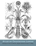
Atlas of Crustacean Larvae
by Joel W. Martin, Jørgen Olesen, and Jens T. Høeg.An illustrated guide to the sweeping diversity of crustacean larval forms.Winner of the CHOICE Outstanding Academic Title of the Choice ACRLCrustaceans—familiar to the average person as shrimp, lobsters, crabs, krill, barnacles, and their many relatives—are easily one of the most important and diverse groups of marine life. Poorly understood, they are among the most numerous invertebrates on earth. Most crustaceans start life as eggs and move through a variety of morphological phases prior to maturity. In Atlas of Crustacean Larvae, more than 45 of the world's leading crustacean researchers explain and illustrate the beauty and complexity of the many larval life stages.Revealing shapes that are reminiscent of aliens from other worlds—often with bizarre modifications for a planktonic life or for parasitization, including (in some cases) bulging eyes, enormous spines, and aids for flotation and swimming—the abundant illustrations and photographs show the detail of each morphological stage and allow for quick comparisons. The diversity is immediately apparent in the illustrations: spikes that deter predators occur on some larvae, while others bear unique specializations not seen elsewhere, and still others appear as miniature versions of the adults. Small differences in anatomy are shown to be suited to the behaviors and survival mechanisms of each species.Destined to become a key reference for specialists and students and a treasured book for anyone who wishes to understand "the invertebrate backbone of marine ecosystems," Atlas of Crustacean Larvae belongs on the shelf of every serious marine biologist.
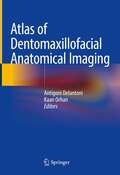
Atlas of Dentomaxillofacial Anatomical Imaging
by Kaan Orhan Antigoni DelantoniThis atlas is a detailed and complete guide on imaging of the dentomaxillofacial region, a region of high interest to a wide range of specialists. A large number of injuries and patient’s treatment involve the facial skeleton. Enriched by radiographic images and illustrations, this book explores the anatomy of this region presenting its imaging characteristics through the most commonly available techniques (MDCT, CBCT, MRI and US). In addition, two special chapters on angiography and micro-CT expand the limits of dentomaxillofacial imaging. This comprehensive book will be an invaluable tool for radiologists, dentists, surgeons and ENT specialists in their training and daily practice.
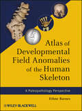
Atlas of Developmental Field Anomalies of the Human Skeleton
by Ethne BarnesWritten by one of the most consulted authorities on the subject, Atlas of Developmental Field Anomalies of the Human Skeleton is the pre-eminent resource for developmental defects of the skeleton. This guide focuses on localized bone structures utilizing the morphogenetic approach that addresses the origins of variability within specific developmental fields during embryonic development. Drawings and photographs make up most of the text, forming a picture atlas with descriptive text for each group of illustrations. Each section and subdivision is accompanied by brief discussions and drawings of morphogenetic development.
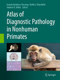
Atlas of Diagnostic Pathology in Nonhuman Primates
by Andrew D. Miller Ivanela Kondova-Perseng Keith G. MansfieldThe Atlas of Diagnostic Pathology in Nonhuman Primates offers the first extensively illustrated collection of classic lesions in nonhuman primate diseases and pathological conditions, compiled by an international team of expert contributors. Organized by infectious and noninfectious conditions, the atlas comprehensively covers viral, bacterial, fungal, and parasitic diseases, as well as nutritional, toxic, and metabolic causes, and genetic, age-related, neoplastic, and noninfectious inflammatory conditions. Since nonhuman primates are an indispensable resource for efficacy and safety evaluation of novel therapeutic strategies targeting clinically important human diseases, research with monkeys is critical to understand how to prevent and treat emerging infectious diseases such as Zika virus disease, Ebola, Middle East Respiratory Syndrome (MERS), COVID-19/SARS-CoV-2/coronavirus, pandemic flu, and many more. This book is intended to serve veterinary practitioners in university facilities, zoos, biotechnological and pharmaceutical companies, as well as clinicians, researchers, and students engaged in nonhuman primate research.
Atlas of Diagnostically Challenging Melanocytic Neoplasms
by Caterina Longo Giuseppe Argenziano Aimilios Lallas Elvira Moscarella Simonetta PianaThis atlas provides a clear, concise overview of the most challenging circumstances faced by clinicians and pathologists when dealing with melanocytic neoplasms. The book is structured as a case series; for each case, the clinical and dermoscopic appearances are presented, accompanied by a brief but comprehensive description and compelling histopathologic images. When available, in vivo confocal microscopy images are also included to highlight additional diagnostic clues. Identification of key messages and selected references will further guide the reader in the diagnosis and management of the neoplasm under consideration. It is well known that melanocytic lesions can be difficult to interpret. Some lesions show an ambiguous combination of morphologic criteria, and in these cases interpretation entails a high degree of subjectivity that results in low interobserver agreement even among expert pathologists. This atlas demonstrates how the addition of clinical information, including that provided by dermoscopy, can assist in reaching a more confident diagnosis.