- Table View
- List View
House (How It's Built)
by Becky HerrickKids are fascinated by how things are constructed, especially big things!This brand-new STEM (science, technology, engineering, and mathematics) series will take readers on visits to various work sites so they can see exactly How It’s Built! Kids will follow along as a small group of fictional characters get to find out exactly what it takes to build things that they probably see all the time, like bridges, houses, boats, and cars, to things that they might dream of being inside, like skyscrapers and rockets.
House Inspector
by Duncan Marshall Nigel DannThis book is a concise and comprehensive guide to building defects and building inspection. Whether, as a practitioner you are employed in buying, selling, managing or maintaining houses or whether, or as a layperson, you are buying a property to invest or live in, this book will help you make sound decisions and avoid costly mistakes. Written by two highly experienced authors, House Inspector is a general and accessible book which describes how and why house construction has changed, identifies some of the more common defects, and provides a series of elemental check lists. Essential reading for trainees and general practice surveyors, maintenance inspectors, housing managers, estate agents, planners, and even private purchasers and investors. This book will improve your knowledge and understanding of potential problems and provide a simple framework for a competent building inspection.
House Out of Factory (John Gloag On Industrial Design Ser.)
by John Gloag Grey WornumOriginally published in 1946, when Britain was facing a post-war housing crisis, this book dealt with the issue of the factory-produced house in being part of the solution for housing people in an affordable manner and a short time-scale. The book, aimed at both lay-people and technicians discusses aspects of pre-fabricated housing such as comfort, standardisation and aesthetics. The book is illustrated with 48pp of black and white plates.
House Rating Schemes
by Maria Kordjamshidi"House Rating Schemes" provides information to students, architects and researchers in the field of the built environment. It reviews current House Rating Schemes (HRS) used in different countries and investigates how these schemes assess the thermal performance of a house. It challenges the way that these schemes assess building energy efficiency and their inability to evaluate free running buildings which do not need an energy load for heating and cooling indoor environments. Finally, the book proposes a new index and method for HRS in which the efficiency of a house design can be evaluated with reference to its thermal performance in both free running and conditioned operation modes. The book deals with various approaches and methods for rating buildings on the basis of different indexes, with implications for both energy efficiency and thermal comfort. It also guides readers through a computer simulation program for developing a rating system that evaluates and ranks building energy efficiency.
House Wiring
by Greg FletcherWith the same innovative "hands-on" approach and task-focused organization that made the first edition so popular, Residential Construction Academy: House Wiring, 2E has been updated to reflect the most current electrical wiring practices in use, including the 2008 National Electrical Code. Designed to highlight the principles and practices used in the installation of a residential electrical wiring system, as well as how to effectively put them into practice, this book provides a valuable resource for lear
House of Robots (House of Robots #1)
by James Patterson Chris GrabensteinIn this highly-illustrated series from James Patterson, an extraordinary robot signs up for an ordinary fifth grade class . . . and elementary school will never be the same!It was never easy for Sammy Hayes-Rodriguez to fit in, so he's dreading the day when his genius mom insists he bring her newest invention to school: a walking, talking robot he calls E-for "Error". Sammy's no stranger to robots; his house is full of a colorful cast of them. But this one not only thinks it's Sammy's brother . . . it's actually even nerdier than Sammy. Will E be Sammy's one-way ticket to Loserville? Or will he prove to the world that it's cool to be square? It's a roller-coaster ride for Sammy to discover the amazing secret E holds that could change family forever . . . if all goes well on the trial run!
House of Robots: Robots Go Wild! (House of Robots #2)
by James Patterson Chris GrabensteinIn book two of the House of Robots series, it's 'bot brains versus 'bot brawn in an all-out war!Sammy Hayes-Rodriguez and his "bro-bot" E are making new friends every day as E works as his bedridden sister Maddie's school proxy. But disaster strikes when E malfunctions just in time to be upstaged by the super-cool new robot on the block-and loses his ability to help Maddie. Now it's up to Sammy to figure out what's wrong with E and save his family!
House-Dreams
by Hugh HowardImagine a house built and tailored to your every need and personal taste. Hugh Howard dreamed of such a house, and when he and his wife, Betsy, learn that they're expecting their second child, he seizes the opportunity to build a home for their growing family. Fifteen months later and just in time for the winter holidays, Howard, exhausted and wildly over his budget, completes their home-a fine 2,500-square-foot Federal-style house. And each piece has a story, from the cut nails that come from Howard's old elementary school janitor to the staircase that comes from a parsonage built just after the Civil War. Howard discovers that all his planning and hard work earn him a house, yes, but he also gains a community of new friends-the people who help him along the way. There's Charlie, whose ancestors helped establish the upstate New York hamlet where they build the house; Ralph, a third-generation mason, who constructs a remarkable Russian heater; and Robbie, an eccentric Irish landscaper who has his own peculiar way of designing a garden. HOUSE-DREAMS is for readers who spend weekends improving their houses, hardware store die-hards, and the millions who regularly tune in to the Home Garden Network and PBS's This Old House.
Household Chemicals and Emergency First Aid
by Jack L. Weddell Betty A. Foden Rosemary S. HappellHousehold Chemicals and Emergency First Aid is an essential manual that covers 386 household chemicals, discusses their hazards when mixed with other chemicals, describes the symptoms of overexposure, and provides instructions for emergency first aid treatment. The book is intended to be used in the event that label instructions on household chemicals have not been followed. It describes what may possibly happen and how to handle the situation if it does occur. Poison control centers are listed by state with phone numbers and addresses.Because household accidents involving chemicals are so prevalent, this manual is a ""must have"" book for all emergency medical technicians, paramedics, and other emergency first aid providers. It is also useful for anyone wanting detailed information regarding emergency situations with household chemicals.
Household Recycling and Consumption Work: Social and Moral Economies (Consumption and Public Life)
by Kathryn Wheeler Miriam GlucksmannConsumers are not usually incorporated into the sociological concept of 'division of labour', but using the case of household recycling, this book shows why this foundational concept needs to be revised.
Household Reusable Rainwater Technology for Developing and Under-Developed Countries
by Jaan H. Pu Chukwuemeka Kingsley JohnHousehold Reusable Rainwater Technology for Developing and Under-Developed Countries provides insight into household techniques for collecting and treating harvested rainwater safely for both potable and nonpotable uses, as well as practices to improve its quality, with numerous realworld case studies and data. It gives a comprehensive, holistic account on the household scale for both developing and under-developed countries. Improvement mechanisms such as the impacts of first flush, household water treatment techniques, and sedimentation in the harvested water are described in depth together with the advantages and disadvantages of their common practices in developing and under-developed societies. Also discussed is a comprehensive survey illustrating the impact of rainwater sources on the daily life of a carefully selected community from the perspective of its residents. The book is ideal for students, researchers, academics, water policy providers, and bodies worldwide such as WHO and DFID.
Housing Decisions
by Evelyn L. Lewis Carolyn S. TurnerLewis (emeritus, home economics, Northern Arizona U. , Flagstaff) and Turner (housing research, North Carolina Agricultural and Technical State U. , Greensboro) address numerous aspects of housing including related careers. This textbook's 7th incarnation (the last being in 2000) features color illustrations, relevant US legislation, energy-saving tips, and a glossary.
Housing Policy, Wellbeing and Social Development in Asia (Routledge Studies in International Real Estate)
by Rebecca Lai Chiu Seong-Kyu HaThis book investigates how housing policy changes in Asia since the late 1990s have impacted on housing affordability, security, livability, culture and social development. <P><P> Using case study examples from countries/cities including China, Hong Kong, India, Japan, Taiwan, Korea, Malaysia, Bangladesh, Singapore, Indonesia, Thailand and Vietnam, the contributors contextualize housing policy development in terms of both global and local socio-economic and political changes. They then investigate how policy changes have shaped and re-shaped the housing wellbeing of the local people and the social development within these places, which they argue should constitute the core purpose of housing policy. <P><P> This book will open up a new dimension for understanding housing and social development in Asia and a new conceptual perspective with which to examine housing which, by nature, is culture-sensitive and people-oriented. It will be of interest to students, scholars and professionals in the areas of housing studies, urban and social development and the public and social policy of Asia.
Housing Reclaimed
by Jessica KellnerHousing is a fundamental human right. For most of human history, our homes were built by hand from whatever local materials were available. However, since the Industrial Revolution, most housing has become little more than quickly constructed, mass-produced, uniform boxes. At the same time, the invention and standardization of the thirty-year mortgage and our ever-increasing reliance on credit has come to mean that most of us never own our homes outright.Housing Reclaimed is a call to arms for nonconventional home builders. It examines how technological advances, design evolution, and resourceful, out-of-the-box thinking about materials and efficiency can help us meet the challenge of building affordable, environmentally friendly, beautiful, and unique homes. Focusing on the use of salvaged and reclaimed materials, this inspirational volume is packed with case studies of innovative projects including:*Phoenix Commotion-working together towards low-income home ownership through sweat equity and 100 percent recycled materials*HabeRae-revitalizing neighborhoods by creating urban infill using modern technology and sustainable and reclaimed materials*Builders of Hope-rescuing and rehabilitating whole houses slated for demolitionThese projects and others like them demonstrate that building one's own home does not have to be an unattainable dream. This beautifully illustrated guide is a must-read for anyone interested in creating quality zero- or low-debt housing, reducing landfill waste, and creating stronger communities.Jessica Kellner is the editor of Natural Home and Garden magazine and a passionate advocate of using architectural salvage to create aesthetically beautiful, low-cost housing.
Housing and Asthma
by Stirling HowiesonAsthma is on the rise in a number of countries, in this volume Howieson asks what role the built environment has to play and what the construction industry can do to either slow the increase or reverse the trend. Based on the findings of a six-year research project, this book considers all aspects of housing to develop new strategies for dealing with the asthma pandemic in Britain and beyond. With the focus on the design and use pattern of our dwellings, the book looks at tackling the problems inherent in existing housing as well as forging guiding principles for the design of new dwellings, together with a financial assessment of the proposals.
Housing and Home Unbound: Intersections in economics, environment and politics in Australia (Routledge Housing Research Series)
by Nicole Cook Aidan Davison Louise CrabtreeHousing and Home Unbound pioneers understandings of housing and home as a meeting ground in which intensive practices, materials and meanings tangle with extensive economic, environmental and political worlds. Cutting across disciplines, the book opens up the conceptual and empirical study of housing and home by exploring the coproduction of the concrete and the abstract, the intimate and the institutional, the experiential and the collective. Exploring diverse examples in Australia and New Zealand, contributors address the interleaving of money and materials in the digital commodity of real estate, the neoliberal invention of housing as a liquid asset and source of welfare provision, and the bundling of car and home in housing markets. The more-than-human relations of housing and home are articulated through the role of suburban nature in the making of Australian modernity, the marketing of nature in waterfront urban renewal, the role of domestic territory in subversive social movements such as Seasteading and Tiny Houses, and the search for home comfort through low-cost energy efficiency practices. The transformative politics of housing and home are explored through the decolonizing of housing tenure, the shaping of housing policy by urban social movements, the lived importance of marginal spaces in Indigenous and other housing, and the affective lessons of the ruin. Beginning with the diverse elements gathered together in housing and home, the text opens up the complex realities and possibilities of human dwelling.
Housing and Technology: Special Focus on Zimbabwe (SpringerBriefs in Environment, Security, Development and Peace #37)
by Innocent Chirisa Abraham R. Matamanda Siphokazi Rammile Mario MaraisThe housing and human settlement sector is fast changing, and technology is making it more complex than ever before. With reference to Zimbabwe, a developing country in Southern Africa, the essence of this book is to bring out housing as an issue within the technology debate and practice. The following themes emerge from the 6 chapters in the book: • The characterisation and conceptualisation of housing and technology and the nexus of both • The complexity of housing challenges and the problems governments face in providing adequate housing, especially for the poor • Diverse practices in housing construction through the application of different typologies of technology • Assessment of the feasibility of technologies in housing development in Zimbabwe by mirroring them against global experiences. • Discussion of alternative policy approaches that may guide technology integration in housing development. This book will excite scholars and practitioners in urban and development studies, construction project management, urban sociology, geography, real estate together with policymakers and government officials.
Housing, Care and Inheritance (Housing and Society Series)
by Misa IzuharaHousing, Care and Inheritance draws on the author’s long-standing research into housing issues surrounding the ageing society, a phenomenon which is now a concern in many mature economies. If an adult child provides care for their elderly parent, should that person be rewarded? If so, should they inherit their parent’s house or a larger share of the assets? The ‘generational contract’ is often influenced by cultural norms, family traditions, social policy and housing market, so it is negotiated differently in different societies and at different times. Such generational contract is however breaking down as a result of socio-economic and demographic changes. Drawn from the two-part study funded by the UK Economic & Social Research Council, Misa Izuhara explores the myth and the changing patterns of the particular exchange of long-term care and housing assets between older parents and their adult children in Britain and Japan. Highly international and comparative in perspectives, this study addresses important sociological as well as policy questions regarding intergenerational relations involving housing wealth, long-term care, and inheritance.
Housing, Health and Well-Being (Routledge Focus on Environmental Health)
by Stephen Battersby Véronique Ezratty David OrmandyHousing is a social determinant of health and this book aims to provide a concise source of the theory and evidence on safe and healthy housing to inform students, academics, public and environmental health practitioners, and policy-makers, nationally and internationally. The book reviews the functions of housing and its relationship with the health and well-being of residents. It examines the implications of failures to satisfy those functions, including the potential impact on individuals, households, and society. It assesses options directed at avoiding, removing, or reducing threats and at promoting healthy indoor environments, particularly for the most susceptible and vulnerable members of society. It is essential reading for students, academics, and professionals within the areas of environmental health, public health, housing, built environment, social policy, housing policy, health policy, and law.
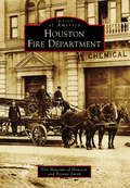
Houston Fire Department (Images of America)
by Tristan Smith Fire Museum of HoustonHouston's firefighting service began in 1838 with the founding of Protection Fire Company No. 1. As the city of Houston grew throughout the early and mid-19th century, volunteer companies formed and grew along with it. By 1895, city leaders decided to form the Houston Fire Department as a city department, culling the volunteer forces for men, stations, apparatus, and horses. The city grew in leaps and bounds, swallowing up neighborhoods, communities, and smaller cities along the way, with the fire department nipping at their heels. This brave force battled devastating fires throughout the years, most notably the Great Fifth Ward Fire in 1912, the 1943 Gulf Hotel Fire, the Woodway Square Apartment Conflagration in 1979 and the Southwest Inn Fire in 2013. What was once a smattering of volunteer fire brigades has grown into an imposing force of over 3,000 firefighters protecting over two million people in the fourth largest city in the United States.
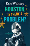
Houston, Is There A Problem?: Teen Astronauts #1 (Teen Astronauts #1)
by Eric WaltersKey Selling Points A young teen earns a scholarship to go to space camp. The first in the Teen Astronauts series featuring Houston at space camp. Examines themes of perseverance, leadership and growth mindset. This is an adventure story with an exciting setting: astronaut training camp. Eric Walters is very well known to librarians and booksellers.
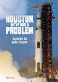
Houston, We've Had a Problem: The Story of the Apollo 13 Disaster (Tangled History)
by Rebecca RissmanIn an immersive, exciting narrative nonfiction format, this powerful book follows a selection of people who experienced the events surrounding the Apollo 13 disaster.

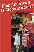
How "American" Is Globalization?
by William H. MarlingWilliam Marling's provocative work analyzes—in specific terms—the impacts of American technology and culture on foreign societies. Marling answers his own question—how "American" is globalization?—with two seemingly contradictory answers: "less than you think" and "more than you know." Deconstructing the myth of global Americanization, he argues that despite the typically American belief that the United States dominates foreign countries, the practical effects of "Americanization" amount to less than one might suppose. Critics point to the uneven popularity of McDonalds as a prime example of globalization and supposed American hegemony in the world. But Marling shows, in a series of case studies, that local cultures are intrinsically resilient and that local languages, eating habits, land use, education systems, and other social patterns determine the extent to which American culture is imported and adapted to native needs. He argues that globalization can actually accentuate local cultures, which often put their own imprint on what they import—from translating films and television into hundreds of languages to changing the menu at a McDonalds to include the Japanese favorite Chicken Tastuta.Marling also examines the unexpected ways in which American technology travels abroad: the technological transferability of the ATM, the practice of franchising, and "shop-floor" American innovations like shipping containers, bar codes, and computers. These technologies convey American attitudes about work, leisure, convenience, credit, and travel, but as Marling shows, they take root overseas in ways that are anything but "American."