- Table View
- List View

Atlas of Living Cell Cultures
by Rosemarie Steubing Toni LindlThe first atlas in many years giving researchers a good visual reference of the status of their cell lines. Given the increasing importance of well defined cellular models in particular in biomedical research this is a sorely needed resource for everyone performing cell culture.
Atlas of Lymph Node Anatomy
by Mukesh G. HarisinghaniDetailed anatomic drawings and state-of-the-art radiologic images combine to produce this essential Atlas of Lymph Node Anatomy. Utilizing the most recent advances in medical imaging, this book illustrates the nodal drainage stations in the head and neck, chest, and abdomen and pelvis. Also featured are clinical cases depicting drainage pathways for common malignancies. 2-D and 3-D maps offer color-coordinated representations of the lymph nodes in correlation with the anatomic illustrations. This simple, straightforward approach makes this book a perfect daily resource for a wide spectrum of specialties and physicians at all levels who are looking to gain a better understanding of lymph node anatomy and drainage. Edited by Mukesh G. Harisinghani, MD, with chapter contributions from staff members of the Department of Radiology at Massachusetts General Hospital.
Atlas of Lymph Node Anatomy
by Mukesh G. HarisinghaniThis book is a comprehensive atlas on lymph node anatomy and drainage to aid in cancer staging and therapy. Nodal drainage is pertinent to all aspects of cancer staging and therapy and is used by radiation oncologists, surgeons, and medical oncologists to increase accuracy. The first edition of this text was the first comprehensive monograph on this topic, allowing physicians across various specialties to utilize this information and easily share that knowledge with residents, fellows, and junior faculty.Detailed anatomic drawings and state-of-the-art radiologic images combine to produce this essential Atlas of Lymph Node Anatomy. Utilizing the most recent advances in medical imaging, this book illustrates the nodal drainage stations in the head and neck, chest, abdomen, and pelvis. Also featured are clinical cases depicting drainage pathways for common malignancies. 2-D and 3-D maps offer color-coordinated representations of the lymph nodes in correlation with the anatomic illustrations. This simple, straightforward approach makes this book a perfect daily resource for a wide spectrum of specialties and physicians at all levels who are looking to gain a better understanding of lymph node anatomy and drainage.This new edition enables physicians to educate themselves on the location of various nodal stations, especially in the context of common primary tumors, so that they are able to detect, localize, and characterize nodes seen with novel new imaging methods and with an increased level of accuracy. Chapters now cover the significant strides made in the imaging realm, such as PET CT, conventional MRI, MRI with novel imaging agents, and multidetector CT, which allows visualization of lymph nodes in various anatomic compartments.
Atlas of Macroscopic Wood Identification: With a Special Focus on Timbers Used in Europe and CITES-listed Species
by Flavio Ruffinatto Alan CrivellaroThis atlas presents macroscopic descriptions, macro cross section pictures, general characteristics and identification keys of 335 wood species currently introduced in the European timber market from all over the world. Overall 292 different genera are represented and CITES-listed timbers are also included. Macroscopic descriptions are based on a recently proposed list of macroscopic features for wood identification. Macroscopic features and their codes are defined and illustrated in the atlas. Wood descriptions also include information about natural durability, physical and mechanical properties, end uses, environmental sustainability and possible related misleading commercial names. Furthermore, each genus is described in terms of number of species, geographical distribution and main commercial timbers, and details are given about to what extent timbers within the genus can be typically identified through macroscopic and microscopic analysis, if any.The atlas will be a valuable guide for all agents in charge for timber verification, those involved in the European Timber Regulation enforcement and CITES inspections, as well as wood scientists, foresters, wood sellers, wood restorers, and any wood worker and wood passionate interested in a fast and reliable tool for wood identification.
Atlas of Mammalian Chromosomes
by Alexander S. Graphodatsky Polina L. Perelman Stephen J. O’BrienTHE UPDATED NEW EDITION OF THE POPULAR COLLECTION OF HIGH-RESOLUTION CHROMOSOME PHOTOGRAPHS—FOR GENETICISTS, MAMMOLOGISTS, AND BIOLOGISTS INTERESTED IN COMPARATIVE GENOMICS, SYSTEMATICS, AND CHROMOSOME STRUCTURE Filled with a visually exquisite collection of the banded metaphase chromosome karyotypes from some 1,000 species of mammals, the Atlas of Mammalian Chromosomes offers an unabridged compendium of the state of this genomic art form. The Atlas??contains the best karyotype produced, the common and Latin name of the species, the published citation, and identifies the contributing authors. Nearly all karyotypes are G-banded, revealing the chromosomal bar codes of homologous segments among related species. The Atlas brings together information from a range of cytogenetic literature and features high-quality karyotype images for nearly every mammal studied to date. When the Atlas was first published, only three mammals were sequenced. Today, that number is over 300. Now in its second edition, this book contains extensive revisions and major additions such as new karyotypes that employ G- and C- banding to represent euchromatin and heterochromatin genome composition, new phylogenetic trees for each order, homology segment chromosome information on published aligned chromosome painting. Summaries of the painting data for some species indicate conserved homology segments among compared species. An invaluable resource for today's comparative genomics era, this comprehensive collection of high-resolution chromosome photographs: Assembles information previously scattered throughout the cytogenetics literature in one comprehensive volume Provides chromosome information and illustrations for the karyotypes of 300 new species Addresses the mandate of the Human Genome Project to annotate the genomes of other organisms Serves as a basis for chromosome-level genome assemblies Offers a detailed summation of three decades of ZooFish (chromosome painting) Presents high-resolution photos of karyotypes that represent more than 1,000 mammal species Written for geneticists, mammalogists, and biologists, the Atlas of Mammalian Chromosomes offers a step forward for an understanding of species formation, of genome organization, and of DNA script for natural selection.
Atlas of Meteor Showers: A Practical Workbook for Meteor Observers (The Patrick Moore Practical Astronomy Series)
by Philip M. BagnallThis atlas contains everything you need to know about meteor showers and how to observe them. It begins with the science behind these celestial fireworks, then equips you with all the practical knowledge you’ll need to make the most of these wonderful astronomical events. The book is rich with illustrations, graphs and resources to assist your hobby. In addition, it includes downloadable radiant charts, report sheets, radiant altitude data, plotting charts and more, to help you locate each shower, record its activity and plot individual meteors during your nights of observation. Intended for amateurs of all levels and requiring no special equipment, this accessible Atlas of Meteor Showers will hone your skills and keep you engaged throughout the year, no matter where you are in the world.

Atlas of Meteorites
by Monica M. Grady Giovanni Pratesi Vanni Moggi Cecchi Monica M. Grady Giovanni PratesiA complete visual reference for meteorite classification, this atlas combines high resolution optical microscope images with detailed descriptions. It provides a systematic account of meteorites and their most important classification parameters, making it an essential resource for meteorite researchers. Each chapter starts with a description of the meteorite class, with a summary of the mineralogical, chemical and isotopic characteristics of the group. The full-color images are taken in plane- and cross-polarized light and reflected light, and arranged to highlight textural variations in meteorites. Specimens are grouped to show the effects of increasing thermal alteration and shock, as well as variations in chondrule size and type. Chapters on iron meteorites, pallasites and mesosiderites are included, photographed as mounts in reflected light, to show the range of textural variations that accompany these meteorites. Images from the book can be downloaded from www. cambridge. org/9780521840354.
Atlas of Musculoskeletal Ultrasound Anatomy
by Mike Bradley Paul O'DonnellAtlas of Musculoskeletal Ultrasound Anatomy provides an essential grounding in normal ultrasound anatomy, enabling the reader to assess whether anatomy is disrupted through injury or disease. The book is structured systematically, with all commonly imaged areas illustrated by high quality ultrasound scans with accompanying concise descriptive text. Features of the second edition: · Over 100 individual anatomical descriptions · Numerous new images from the latest generation ultrasound machines · Improved surface anatomy diagrams indicating limb and probe optimal positions for each area of anatomy · Numerous radiographic anatomical diagrams showing ultrasound probe overlying the anatomical structure for improved visual understanding Atlas of Musculoskeletal Ultrasound Anatomy appeals to a wide range of practitioners who need to visualize the musculoskeletal system to diagnose injuries or locate blood vessels or nerves while undertaking clinical procedures. Radiologists, sonographers, anaesthetists, physiotherapists, rheumatologists, and orthopaedic surgeons will find this an invaluable practical reference.
Atlas of Nuclear Cardiology (Atlas Of Ser.)
by Vasken Dilsizian Jagat NarulaThe aim of the 4th edition of the Atlas of Nuclear Cardiology is to provide physicians and students in cardiology, radiology, and nuclear medicine who want the latest information in the field of cardiovascular nuclear medicine up-to-date and comprehensive information on advances in instrumentation, radiotracers, protocols, and clinical studies. Unlike other books that are narrow in their scope of either technology and technique or clinical studies, the 4th edition of the Atlas will present diagnostic algorithms and schematic diagrams integrated with nuclear cardiology procedures generously interspersed with color illustrations to emphasize key concepts in cardiovascular physiology, pathology, and metabolism relevant for the clinical practice of cardiology. The atlas emphasizes today's most current information, meeting the requirements for those who will be using the book as a reference source for certifying or re-certifying in cardiology, nuclear cardiology, nuclear medicine or radiology. Hybrid PET/CT and SPECT/CT represent new technologies that were introduced recently in clinical medicine and are evolving rapidly with several improvements in instrumentation, imaging procedures as well as in clinical trials that support the expanded role of these technologies in clinical practice. As such, an updated 4th edition of the Atlas is critical in order for the clinicians remain current with the imaging field and maintain their skills. Imaging protocols with these technologies have to be updated and/or expanded in order to acquire high quality images at a reduced radiation burden to the patient while advancing the application of these techniques for more advanced disease detection. Accordingly, beyond significantly updating the chapters from the 3rd edition, 2 new chapters will be introduced in the 4th edition, which reflects the expanded clinical applications of the technologies in the past 3 years. The new chapters are as follows: "Hybrid SPECT/CT and PET/CT Imaging" and a dedicated chapter on "Radiation Safety and Exposure: Clinical Decision-Making and the Risk-Benefit Ratio". Chapter 7 from the 3rd edition will be deleted. The updated Atlas will serve as a reference source for all cardiologists, radiologists, and nuclear medicine physicians interested in the most up-to-date approaches to noninvasive diagnostic cardiovascular nuclear imaging techniques for the evaluation of patients with known or suspected coronary artery disease as well as non-coronary heart disease. It will also serve as a ready reference textbook for medical students and residents interested in the practice of cardiovascular medicine.
Atlas of Nuclear Cardiology (Atlas Of Ser.)
by Vasken Dilsizian Jagat NarulaThe fifth edition of this book presents clinical data, image acquisition, and interpretation of nuclear cardiology procedures through high quality illustrative image examples. It includes up-to-date and comprehensive coverage of advances in instrumentation, radiotracers, protocols, and clinical studies. New content includes indications in imaging cardiac sarcoidosis, amyloidosis, and device infections as well as recent advances in instrumentation (Hybrid PET/MR). It also provides fresh chapters on the history of nuclear cardiology imaging, radionuclide handling techniques and radiation safety, PET-based myocardial perfusion imaging, and vascular imaging. The entire field is presented in pictographic form that is visually pleasing and conforming to current trends of medical education. The fifth edition of the Atlas of Nuclear Cardiology is an essential reference for cardiologists, radiologists, and nuclear medicine physicians interested in the latest approaches to noninvasive diagnostic cardiovascular nuclear imaging techniques. It also serves as a ready reference textbook for medical students and residents as well as nuclear physicists, nuclear medicine technologists, and radiopharmacists.
Atlas of Ocular Optical Coherence Tomography
by Fedra HajizadehThis book provides a collection of optical coherence tomographic (OCT) images of various diseases of posterior and anterior segments. It covers the details and issues of diagnostic tests based on OCT findings which are crucial for ophthalmologists to understand in their clinical practice. Throughout the chapters all aspects of this non-invasive, popular imaging technique, known for ingenuity and accuracy, is clearly illustrated. Atlas of Ocular Optical Coherence Tomography has been categorized into eleven sections, discussing and illustrating distinct OCT features, as well as showing other image modalities such as fluorescein angiography, fundus autofluorescence, perimetry and laboratory examination. This book also covers choroidal pathologies and vitreous abnormalities. The last section has been allocated to anterior segment disease, including cornea, angle, iris and conjunctival abnormalities. Above all, the numerous images, and detailed descriptions of diseases, make this book an essential guide for general ophthalmologists and ophthalmology residences.
Atlas of Ocular Optical Coherence Tomography
by Fedra HajizadehThis book provides a collection of optical coherence tomographic (OCT) images of various diseases of posterior and anterior segments. It covers the details and issues of diagnostic tests based on OCT findings which are crucial for ophthalmologists to understand in their clinical practice. Throughout the chapters all aspects of this non-invasive, popular imaging technique, known for ingenuity and accuracy, is clearly illustrated. Atlas of Ocular Optical Coherence Tomography, 2nd Edition has been fully revised to include updates optic disc disease and advancements in OCT for the diagnosis and monitoring of glaucoma. In addition, many other recent developments in CSCR, ARMD and OCT-A are highlighted throughout the book with new image modalities featured throughout. This book is an essential guide for general ophthalmologists and ophthalmology residences seeking an easy to use resource with numerous images and detailed descriptions of diseases.
Atlas of Ocular Trauma (Ocular Trauma)
by Hua YanThis book aims to provide comprehensive pictures of ocular trauma illustrating signs, examinations and surgical procedures to clinical practitioners including the nurses, medical students, residents, fellows and ophthalmologists, and help them make the appropriate decision on the diagnosis and management of such patients. <p><p> The first chapter gives a general introduction of ocular trauma which helps clinical practitioners generate the basic ideas of classification of ocular trauma and understand general principles of examination and first-aid management of such patients. The following chapters cover all types of ocular trauma with the comprehensive pictures combined with brief case studies. For each disease, a brief introduction, explanation as well as management are offered to the readers. With the illustrative figures, making the right diagnose, offering the best advice or treatment to the patients, and understanding surgical procedures would be easily achieved. The highlight of this book is that the diagnosis and treatment of each disease are concentrated on the pictures and practitioners would understand a sign or even a disease in one visual sweep. Since ophthalmology is such an imaging-heavy specialty, and ocular trauma comes as an emergency condition at most of the time, making the right decision for ocular traumatic patients the first glance is necessary for daily clinical practice. Hopefully this book may help the audiences to be prepared for any challenge of ocular traumatic cases.
Atlas of Oocytes, Zygotes and Embryos in Reproductive Medicine
by Marc Van den Bergh Thomas Ebner Kay ElderPractitioners of reproductive medicine Van den Bergh (Switzerland), Thomas Ebner (Austria) and Kay Elder (Britain) compile images of oocyes, zygotes, and embryos that led to embryo transfer in 111 cases in their clinics. The photographs are accompanied by clinical details as well as access to a complete image database to provide insight into the daily practical approach to controlled ovarian stimulation, gamete culture, and selection as performed by experienced and successful in vitro fertilization teams. The material could also be used in teaching contexts. The accompanying disk contains over 2000 photographs. Annotation ©2012 Book News, Inc. , Portland, OR (booknews. com)
Atlas of Oral Microbiology: From Healthy Microflora To Disease
by Xuedong Zhou Yuqing LiThis book is the second edition of Atlas of Oral Microbiology: From Healthy Microflora to Disease (ISBN 978-0-12-802234-4), with two new features: we add about 60 pictures of 14 newly isolated microbes from human dental plaque, at the same time, we re-organize the content of this book and provide more research progress about the oral microbiome bank of China, the invasion of oral microbiota into the gut, and the relationships between Oral Microflora and Human Diseases. This book is keeping up with the advanced edge of the international research field of oral microbiology. It innovatively gives us a complete description of the oral microbial systems according to different oral ecosystems. It collects a large number of oral microbial pictures, including cultural pictures, colonies photos, and electron microscopy photos. It is by far the most abundant oral microbiology atlas consists of the largest number of pictures. In the meantime, it also described in detail a variety of experimental techniques, including microbiological isolation, culture, and identification. It is an atlas with strong practical function. The editors and writers of this book have long been engaged in teaching and research work in oral microbiology and oral microecology. This book deserves a broad audience, and it will meet the needs of researchers, clinicians, teachers, and students major in biology, dental medicine, basic medicine, or clinical medicine. It can also be used to facilitate teaching and international academic exchanges.
Atlas of Paediatric Surgical Imaging: A Clinical and Diagnostic Approach
by Robert CarachiThis book is a comprehensive compendium of paediatric conditions, and covers clinical and diagnostic imaging for most diseases affecting neonates and children. Detailed descriptions of radiological signs aim to aid the diagnosis and identification of clinical symptoms. The book contains a large number of images taken from a collection of current and archival photos obtained from three generations of paediatric surgeons and radiologists which further illustrate the points made in the text. This book will act as a reference manual for any person in training who has to care for neonates and children in a hospital setting.
Atlas of Pediatric Cardiac CTA: Congenital Heart Disease
by Randy Ray RichardsonAtlas of Pediatric Cardiac CTA is a concise visual guide to the imaging of congenital heart disease in infants and children. The book focuses on the utilization of cardiac CTA imaging for pediatric patients as distinct from adult patients, with an emphasis on techniques for retrospective and prospective scanning, reduction of the radiation dose, and CT data processing and analysis. It also describes cardiac CTA evaluation search patterns to assess the complex anatomy in congenital heart patients. As pediatric patients often present with multiple findings, separate chapters are devoted to the major structures of the cardiovascular system, accompanied with extensive imaging examples of the atria, ventricles, great vessels, coronary arteries, lungs and airways, and the situs. The book concludes with a review of shunts, procedures, and surgeries used in the management of this disorder. Atlas of Pediatric Cardiac CTA is a valuable resource for radiologists, cardiologists, and other clinicians involved in the care of pediatric patients with congenital heart disease.
Atlas of Peripheral Nerve Ultrasound: With Anatomic and MRI Correlation
by Hannes Gruber Siegfried PeerIn recent years, sonography of the peripheral nervous system has gained widespread acceptance. New diagnostic applications have emerged, and the field of ultrasound-guided interventions has expanded significantly: regional anesthesia, peripheral nerve blocks, and similar techniques are now frequently performed under ultrasound guidance by anesthesiologists and pain physicians alike. This atlas of peripheral nerve ultrasound is designed to meet the daily needs of both radiologists and clinicians by allowing rapid review of typical features, knowledge of which is important for successful diagnosis and intervention. The side by side presentation of ultrasound images with anatomical cryosections and photographs of transducer positions allows for reliable sonographic identification of even tiny nerves in regions of complex topography. The practical value of the atlas is further enhanced by correlations with high-resolution MRI scans.
Atlas of PET/MR Imaging in Oncology
by Osman Ratib Markus Schwaiger Thomas BeyerThis new project on PET-MR imaging in oncology includes digital interactive software matching the cases in the book. The interactive version of the atlas is based on the latest web standard, HTML5, ensuring compatibility with any computer operating system as well as a dedicated version for Apple iPad and iPhone. The book opens with an introduction to the principles of hybrid imaging that pays particular attention to PET/MR imaging and standard PET/MR acquisition protocols. A wide range of illustrated clinical case reports are then presented. Each case study includes a short clinical history, findings, and teaching points, followed by illustrations, legends, and comments. The multimedia version of the book includes dynamic movies that allow the reader to browse through series of rotating 3D images (MIP or volume rendered), display blending between PET and MR, and dynamic visualization of 3D image volumes. The movies can be played either continuously or sequentially for better exploration of sets of images. The editors of this state-of-the-art publication are key opinion leaders in the field of multimodality imaging. Professor Osman Ratib (Geneva) and Professor Markus Schwaiger (Munich) were the first in Europe to initiate the clinical adoption of PET/MR imaging. Professor Thomas Beyer (Zurich) is an internationally renowned pioneering physicist in the field of hybrid imaging. Individual clinical cases presented in this book are co-authored by leading international radiologists and nuclear physicians experts in the use of PET and MRI.
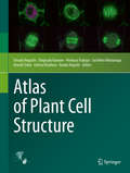
Atlas of Plant Cell Structure
by Tetsuko Noguchi Shigeyuki Kawano Hirokazu Tsukaya Sachihiro Matsunaga Atsushi Sakai Ichirou Karahara Yasuko HayashiThis atlas presents beautiful photographs and 3D-reconstruction images of cellular structures in plants, algae, fungi, and related organisms taken by a variety of microscopes and visualization techniques. Much of the knowledge described here has been gathered only in the past quarter of a century and represents the frontier of research. The book is divided into nine chapters: Nuclei and Chromosomes; Mitochondria; Chloroplasts; The Endoplasmic Reticulum, Golgi Apparatuses, and Endocytic Organelles; Vacuoles and Storage Organelles; Cytoskeletons; Cell Walls; Generative Cells; and Meristems. Each chapter includes several illustrative photographs accompanied by a short text explaining the background and meaning of the image and the method by which it was obtained, with references. Readers can enjoy the visual tour within cells and will obtain new insights into plant cell structure. This atlas is recommended for plant scientists, students, their teachers, and anyone else who is curious about the extraordinary variety of living things.
Atlas Of Plant Viruses: Volume II
by Robert G. Francki R.I.B; MilneThis book assembles a comprehensive collection of plant virus electron micrographs of good quality, offers a consistent treatment, and backs the visual data with a consistent and comprehensive text. Although this book is primarily about the structure of virus particles and infected cells, the results of biochemical experiments are referred too when relevant, so that the virus particles described appear as part of a replicating complex. Similarly, infected cells are portrayed as active rather than static structures.
Atlas Of Plant Viruses: Volume I
by Robert G. Francki R.I.B; MilneThis book assembles a comprehensive collection of plant virus electron micrographs of good quality, offers a consistent treatment, and backs the visual data with a consistent and comprehensive text. Although this book is primarily about the structure of virus particles and infected cells, the results of biochemical experiments are referred too when relevant, so that the virus particles described appear as part of a replicating complex. Similarly, infected cells are portrayed as active rather than static structures.
Atlas of Quaternary Pollen and Spores in China
by Lingyu Tang Limi Mao Junwu Shu Chunhai Li Caiming Shen Zhongze ZhouThis book provides an important reference guide to pollen and spore identification for Chinese Quaternary palynological studies. Presenting and describing more than 400 color photomicrographs of pollen grains and spores retrieved from sediments in China, it offers a unique asset for researchers, graduate students, and newcomers to the field of Quaternary palynology, which constitutes a major aspect of Quaternary paleoecology, paleoclimatology, and paleogeography.
An Atlas of Radioscopic Catheter Placement for the Electrophysiologist
by Andrea Natale Michela Casella Antonio Dello Russo P. Della BellaAn Atlas of Radioscopic Catheter Placement is unique, and has been conceived as a handy reference guide for students, interventional cardiologists, nurses and electrophysiology technicians. It includes plenty of schemes and X-ray images, and every EP correct catheter positioning is explained step by step through detailed descriptions of the necessary manoeuvres, including some "trucks" brought about by the experience.
Atlas of Rangeland Plants in Hulun Buir
by Xiaohui Yang Yuanjun Zhu Baizhu Wang Yanshu LiuThis book includes description of main morphological characteristics of 435 species (including varieties and subspecies) belonging to 57 families and 233 genera of endemic and endangered plants of Hulun Buir Rangeland in China. A brief description of the morphological characteristics of each plant, flowering period, zoning, habitat, and the usage habits of most plants, together with 1 to 4 photographs taken in the field are provided. This work is designed not only for researchers working in rangeland science, ecological restoration and protection but also for professionals working in rangeland and related fields. The work is a result of many years of rangeland plant collection and specimens identification.