- Table View
- List View
Atlas of Endocrine Pathology (Atlas of Anatomic Pathology)
by Lori A. EricksonAtlas of Endocrine Pathology provides a comprehensive compendium of photomicrographs of common and uncommon entities in endocrine pathology. The volume includes histologic features of normal features, reactive conditions, hyperplasia and tumors. The most helpful diagnostic features are illustrated to provide direction and clues to the diagnosis of endocrine tumors. Furthermore, photomicrographs highlight the most pertinent diagnostic features in problematic diagnoses in endocrine pathology. Authored by a nationally and internationally recognized pathologist, Atlas of Endocrine Pathology is an important learning tool for those becoming familiar with the diverse entities encountered in endocrine pathology and a valuable reference for practicing pathologists faced with challenging diagnoses in endocrine pathology.
Atlas of Endometrial Histopathology
by Dietmar Schmidt Gisela Dallenbach-Hellweg Friederike DallenbachThe prime purpose of this atlas is to help the pathologist find, classify and differentially diagnose the changes he is observing. Information on the daily changes during the menstrual cycle and how to date them is essential for recognizing changes caused by abnormal endogenic and iatrogenic hormonal stimuli in functional endometrial disturbances. To provide their patients with the proper hormonal therapy, gynecologists cannot rely on blood samples only because of the constantly fluctuating hormonal levels. A precise functional diagnosis of the endometrial biopsy is the most accurate and best means for assessing hormonal action. Numerous microphotographs explain in detail how to recognize the normal and pathological changes that can develop in the endometrium. The subject of endometritis and the complex endometrial neoplasms and their precursors, with their differential diagnosis as well as their clinical prognosis are covered in detail. To complete the endometrial histopathology, gestational changes are reviewed and illustrated in the last chapter.
Atlas of Endometriosis (Encyclopedia of Visual Medicine Series)
by Colin Davis Robert W. Shaw Caroline Overton Lindsay McMillanEndometriosis affects women in the reproductive years, is associated with pelvic pain and infertility, and - although not life threatening - can seriously impair health, with huge economic and social consequences. It is arguably the most frequent problem encountered in contemporary More...gynecology and is the subject of much ongoing research and innovation in management. This beautifully and comprehensively illustrated Atlas, now in its third edition, provides a useful educational tool for trainees and general obstetricians and gynecologists who may not be up-to-date with the most important recent research on the diagnosis and management of the condition; particularly expanded for this edition are the chapters on ultrasound imaging and the nutritional aspects of the subject.
Atlas of Endoscopic Major Pulmonary Resections
by Dominique GossotIt is my greatest honor to be asked to write this foreword for the first edition of the Atlas of Endoscopic Major Pulmonary Resections by Dr Dominique Gossot. I have known Dr Gossot for over 15 years and have worked with him for many workshops and thoracic meetings. He is a pioneer in video-assisted thoracic surgery, and one of the most innovative thoracic surgeons I have known. Minimally invasive surgery has set a new standard of care for all surgical disciplines. Video-assisted thoracic surgery (VATS) offers a much kinder approach to the management of a wide variety of surgical conditions c- pared with conventional thoracotomy for these patients. Anatomical or major lung resections are a complex set of procedures commonly performed by thoracic s- geons. The adoption of the VATS approach for these procedures has received increasing acceptance by the thoracic surgical community, our pulmonologist and oncology colleagues, as well as the patients over the past two decades. There is now a growing body of evidence in the literature showing that the VATS approach is safe, oncologically sound, and associated with much lower morbidity compared with its conventional counterparts in the management of early lung cancers and benign conditions. Although there have been other books and atlases on VATS, this volume distinguishes itself in two respects.
Atlas of Endoscopic Major Pulmonary Resections
by Dominique GossotThis third edition Atlas of Endoscopic Major Pulmonary Resections describes in detail the totally thoracoscopic approach to major pulmonary resections, in which only endoscopic instruments and monitor control are used. Pulmonary lobectomies and segmentectomies are presented step by step, using brief technical notes and high-quality, clearly labeled still images. Each chapter begins with information on the anatomical background, which is illustrated in three-dimensional reconstructions. In turn, technical ‘tricks’ and specific pitfalls are explained in pictograms. The technical descriptions presented here are based on the author’s own technique, which in some cases differs from other video-assisted approaches. The goal is for surgeons embarking on video-assisted major pulmonary resections – regardless of which approach they use – to find helpful hints and guidance on this totally new vision of pulmonary and mediastinal anatomy.The first edition of this atlas was released at a time when video-assisted major pulmonary resections were just emerging as a valid alternative to conventional techniques. In the second edition, chapters on sublobar resections, as a new alternative to lobectomy in selected patients, were added. In this third edition, as the interest in sublobar resections is growing, and because they are challenging, the technique is dealt with in depth. In particular, readers will be introduced to new imaging technologies to support these techniques.
Atlas of Endoscopic Neurosurgery of the Third Ventricle: Basic Principles for Ventricular Approaches and Essential Intraoperative Anatomy
by Roberto Alexandre DezenaThis book describes in practical terms the endoscopic neurosurgery of the third ventricle and surrounding structures, emphasizing aspects of intraoperative endoscopic anatomy and ventricular approaches for main diseases, complemented by CT / MRI images. It is divided in two parts: Part I describes the evolution of the description of the ventricular system and traditional ventricular anatomy, besides the endoscopic neurosurgery evolution and current concepts, with images and schematic drawings, while Part II presents a collection of intraoperative images of endoscopic procedures, focusing in anatomy and main pathologies, complemented by schemes of the surgical approaches and CT / MRI images. The Atlas of Endoscopic Neurosurgery of the Third Ventricle offers a revealing guide to the subject, addressing the needs of medical students, neuroscientists, neurologists and especially neurosurgeons.
Atlas of Endoscopic Plastic Surgery
by Edoardo RaposioConcentrating on technique, which is explained and illustrated in detail, this book is written by worldwide experts and provides detailed, step-by-step instructions on how to perform state-of-the-art endoscopic surgical techniques in the complex Plastic Surgery field. More than 300 high-quality photos help clarify complex techniques throughout the book. Atlas of Endoscopic Plastic Surgery represents a comprehensive description of the current endoscopic techniques in the plastic, reconstructive an aesthetic field. It supplies surgeons with all the information necessary to successfully accomplish an endoscopic approach to vary plastic surgery procedures, from carpal and cubital tunnel release, breast augmentation and reconstruction, migraine surgery, hyperhidrosis management, to facial aesthetic surgery, flap and fascia lata harvesting, and mastectomy and abdominal wall surgery.
Atlas of Endoscopic Ultrasonography
by John C. Deutsch Frank Gress Thomas Savides Brenna C. BoundsThe Atlas of Endoscopic Ultrasonography provides readers with a large collection of excellent images obtained from both diagnostic and therapeutic procedures. The Atlas includes a DVD which will be an invaluable addition to the library of trainee and practising gastroenterologists with video clips and searchable database of images. Together the book and DVD offer a first class collection of images to give a highly integrated introduction to endoscopic ultrasonography. The Atlas is an ideal companion to Dr Gress et al’s Endoscopic Ultrasonography, Second Edition.
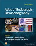
Atlas of Endoscopic Ultrasonography
by Frank Gress Thomas Savides Brenna Casey Everson L. A. ArtifonAtlas of Endoscopic Ultrasonography Atlas of Endoscopic Ultrasonography Atlas of Endoscopic Ultrasonography, Second Edition offers an outstanding visual guide to this very common diagnostic and therapeutic endoscopic tool. With contributions from noted experts in the field, the Atlas contains 400 high-quality color and black and white images obtained from real cases, each accompanied by detailed annotation to aid readers in their understanding of this popular technical procedure. In addition, there is a companion website featuring 50 video clips of real-life procedures in action, as well as the entire collection of images from within the book. Updated throughout to include the most recent advances in interventional Endoscopic Ultrasound (EUS) guided therapies Contains a large collection of color images obtained from both diagnostic and therapeutic procedures, also available on the companion website image bank Provides a highly integrated and accessible multimedia introduction to endoscopic ultrasonography Includes a companion website offering insightful videos Written for gastroenterologists, students, residents, and radiologists, Atlas of Endoscopic Ultrasonography, Second Edition is an essential introduction to endoscopic ultrasonography.
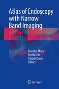
Atlas of Endoscopy with Narrow Band Imaging
by Manabu Muto Kenshi Yao Yasushi SanoWith its focus on narrow band imaging, this book is an excellent reference for new as well as experienced practitioners in the field of endoscopy. Narrow band imaging has brought about a revolutionary improvement in diagnostic endoscopy, enabling objective diagnosis and precise detection of lesions. It has enhanced the capability of endoscopy to facilitate qualitative diagnoses for the great benefit of patients who undergo endoscopic examinations. However, a standardized system of classification has not yet been established and many clinicians and researchers are not yet highly skilled in utilizing the technique or interpreting the images that are produced. This atlas addresses those issues, providing clear, simple and easy-to-understand descriptions illustrated with generous use of endoscopic images.
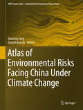
Atlas of Environmental Risks Facing China Under Climate Change (IHDP/Future Earth-Integrated Risk Governance Project Series)
by Qiuhong Tang Quansheng GeThis atlas provides the most comprehensive and accurate overview of environmental risks relating to climate change vulnerability and adaptation in China. It addresses the agricultural, ecosystem and heat wave health risk posed by climate change and presents the projected environmental risks in the 21st century under climate change and socioeconomic scenarios. The detailed and concise risk assessments are mapped in grid units, allowing easy environmental risk assessment for specific locations. The atlas contributes significantly to the knowledge base for climate change adaptation in China and is a valuable resource for students and professionals in the fields of geographic sciences and climate change.
Atlas of Equine Ultrasonography
by Michele L. Frazer Kristina G. Lu Jessica A. KiddThe only visual guide to equine ultrasonography based on digital ultrasound technology. Atlas of Equine Ultrasonography provides comprehensive coverage of both musculoskeletal and non-musculoskeletal areas of the horse. Ideal for practitioners in first opinion or referral practices, each chapter features normal images for anatomical reference followed by abnormal images covering a broad range of recognised pathologies. The book is divided into musculoskeletal, reproductive and internal medicine sections and includes positioning diagrams demonstrating how to capture optimal images. With contributions from experts around the world, this book is the go-to reference for equine clinical ultrasonography.Key features include:Pictorially based with a wealth of digital ultrasound images covering both musculoskeletal and non-musculoskeletal areas and their associated pathologies.Each chapter begins with a discussion of normal anatomy and demonstrates how to obtain and interpret the images presented.A video library of over 50 ultrasound examinations is available at www.wiley.com/go/kidd/equine-ultrasonography for streaming or download and viewing on-the-go.
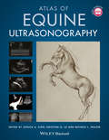
Atlas of Equine Ultrasonography
by Michele L. Frazer Kristina G. Lu Jessica A. KiddATLAS OF EQUINE ULTRASONOGRAPHY A THOROUGH EXPANSION TO THE FIRST ATLAS OF ULTRASONOGRAPHY IN THE HORSE, WITH NEW AND SIGNIFICANTLY IMPROVED IMAGES Ultrasonography is a vital diagnostic tool that can be applied in numerous functions in a veterinary practice. In conjunction with relevant clinical information—patient history and physical examination findings, for example—it can act as an important aid in the veterinarian’s decision-making process. Many vets in equine practice rely upon ultrasonography as a mainstay of equine diagnostic imaging on a wide range of structures and body systems. Ultrasonography is a useful procedure that is non-invasive and acts in complement to radiography to successfully diagnose the animal’s condition. This book’s aim is to encourage the clinician to rely further on the use of ultrasonography in their practice. The second edition of Atlas of Equine Ultrasonography provides an updated and expanded revision of the first atlas of ultrasonography in the horse. The first edition of this important resource was the first pictorially-based book to cover ultrasonography in the horse, and remains the only book currently available on the subject. The current version offers 450 additional images with greater clarity and precision in the images throughout and demonstrates how to obtain images in each body region while offering clinical ultrasonograms that show pathology. Atlas of Equine Ultrasonography readers will also find: High-quality clinical ultrasonograms for important musculoskeletal, reproductive, and medical conditions in the horse More than 1,500 images, with accompanying concise text describing the images A companion website that provides video clips showing dynamic ultrasound exams Atlas of Equine Ultrasonography is an invaluable reference to any veterinarian evaluating ultrasonograms in equine patients. As a result, this book will be of particular interest to equine specialists, veterinary radiologists, equine practitioners, and veterinary students.
Atlas of Esophageal Disease and Intervention: A Multidisciplinary Approach
by Shanda H. Blackmon Min P. Kim Karen J. DickinsonThis atlas provides a comprehensive, state-of-the-art review of all interventions that pertain to the esophagus. It includes a review of the current staging modalities, ablation technologies, resection and reconstruction techniques, and disease classification. Evidence-based guidelines regarding how each intervention is chosen are also included. With color illustrations and photographs for each surgery, the atlas details specific anatomic topics such as micro-anatomy of Barrett's and Dysplasia, EMR pathology, endoscopic ultrasound, and conventional surgical anatomy. Each intervention is presented in task format as a task list to be checked-off as each step is completed. Written by experts in the field, Atlas of Esophageal Disease and Intervention: A Multidisciplinary Approach serves as a valuable resource for any practitioner who performs esophageal intervention and will guide new surgeons and gastroenterologists into the hybrid multidisciplinary approach to this disease.
Atlas of Esophageal Surgery
by Marco G. Patti Fernando A. M. HerbellaThis Atlas focuses on surgical techniques used to treat the entire spectrum of esophageal diseases. Surgical “pearls” and tips on how to select and perform the correct operation are included and based both on evidence-based data and the experience of the Editors.Step-by-step descriptions of operative and endoscopic procedures in esophageal surgery are provided. Each chapter describes the current indications, perioperative management strategies, and a detailed operative approach with relevant technical considerations.The description of approaches and surgical techniques used in esophageal surgery are outlined in an easily understandable manner for the specific target audience. This book is written by world-class internationally renowned esophageal surgeons and gastroenterologists
Atlas of Esophageal Surgery
by P. Marco Fisichella Marco G. PattiThis Atlas focuses on the description of approaches and surgical techniques used to treat the entire spectrum of esophageal diseases. Surgical "pearls" and tips on how to select and perform the correct operation are included and based both on evidence-based data and the experience of the Editors. Step-by-step descriptions of 14 operative procedures in esophageal surgery are provided. Each chapter describes the current indications, perioperative management strategies, and a detailed operative approach with relevant technical considerations. The description of approaches and surgical techniques used in esophageal surgery are outlined in an easily understandable manner for the specific target audience.
Atlas of Esophagus and Stomach Pathology (Atlas of Anatomic Pathology)
by Henry D. Appelman Scott R. OwensAtlas of Esophagus and Stomach Pathology provides an image-based resource for those studying normal histology of the upper gastrointestinal tract, as well as the microscopic manifestations of developmental abnormalities, toxic insults, infectious diseases, inflammatory and autoimmune conditions, and neoplasia in the esophagus and stomach. Because modern gastrointestinal pathology practice centers on specimens obtained during endoscopic examination, the atlas focuses on biopsy pathology, providing "real-world" microscopic images and ancillary diagnostic studies for most commonly-encountered abnormalities and diseases affecting these two organs. The book is supplemented with endoscopic and special study images. Authored by nationally and internationally recognized pathologists, Atlas of Esophagus and Stomach Pathology is a valuable tool for both pathologists-in-training seeking to make "new acquaintances", and practicing surgical pathologists in need of a quick visual reference in recalling "old friends" in the world of diagnostic gastrointestinal pathology.
Atlas of Essential Dermatopathology
by Andrea Primiani Lyn M. Duncan Kasia S. MasterpolThe book is not intended to be an all-encompassing atlas or textbook but rather a foundation of principles in dermatopathology, highlighting key elements in the field for trainees and will also serve as a basic resource for the pathologist in general practice. In addition to the sketches and minimal text, we envision accompanying high resolution histopathologic micrographs for ultimate correlation as well.
Atlas of Essential Orthopaedic Procedures, Second Edition
by Alexis C. Colvin Evan FlatowCovering more than 100 fundamental orthopaedic techniques, Atlas of Essential Orthopaedic Procedures, 2nd edition offers a highly illustrated, step-by-step guide to the wide variety of conditions you’re most likely to see in practice. The easy-to-follow format begins with patient selection, walks you through a detailed, step-by-step description of the procedure, and concludes with the author’s surgical pearls—all heavily illustrated with radiographs, intraoperative photographs, and line drawings for optimal visualization of the procedure. This technique-focused reference is an essential resource for busy orthopaedic surgeons and a must-have reference for orthopaedic residency.
Atlas of Extraterrestrial Zones (Atlas Ser. #2)
by Bruno Fuligni32 unusual and fascinating extraterrestrial encounters that explores the places and portals between our world and theirs, with illustrated maps for each location (Book 2 of the bestselling Atlas series) Where is the best place to meet kind and peaceful aliens? How do we communicate with intelligent interstellar life forms, socialize with Martians, and avoid unwanted close encounters of the third kind? These and other answers to ET mysteries are captured in the thirty-two stories in the Atlas of Extraterrestrial Zones, where maps direct readers to the portals that open up between the worlds, a geography of strange events that covers the entire earth, demonstrating how widespread the phenomenon is. Learn about locations of extraterrestrial sightings, hidden bases, secret embassies, and long-lost traces of thousand-year-old passages. From the UFO port of Arès to the underground center of Area 51, from the crash at Roswell to setting up the SETI program, this atlas lists, for the first time, the meeting points between earthlings and these mysterious extraterrestrial biological entities (EBEs).
Atlas of Extreme Facial Cancer: Challenges and Solutions
by Ian Burton Michael F. KlaassenThis book shares an expert experience in managing difficult facial skin cancer and presents rare cases from the authors's own practice. The content follows an anatomical approach based on the complex and often staged reconstruction of extreme facial skin cancer. Written in a clear and logical format, it is intended as a guideline atlas and surgical handbook, defining the principles of CLEAR (Complete Local Excision of the cancer + Aesthetic Reconstruction) and DRAPE (Delayed Reconstruction After Pathological Examination) for reconstructing the face. The book incorporates a wealth of multi-disciplinary knowledge from surgeons, anaesthetists, research scientists, speech / swallowing therapists, pathologists, radiologists and prosthetic rehabilitation specialists. The surgical techniques presented here require a high degree of expertise. Accordingly, the book will be of interest to professionals in the fields of Plastic and Oral and Maxillofacial Surgery, as well as Dermatology.
Atlas of FFR-Guided Percutaneous Coronary Interventions
by Tommaso Gori Massimo FineschiThis book reviews the theory and practice of fractional flow reserve (FFR)-guided coronary intervention, a technique that gives sense and a rationale to daily decisions in the interventional suite. FFR guidance provides detailed information on coronary hemodynamics for the interventional cardiologist. This technique has profound practical implications for therapeutic decisions and for the prognosis of patients. The Atlas of FFR-Guided Percutaneous Coronary Interventions provides practicing physicians clear information to understand both the complexity of the technique and the correct way to apply it. It is designed both to assist younger faculty and those in training, and to act as a clinical resource for more experienced practitioners. Using the clinical cases outlined, the reader can learn to appreciate the pitfalls, tips and tricks that simplify the performance and interpretation of FFR and iFR.
Atlas of Fallen Dust in Kuwait
by Ali Al-DousariThis open access book serves as an atlas of deposited dust and dust storms in Kuwait in relation to local and global regions. It features a wealth of maps and images of dust storm trajectories in the region, together with detailed descriptions of the chemical and physical properties of fallen dust, including the amount, particle size, statistical parameters, spectra absorption, dust mineralogy, trace and major elements, organic matter, associated pollen, and radionuclides and connected pollutants. Given its scope, the book is a valuable resource for a broad range of researchers, including geologists, chemists, environmentalists, botanists, air quality specialists, nanotechnology scientists, and solar energy experts.
Atlas of Feline Ophthalmology
by Kerry L. Ketring Mary Belle GlazeSuccessful management of eye disease relies on the veterinarian’s ability to identify ocular features and distinguish pathologic changes. Atlas of Feline Ophthalmology, Second Edition is an invaluable diagnostic reference, providing high-quality color photographs for comparison with a presenting complaint. Presenting 394 photographs illustrating both normal and pathologic ocular conditions, this Second Edition offers a current, complete reference on ocular diseases, adding conditions recognized since publication of the first edition, a broader geographic scope, and many new images with improved quality. Carefully designed for easy reference, the contents are divided into sections corresponding to specific anatomical structures of the eye. A useful appendix new to this edition groups figures by etiology, making it easy to find every image associated with a specific agent or disease. Atlas of Feline Ophthalmology, Second Edition is a useful tool aiding general practitioners in diagnosing eye disease in cats.
Atlas of Fetal Imaging: Abdomen
by S. Boopathy VijayaraghavanThis atlas presents the sonographic features of the normal fetal abdomen at different gestational ages, and describes the anomalies of different organ systems in the fetal abdomen. It covers the gastrointestinal tract, genitourinary tract, and abdominal wall defects with the help of numerous ultrasound images, and also addresses differential diagnosis using various sonographic images of the fetal abdomen, as well as effective diagnostic approaches for these conditions.