- Table View
- List View
Atlas of Comparative Diagnostic and Experimental Hematology
by Alfred Jarecki Clifford SmithA vital resource on blood and bone marrow cell morphology in laboratory animal medicine. This fully revised new edition is an essential reference for clinical pathologists in diagnostic laboratories, and medical or veterinary research. The atlas contains over 400 color images of cells from the peripheral blood and bone marrow from a variety of animals encountered in laboratory animal medicine, in health and disease. Key features: New chapter on flow cytometry and its application in terms of routine analyses as a means of identifying abnormalities in cell marker expression, which is of particular relevance for pre-clinical safety assessment Covers the most recent developments in laboratory animal hematology, including parameters measured by the latest generation of analyzers Coverage of a wide range of laboratory animal species, as well as those used in clinical veterinary trials Photomicrographs present normal and abnormal blood cells from a variety of hematological conditions along with descriptive text
Atlas of Conducted Electrical Weapon Wounds and Forensic Analysis
by Jeffrey D. Ho Mark W. Kroll Donald M. DawesAtlas of Conducted Electrical Weapon Wounds and Forensic Analysis provides a comprehensive publication on the subject of Conducted Electrical Weapon (CEW) wounds and signature markings created by this class of weapon. This volume will serve as a very useful resource for all professions tasked with assisting persons that have allegedly been subjected to a CEW exposure. The volume provides an introduction to basic CEW technology and the types of CEWs currently available. It also serves as a comprehensive pictorial atlas of signature markings that CEW exposures make in the immediate and more remote post-exposure periods. Also, it discusses the ability of forensic specialty examinations of the CEW itself to aid in the determination of whether the alleged CEW exposure is consistent with the objective evidence and the subjective statements. Finally, this text addresses the important and growing area of factitious CEW markings that will be useful for consideration by investigators and litigators. Atlas of Conducted Electrical Weapon Wounds and Forensic Analysis provides an objective atlas of evidence for reference that will benefit those professionals who often must make diagnostic, treatment or legal judgments on these cases including Emergency and Primary-Care Physicians, Medical Examiners, Forensic Pathologists, Coroners, Law Enforcement Investigators, and Attorneys.
Atlas of Cone Beam Computed Tomography
by Ghassem Ansari Yaser Safi Mitra Ghazizadeh Ahsaie Ingrid Różyło-Kalinowska Mahsima Tayefi Nasrabadi Maryam FazlalipourA comprehensive collection of oral and maxillofacial cases using cone beam CT imaging Atlas of Cone Beam Computed Tomography delivers a robust collection of cases using this advanced method of imaging for oral and maxillofacial radiology. The book features over 1,500 high-quality CBCT scans with succinct descriptions covering a wide range of maxillofacial region conditions, including normal anatomy, anomalies, inflammatory diseases, and degenerative diseases. Easy to navigate and featuring multiple images of normal variation and pathologies, the book offers readers guidance on the diagnostic values of CBCT, as well as CBCT images of the inferior alveolar nerve canal, dental implants, temporomandibular joint evaluations, and surgical interventions. The book also includes: A thorough introduction to cone beam computed tomography, including in vivo and in vitro preparation and evaluation, indications in dentistry, and indications in medicine Comprehensive explorations of cone beam computed tomography artefacts and anatomic landmarks Practical discussions of cone beam computed tomography of dental structure, including normal anatomy, anomalies, and the difficulties of eruption In-depth examinations of cone beam computed tomography of pathological growth and development, including maxillofacial congenital and developmental anomalies Perfect for graduate dental students and postgraduate dental students in oral and maxillofacial radiology, Atlas of Cone Beam Computed Tomography is also useful to general dentists, oral and maxillofacial radiologists, head and neck maxillofacial surgeons, head and neck radiologists, general radiologists, and ENT surgeons.
Atlas of Contraception
by Malcolm Potts Pramilla SenanayakeThis revised and updated Atlas provides a comprehensive guide to modern contraceptive practice. The book is heavily illustrated with color photographs and line drawings that guide the reader through the various options available and provide a valuable educational resource. The supporting text offers a concise description of family planning in today
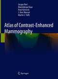
Atlas of Contrast-Enhanced Mammography
by Jacopo Nori Maninderpal Kaur Anat Kornecki J. Devi Meenal Martin J. YaffeThis superbly illustrated atlas serves as a basic introduction to contrast-enhanced mammography (CEM), a breakthrough functional breast imaging modality, which is rapidly growing. This book is an essential guide for the latest developments with correlative findings, practical interpretation tips, physics, and information on how contrast mammography differs from conventional 2D and 3D Full Field of View digital mammography (FFDM). It includes: · over 1000 high-quality 2D, 3D and recombined contrast mammography images representing the spectrum of breast imaging · findings obtained in the full range of benign, pre-malignant and malignant conditions, including artefacts and postoperative changes, presented with high-quality illustrations from case examples · image interpretation tips using mammographic and DCE-MRI descriptors of the BI-RADS lexicon to effectively read and interpret this advanced imaging modality · practical tips to interpret this new modality and how it is used as an adjunct to 2D mammography · details on how integration of contrast-enhanced mammography drastically changes lesion work-up and overall workflow in the department · "imaging pearls" boxes offering interpretation tips for expert clinical guidance · a case on the recently introduced CEM guided biopsy procedure The book’s target audience consists of diagnostic radiologists, residents, fellows, technologists and clinicians involved in the care of breast cancer patients, including surgeons and oncologists. The goal is to provide a concise introduction to CEM and to lead to enhanced interpretation and better patient staging prior to surgery.
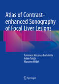
Atlas of Contrast-enhanced Sonography of Focal Liver Lesions
by Massimo Midiri Tommaso Vincenzo Bartolotta Adele TaibbiThis book offers an image-based, comprehensive quick reference guide that will assist in the interpretation of contrast-enhanced ultrasound (CEUS) examinations of the liver in daily practice. It describes and depicts typical and atypical behavior of both common and less frequently observed focal liver lesions. For each type of lesion, the findings on pre- and post-contrast images are presented and key characteristics are highlighted. Individual chapters also focus on the assessment of response to locoregional and systemic treatment and the impact of European guidelines on CEUS. The Atlas of Contrast-Enhanced Sonography of Focal Liver Lesions will serve as an invaluable hands-on tool for practitioners who need to diagnose liver lesions using CEUS in the major clinical settings: oncology patients, cirrhotic patients, and patients with incidental focal liver lesions.
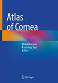
Atlas of Cornea
by Bharat Gurnani Kirandeep KaurThis atlas provides an overview of corneal pathologies. It includes over 300 images of several corneal pathologies in daily clinical practice. It provides a comprehensive overview of corneal pathologies by senior cornea experts across the globe. The chapters offer guidance on numerous corneal pathologies such as microbial keratitis, corneal ectasia, keratoconus, dry eyes, ocular corneal trauma, refractive surgery, keratoconjunctivitis, ocular surface disorders, limbal stem cell deficiency, pterygium, ocular surface squamous neoplasia (OSSN), pinguecula, degenerations, dystrophies, conjunctivitis, etc. The book guides ophthalmologists in practice and training for diagnosing corneal pathologies.
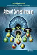
Atlas of Corneal Imaging
by J. Bradley RandlemanA comprehensive reference for physicians, surgeons, and trainees, Atlas of Corneal Imaging covers all aspects of corneal imaging from basic map interpretation to advanced diagnostic uses and features over 1200 illustrative images and figures representing a wide variety of devices and techniques.Drs. J. Bradley Randleman, Marcony Santhiago, and William J. Dupps Jr guide readers through the process of analyzing corneal images using a multitude of different techniques, technologies, and individual devices. This creates a complete picture of the cornea’s basic methods and pathological processes and allows readers to directly visualize how their technology would display the pathology in question.Atlas of Corneal Imaging is designed to help practitioners recognize subtle findings and evaluate signs of weakening or pathology, no matter how they present or what device is being used. Multiple iterations of the same clinical condition are shown with numerous complementary images for the same eye to provide a comprehensive presentation of each case.Chapters feature information on: Topographic patterns and mapping Corneal ectasia evaluations Cornea and refractive surgery evaluations Clinical correlations with corneal disorders Cornea and refractive surgery complications Evaluation for cataract surgery Atlas of Corneal Imaging fills a significant void in corneal imaging resources available today by presenting an image-first approach to understanding all the many different technologies for imaging the cornea.
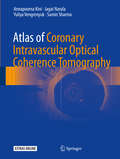
Atlas of Coronary Intravascular Optical Coherence Tomography
by Jagat Narula Annapoorna Kini Samin Sharma Yuliya VengrenyukThis atlas is a practical and fully illustrated guide to the use of intravascular OCT in diagnosis and treatment of coronary artery disease. It consists of two parts. The first part of the book provides a systematic introduction to coronary imaging with OCT. It describes how to interpret images and describes abnormal findings seen in atherosclerosis, complications after intervention, and stent assessment. The second part of the book presents real-life case studies that show how OCT is used in clinical practice in Mount Sinai to assess the disease, select appropriate treatment, and evaluate complications and results. Each case includes a brief clinical history, procedure summary, angiography and OCT images, including video material, and a discussion of how OCT affected the clinical decision-making process.
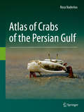
Atlas of Crabs of the Persian Gulf
by Reza NaderlooThis illustrated atlas describes 256 extant brachyuran crab species in the Persian Gulf and the Gulf of Oman. Identification keys are provided for 37 brachyuran families, 142 genera and 256 species on the basis of their main synapomorphies. Brief but precise descriptions highlighting the main characteristics are also provided for every family. The atlas displays features high-quality color photos, offering a hands-on guide and equipping readers to readily diagnose crab species in the region. Importantly, a line drawing of the first male gonopod, as well as its main diagnostic characteristics, are provided for all species. Further, every species is supplemented with synonymies that encompass the original descriptions, overall revision of the given taxa, monographs and all records from the northwestern Indian Ocean including the Persian Gulf and the Gulf of Oman. For each species, the book provides detailed local and global distribution maps, together with important ecological data including habitat preference. Further, it includes a general introduction to the brachyuran crabs with schematic drawings of their external morphology, as well as a comprehensive introduction to the Persian Gulf and the Gulf of Oman as marine ecoregions (geography, hydrology, biology, and environmental condition). The book offers an indispensable guide for all professionals, researchers, and students interested in brachyuran crabs around the globe and particularly in the Persian Gulf and the Gulf of Oman.
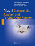
Atlas of Craniocervical Junction and Cervical Spine Surgery
by Livio Presutti Francesco Mattioli Stefano Boriani Alessandro GasbarriniThis atlas documents current surgical approaches to the craniocervical junction and the cervical spine, providing step-by-step guidance on procedures and cervical spine stabilization techniques. Opening chapters present essential information on anatomy, depict pathologies with the aid of illustrative cases, describe the role of imaging techniques in patient evaluation, and discuss surgical instrumentation and patient positioning. The different techniques employed in this delicate anatomic region, including transnasal and transoral endoscopic approaches to the craniocervical junction and posterior and anterior approaches to the cervical spine, are then explained and illustrated with a view to providing the surgeon with a clear reference that can be used in the operating room. In addition, practical advice is offered on the treatment of potential complications, postoperative management, and rehabilitation. This book will be of value not only to neurosurgeons but also to orthopedists, ENT surgeons, neurologists, and physiatrists.
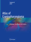
Atlas of Craniopharyngioma: Pathology, Classification and Surgery
by Songtao QiThis book aims to facilitate readers to understand the origin, growth pattern and relationship between tumor and adherent structure of craniopharyngioma, so as to improve the cure rate and safety of surgery. It’s contributed by Neurosurgery Department of Nanfang Hospital, Southern Medical University, China, which focuses on the management of craniopharyngioma. This book covers histoembryology of craniopharyngioma, together with anatomical morphology and abundant clinical data, systematically showing an innovative classification method, i.e. QST classification. This classification method can better reflect the different origin and growth pattern of craniopharyngioma,the relationship between tumors and surrounding structure of the tumor growth pattern, and clinical significance in surgery. The 70 clinical cases with different classification and treatment history are discussed as an important reference for surgical treatment of craniopharyngioma. The anatomy, morphology and pathology of sellar region also have great reference value for researchers in the field of neural science. The underlying intention of this book is to help bring a change in the concept that “craniopharyngioma is an incurable benign tumor only due to its anatomical location”.
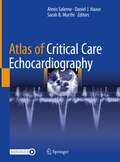
Atlas of Critical Care Echocardiography
by Alexis Salerno Daniel J. Haase Sarah B. MurthiThis atlas covers the basics of ultrasound physics, provides the necessary background to successfully perform TEE and TTE procedures, and provides tips and tricks that will prove invaluable in the critical care environment. It also features an abundance of high quality photographs, illustrations and videos as well as numerous case studies to test the reader's ability to apply knowledge to real-life clinical situations. Written by experts in the field, The Atlas of Critical Care Echocardiography is a concise, visual guide designed for use by all physicians who see cardiac patients in the ICU.
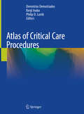
Atlas of Critical Care Procedures
by Demetrios Demetriades Kenji Inaba Philip D. LumbThis atlas is designed to provide an all-inclusive resource that describes step by step how to perform the essential bedside procedures required to provide optimal care to the critically ill patient. Detailed descriptions of these procedures, with sequential photographs depicting each critical step, provide an in depth understanding of the anatomy, critical technical steps and common pitfalls to both military and civilian trainees. For the advanced professional operator, this atlas will provide a rapid refresher of the critical steps and is an excellent teaching resource, based on the decades of collective experience accumulated by the authors. The top quality photographs of perfused and ventilated cadavers makes this atlas a standard reference for surgeons, critical care physicians, nurses and all critical care professionals.
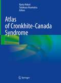
Atlas of Cronkhite-Canada Syndrome
by Tadakazu Hisamatsu Ryota HokariThis manual treats both conceptual and practical aspects of gastrointestinal polyposis syndrome, which is characterized by dermatologic manifestations. The book offers both detailed and comprehensive analysis of the endoscopic findings achieved from 88 cases, which led to novel concepts and classification of the dermatologic manifestations according to distribution pattern, the thickness of mucosa, and intervening inflamed mucosa between polyps. The book also offers comparisons of the endoscopic features to pathological features. Abundant illustrations show the observations of the malignant lesions using special lights, such as NBI and BLI, to provide a comprehensive view of the Cronkhite-Canada syndrome (CCS) polyp.Atlas of Cronkhite-Canada Syndrome is essential guidance for gastroenterologists and pathologists who pursue the new diagnostic approach and treatment for CCS. It also attracts clinicians interested in the latest updates on this field and those who encountered the CCS patients with refractory to corticosteroid treatment. The risk of colorectal cancer in patients with CCS may increase because multiple inflammatory pseudopolyps make it more difficult to detect premalignant adenomas. Additionally, as endoscopy for GI disease becomes widespread in developing countries, the number of diagnosed patients will likely increase. Thus, it is crucial to have a broader understanding of the disease and the treatment strategy, and this book aims to simplify a complicated picture of the rare disease.
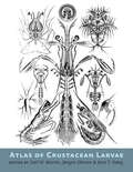
Atlas of Crustacean Larvae
by Joel W. Martin, Jørgen Olesen, and Jens T. Høeg.An illustrated guide to the sweeping diversity of crustacean larval forms.Winner of the CHOICE Outstanding Academic Title of the Choice ACRLCrustaceans—familiar to the average person as shrimp, lobsters, crabs, krill, barnacles, and their many relatives—are easily one of the most important and diverse groups of marine life. Poorly understood, they are among the most numerous invertebrates on earth. Most crustaceans start life as eggs and move through a variety of morphological phases prior to maturity. In Atlas of Crustacean Larvae, more than 45 of the world's leading crustacean researchers explain and illustrate the beauty and complexity of the many larval life stages.Revealing shapes that are reminiscent of aliens from other worlds—often with bizarre modifications for a planktonic life or for parasitization, including (in some cases) bulging eyes, enormous spines, and aids for flotation and swimming—the abundant illustrations and photographs show the detail of each morphological stage and allow for quick comparisons. The diversity is immediately apparent in the illustrations: spikes that deter predators occur on some larvae, while others bear unique specializations not seen elsewhere, and still others appear as miniature versions of the adults. Small differences in anatomy are shown to be suited to the behaviors and survival mechanisms of each species.Destined to become a key reference for specialists and students and a treasured book for anyone who wishes to understand "the invertebrate backbone of marine ecosystems," Atlas of Crustacean Larvae belongs on the shelf of every serious marine biologist.
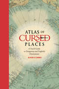
Atlas of Cursed Places: A Travel Guide to Dangerous and Frightful Destinations
by Olivier Le CarrerOliver Le Carrer brings us a fascinating history and armchair journey to the world's most dangerous and frightful places, complete with vintage maps and period illustrations in a handsome volume. This alluring read includes 40 locations that are rife with disaster, chaos, paranormal activity, and death. The locations gathered here include the dangerous Strait of Messina, home of the mythical sea monsters Scylla and Charybdis; the coal town of Jharia, where the ground burns constantly with fire; Kasanka National Park in Zambia, where 8 million migrating bats darken the skies; the Nevada Triangle in the Sierra Nevada mountains, where hundreds of aircraft have disappeared; and Aokigahara Forest near Mount Fuji in Japan, the world's second most popular suicide location following the Golden Gate Bridge.
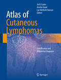
Atlas of Cutaneous Lymphomas: Classification and Differential Diagnosis
by Joi B. Carter Amrita Goyal Lyn Mcdivitt DuncanThis atlas contains excellent clinical and histopathologic images and text of each of the types of cutaneous lymphoma (around 25 entities). It is the first go-to text for those who are considering a diagnosis of cutaneous lymphoma in their differential diagnosis. The text also includes diagnostic mimics of lymphoma and differential diagnosis tables and algorithms. The target audience is general practitioners, dermatologists, pathologists and students, residents and fellows. The diagnosis of lymphoma in the skin is confounded by the myriad of disorders that can mimic lymphoma clinically and histopathologically and by inconsistencies in the diagnostic classification that have only recently been resolved. In the last decade the European Organization for Research and Treatment of Cancer (EORTC) Cutaneous Lymphoma Group and the World Health Organization (WHO) Collaborated in a series of workshops and consensus meetings to arrive at a definitive classification scheme for cutaneous lymphoma. Unfortunately, the publication by the WHO that described this schema included all lymphomas and has the skin tumors scattered throughout the volume. There is currently no go to text for those who are considering a diagnosis of cutaneous lymphoma in their differential diagnosis. As a result there continues to be confusion about the diagnosis of cutaneous lymphoma, although this classification scheme was published in 2008.
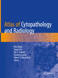
Atlas of Cytopathology and Radiology
by Ritu Nayar Xiaoqi Lin Ajit S. Paintal Ramona Gupta Albert A. NemcekThe aim of this atlas is to guide pathologists and radiologists in the accurate triage and diagnosis of deep seated mass lesions biopsied under ultrasound, CT Scan or fluoroscopic guidance. Fine needle aspiration cytology has become the foremost diagnostic modality in recent years for the diagnosis of mass lesions, including primary and recurrent neoplasms and masses of non-neoplastic and infectious etiology. An essential requirement in the accurate diagnosis of these masses is the correlation of cytomorphology with the radiological findings and adequate triage of acquired material during the biopsy procedure. The cytologic appearance (fine needle aspiration smears, touch preparations, cell blocks and core biopsy), gross surgical resected specimen (where available on follow-up) and imaging findings will be illustrated to provide a complete pathologic-radiologic correlation of the entities discussed. Collection methods and correlation with ancillary studies such as flow cytometry, microbiologic cultures, cytogenetics and immunohistochemistry will be described. The importance of specimen type and cytologic and radiologic techniques will be emphasized.
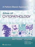
Atlas of Cytopathology: A Pattern-based Approach
by Christopher J. VandenBussche Erika F. Rodriguez Derek B. Allison M. Lisa ZhangAtlas of Cytopathology: A Pattern Based Approach is the latest installment in a unique new series designed to present diagnostic processes in a way similar to how clinicians actually review specimens. The book is image-rich, with scores of illustrations and tables, and filled with checklists, FAQs, and other tools to support fast, easy comprehension of material. Highlighted are common rather than obscure diseases and conditions, and “normal” cytology is presented first to give you a benchmark for subsequent discussions.
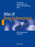
Atlas of Deep Endometriosis: MRI And Laparoscopic Correlations
by Alice Brandão Claudio Peixoto Crispi Marco Aurelio Pinho OliveiraAn MRI-based guide, followed by laparoscopic and histopathological correlations.<P><P>Intended for radiologists, gynaecologists and all other professionals interested in deep endometriosis.<P> Features a wealth of images, addressing the most important radiological aspects of deep endometriosis.<P>This Atlas presents an MRI-based guide to the diagnosis, treatment and follow up of deep endometriosis. Developed by professionals with a extensive clinical experience in the diagnosis and treatment of deep endometriosis, it provides a global overview of the disease, from basic clinical aspects of imaging diagnosis, to the correlation with surgical findings and histopathological results. Deep endometriosis is a serious gynecological condition, which can severely impact on women’s quality of life. It shares the main features of regular endometriosis, but also displays a highly infiltrative pattern, involving multiple organs and leading to severe symptoms such as dysmenorrhea, chronic pelvic pain and dyspareunia. Atlas of Deep Endometriosis – MRI and Laparoscopic Correlations is a complete guide, intended for radiologists, gynecologists and all other medical professionals interested on the diagnosis and treatment of deep endometriosis. (NOTE: This title was previously published in 2014 in Portuguese and Spanish and comes from Springer's partnership with Brazilian publisher, Revinter.)
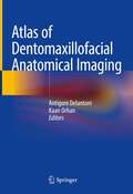
Atlas of Dentomaxillofacial Anatomical Imaging
by Kaan Orhan Antigoni DelantoniThis atlas is a detailed and complete guide on imaging of the dentomaxillofacial region, a region of high interest to a wide range of specialists. A large number of injuries and patient’s treatment involve the facial skeleton. Enriched by radiographic images and illustrations, this book explores the anatomy of this region presenting its imaging characteristics through the most commonly available techniques (MDCT, CBCT, MRI and US). In addition, two special chapters on angiography and micro-CT expand the limits of dentomaxillofacial imaging. This comprehensive book will be an invaluable tool for radiologists, dentists, surgeons and ENT specialists in their training and daily practice.
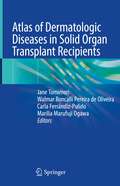
Atlas of Dermatologic Diseases in Solid Organ Transplant Recipients
by Jane Tomimori Walmar Roncalli Pereira de Oliveira Carla Ferrándiz-Pulido Marilia Marufuji OgawaThe number of solid organ transplant recipients is increasing worldwide every year, which has meant a significant improvement of recipient’s survival and quality of life. To prevent graft rejection, patients require long-term immunosuppression, which is responsible for their increased risk of neoplastic and infectious diseases. This practical and concise atlas presents the most important dermatoses in solid organ transplant recipients. Providing a guide to diagnosis and appropriate therapy, it helps dermatologists, general practitioners and physicians manage the dermatoses found in organ transplant recipients. The first three chapters discuss immunosuppressive regimens and the prevalence of dermatoses, while the other chapters approach the main diseases didactically, providing a large number of illustrations.
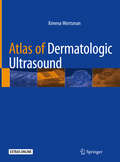
Atlas of Dermatologic Ultrasound
by Ximena WortsmanThis atlas presents a practical and systematic approach for performing dermatologic ultrasound. In recent years, the use of this imaging modality for diagnosing pathologic conditions of the skin, hair, nails, scalp, and soft tissues has grown dramatically and there is a demonstrated need for quick access to this information. For common dermatologic entities, richly-illustrated figures and drawings describe the ultrasound normal anatomy, technical guidelines, common findings, variants, key points, and tips and pitfalls. The extensive collection includes clinical and ultrasonographic correlations with 3D color Doppler ultrasound images and high-definition videos produced with state-of-the-art technology and relevant topics such as benign cutaneous and nail tumors and pseudotumors, skin cancer, vascular anomalies, facial ultrasound anatomy for cosmetic purposes, aesthetic complications, inflammatory diseases, etc. The Atlas of Dermatologic Ultrasound is a valuable resource and a must-have book for radiologists, dermatologists, plastic surgeons, sonographers, residents, and medical professionals who wish to strengthen their knowledge of the wide spectrum of sonographic presentations of dermatologic conditions and successfully integrate this field of ultrasound into their clinical practice.
Atlas of Dermatological Conditions in Populations of African Ancestry
by Andrew F. Alexis Claudia M.Y.A. Donkor Jeannette Aryee-Boi Itohan Roseline Osazuwa Francis Kwame AffluThis book presents the nuances of dermatology from the African diaspora and the tropics. It not only addresses the dark pigmentation of the patient’s skin and the occurrence of tropical infections, but also the socioeconomic conditions which lead to unique features and the development of skin diseases. Chapters present numerous dermatological cases, with clear/relevant pictures, to serve as illustration of how skin conditions present in African/dark skin. Of these specific conditions, the book includes chapters on eczema, bullous diseases, hair disorders, acne, and papulosquamous disorders. Additionally, chapters address emotionally sensitive and socioeconomic-related issues such as skin bleaching and dermatological manifestations of HIV/AIDS, an infectious condition that disproportionately affects those residing in sub-Saharan Africa. Expertly written text is supplemented by hundreds of high-quality, real patient photos. Written by doctors living and treating patients in the tropical environment of Africa, Atlas of Dermatological Conditions in Populations of African Ancestry is an essential tool in broadening the scope of care for professionals and residents in dermatology alike.