- Table View
- List View
Assessing and Developing Communication and Thinking Skills in People with Autism and Communication Difficulties: A Toolkit for Parents and Professionals
by Paul DobsonThis fully photocopiable resource offers a flexible framework for the assessment and measurement of the communication skills of children with autistic spectrum disorders (ASDs). Packed with practical assessment and planning sheets, it enables teachers, educators and other professionals to observe and record how children use and understand language, and to follow their progress over time. The completed assessment record is an accessible summary of a child's individual communication style, identifying strengths and weaknesses and the ways in which he or she is best assisted and motivated to communicate. It focuses on how children express themselves in everyday situations - for example, how they make requests or gain attention, the words they use most frequently, and how their communication is affected by different people and places. Most importantly, it provides a diagnosis of where communication skills can be developed and improved. Using the communication curriculum, educators can set appropriate targets, linked to work in other areas, such as literacy and science. A separate thinking skills curriculum aims to develop the skills and confidence necessary for social interaction, from making simple choices to understanding humour and abstract ideas. Originally devised for use with children with ASDs, this toolkit is equally effective in identifying communication problems in other children, and is an invaluable resource for teachers and speech and language therapists.
Assessing and Diagnosing Speech Therapy Needs in School: Pedagogical Diagnostics in Theory and Practice
by Małgorzata Przybysz-Zaremba Aleksandra Siedlaczek-Szwed Krzysztof PolokAssessing and Diagnosing Speech Therapy Needs in School is a unique text that offers practical guidance in pedagogical diagnosis of speech and communication difficulties within educational settings It outlines theoretical assumptions of the diagnosis process and presents hands-on solutions for pedagogical and speech therapy. Underpinned by theoretical knowledge and written by experienced practitioners, the book equips its readers with tools to understand the diagnostic process and make accurate diagnoses based on each child’s individual circumstances. It starts by clearly distinguishing between pedagogy and speech therapy and outlines issues and theoretical considerations in diagnosing these disorders. To contextualize the theorical observations, it goes on to present case studies, and touches upon crucial topics including readiness to start education, tendency toward aggressive behavior, aphasia and hearing loss. The authors also elaborate on a range of selected diagnostic tools to assess specific difficulties in speech and language therapy. Finally, a list of resources, including games and exercises that can target reading, writing and articulation skills to help children develop, are also featured in the book. Highlighting the importance of practical and theoretical knowledge for those who work with children, this will be a valuable aid for teachers, special educators and speech and language therapists working within school settings. The book will also be of interest to students, teachers and trainee practitioners in the fields of speech therapy and special educational needs.
Assessing and Diagnosing Young Children with Neurodevelopmental Disorders: A DSM-5-TR Compliant Guide
by Neil NicollNow in its second edition, this practical handbook assesses global developmental delay and other neurodevelopmental disorders in young children. Explaining diagnostic, support, and treatment services available for children and their families, this volume clarifies psychological and medical terminology, and global legislative and societal factors relating to assessment. Fully updated, this new edition incorporates the transition from DSM-5 to DSM-5-TR and has an increased emphasis on cross-cultural and ethnic diversity aspects of assessing and diagnosing neurodevelopmental disorders in young children.Designed as a comprehensive compendium for student and practicing psychologists, it offers an introduction to historical perspectives around child development and developmental disorders, and how these have affected our understanding of neurodevelopmental disorders. It explains professional and ethical considerations surrounding the clinical practice of developmental assessments and focuses on the crucial importance of understanding and supporting the parental experience of assessment and diagnosis. Key topics covered include definitions and descriptions of genetic and chromosomal disorders and neurodevelopmental disorders; eligibility criteria for support and assistance; the Griffiths Scales, Bayley Scales, and other notable assessments for young children; autism spectrum disorder; the process of assessment and diagnosis, diagnostic tools, and report writing.Including a chapter of illustrative case studies of children with developmental disorders, this book is essential reading for educational, clinical, and developmental psychologists working with children and their families, as well as postgraduate students training in the field.

Assessing and Enhancing Student Experience in Higher Education
by John T. E. Richardson Anja Pabel Mahsood Shah Beverley OliverThe book makes an important contribution to the discourse on student experience in higher education. The book includes chapters that cover important aspects of the 21st century student experience. Chapters cover issues such as: new trends and insights on the student experience; the changing profile of students in higher education and performance measures used to assess the quality of student experience, institutional approaches in engaging students, using student voice to improve the quality of teaching, COVID-19 and its impact on international students, innovative partnerships between students and academic staff, student feedback and raising academic standards, the increased use of qualitative data in gaining insights into student experience, the use of innovative learning spaces and technology to enhance the learning experience, and the potentially disrupting nature of student feedback and its impact on the health and wellbeing of academic staff, and the increased use of social media reviews by students.
Assessing and Evaluating Early Childhood Education Systems (Early Childhood Research and Education: An Inter-theoretical Focus #2)
by Susanne Garvis Heidi Harju-Luukkainen Jonna KangasThis book provides global perspectives on assessment and evaluation practices with young children in contemporary times within early childhood education systems. It critiques and evaluates current evaluation and assessment goals and tools in early childhood settings. The book also compares the different approaches to educational evaluations from different countries in early childhood education and care. It provides insights into different approaches, techniques as well as perspectives of micro and macro-levels of analysis. This book aims to create an international understanding about the thematic conceptions of assessment for early childhood education and care.
Assessing and Improving Data Quality
by Thomas C. RedmanOrganizations with the best data invest their data quality efforts toward preventing errors at their sources. This chapter examines the cases of Tele-Tech Services, Interactive Data, and Morningstar, outlining ten data quality habits that organizations should adopt, and providing tools and role models that will enable managers to baseline their organization's current data quality efforts.
Assessing and Improving Prediction and Classification: Theory and Algorithms in C++
by Timothy MastersThis book begins by presenting methods for performing practical, real-life assessment of the performance of prediction and classification models. It then goes on to discuss techniques for improving the performance of such models by intelligent resampling of training/testing data, combining multiple models into sophisticated committees, and making use of exogenous information to dynamically choose modeling methodologies. Rigorous statistical techniques for computing confidence in predictions and decisions receive extensive treatment. Finally, a hundred pages are devoted to the use of information theory in evaluating and selecting useful predictors. Special attention is paid to Schreiber's Information Transfer, a recent generalization of Grainger Causality. Well commented C++ code is given for every algorithm and technique. The ultimate purpose of this text is three-fold. The first goal is to open the eyes of serious developers to some of the hidden pitfalls that lurk in the model development process. The second is to provide broad exposure for some of the most powerful model enhancement algorithms that have emerged from academia in the last two decades, while not bogging down readers in cryptic mathematical theory. Finally, this text should provide the reader with a toolbox of ready-to-use C++ code that can be easily incorporated into his or her existing programs.
Assessing and Improving Student Organizations: A Guide for Students (Higher Education Ser.)
by Brent D. Ruben Tricia NolfiThis Assessing and Improving Student Organization (AISO) program is intended as a guide for leaders of student-led college organizations. It is designed to promote the assessment of their organization by leaders and members, help them with planning and improvement, and assist them in responding to reviews by governing bodies and national chapters. Apart from affording their members a structure for engaging with peers in activities of mutual interest, collegiate organizations provide them with hands-on opportunities for enhancing understanding of groups and organizations, and how they operate, and for acquiring and practicing the leadership, communication and collaborative skills that are so important for personal and professional effectiveness throughout life. This Guide provides you with a structure for analyzing the workings of your organization. It will generate insights to help you determine how effectively the organization is functioning, identify strengths and weaknesses, devise priorities and plans for future improvement, and in the process, promote your reflective learning.The AISO process constitutes an ideal laboratory to practice and refine your capabilities for analyzing and improving groups and organizations.Purpose and Elements of the AISO ProgramThe Assessing and Improving Student Organization (AISO) program is intended as a guide for leaders of student-led college organizations. It is designed to promote the assessment of student organizations by their leaders and their members, to help them with planning and improvement, and assist them in responding to reviews by governing bodies and national chapters. Apart from affording their members a structure for engaging with peers in activities of mutual interest, collegiate organizations provide them with hands-on opportunities for enhancing understanding of groups and organizations, and how they operate, and for acquiring and practicing the leadership, communication and collaborative skills that are so important for personal and professional effectiveness throughout life.In addition, the AISO leadership process – unlike comparable programs – provides students with immediate and authentic feedback to evaluate their leadership, and how they impact their organization, community, and campus. The program consists of three elements: a Guide for Students, a Student Workbook, and a CD-ROM for facilitators.AISO has been developed under the auspices of NACA and ACPA by two authors who are experts in organizational and leadership development, student affairs, and human resources management.This is a unique, easy to use, and effective process that reflects input from student leaders.An ACPA Publication
Assessing and Improving Student Organizations: Student Workbook
by Brent D. Ruben Tricia NolfiThis Assessing and Improving Student Organization (AISO) program is intended as a guide for leaders of student-led college organizations. It is designed to promote the assessment of their organization by leaders and members, help them with planning and improvement, and assist them in responding to reviews by governing bodies and national chapters. Apart from affording their members a structure for engaging with peers in activities of mutual interest, collegiate organizations provide them with hands-on opportunities for enhancing understanding of groups and organizations, and how they operate, and for acquiring and practicing the leadership, communication and collaborative skills that are so important for personal and professional effectiveness throughout life. This Guide provides you with a structure for analyzing the workings of your organization. It will generate insights to help you determine how effectively the organization is functioning, identify strengths and weaknesses, devise priorities and plans for future improvement, and in the process, promote your reflective learning.The AISO process constitutes an ideal laboratory to practice and refine your capabilities for analyzing and improving groups and organizations.Purpose and Elements of the AISO ProgramThe Assessing and Improving Student Organization (AISO) program is intended as a guide for leaders of student-led college organizations. It is designed to promote the assessment of student organizations by their leaders and their members, to help them with planning and improvement, and assist them in responding to reviews by governing bodies and national chapters. Apart from affording their members a structure for engaging with peers in activities of mutual interest, collegiate organizations provide them with hands-on opportunities for enhancing understanding of groups and organizations, and how they operate, and for acquiring and practicing the leadership, communication and collaborative skills that are so important for personal and professional effectiveness throughout life.In addition, the AISO leadership process – unlike comparable programs – provides students with immediate and authentic feedback to evaluate their leadership, and how they impact their organization, community, and campus. The program consists of three elements: a Guide for Students, a Student Workbook, and a CD-ROM for facilitators.AISO has been developed under the auspices of NACA and ACPA by two authors who are experts in organizational and leadership development, student affairs, and human resources management.This is a unique, easy to use, and effective process that reflects input from student leaders.An ACPA Publication
Assessing and Improving Student Writing in College: A Guide for Institutions, General Education, Departments, and Classrooms
by Barbara E. WalvoordStep-by-step guidance for shaping better writers while keeping faculty workloads manageable Effective communication is a critical skill for many academic disciplines and careers, and so colleges and universities and their faculty members are rightfully committed to improving student writing across the curriculum. Guiding and assessing student writing in classrooms, general education, and departments takes knowledge, planning, and persistence, but it can be done effectively and efficiently. Written in the concise, accessible style Barbara Walvoord is known for, Assessing and Improving Student Writing in College: A Guide for Institutions, General Education, Departments, and Classrooms offers administrators, program chairs, general education leaders, and classroom instructors the guidance they need. The book provides concrete suggestions for how to: Articulate goals for student writing Measure student writing Improve student writing Document that improvement The book begins by addressing four basic concepts: what we mean by writing, what we mean by "good" writing, how students learn to write, and the purposes of assessment. Next, Walvoord explains the various approaches and methods for assessing writing, urging a combination of them adapted to the institution's purposes and political context. After this introduction, successive chapters offer realistic, practical advice to institution-wide and general education leaders, department members, and classroom instructors. Walvoord addresses issues such as how to engage faculty, how to use rubrics, how to aggregate assessment information at the department and institutional levels, and how to report assessment information to accreditors. The chapter for classroom instructors offers practical suggestions: how to add more writing to a course without substantially increasing the grading load; how to construct writing assignments, how to make grading and responding more effective and time-efficient, how to address grammar and punctuation, and how to support students whose native language is not English. The book also includes four helpful appendices: a taxonomy of Writing Across the Curriculum (WAC) and Writing in the Disciplines (WID) programs; sample outlines for faculty development workshops; a student survey on teaching methods instructors can use to inform their choices in the classroom; and a student self-check cover sheet designed to help students take ownership of their own learning and responsibility for turning in complete, correct assignments. Practical, step-by-step guidance for each point in the assessment and improvement process creates a cohesive, institution-wide system that keeps students, faculty, and administrators on the same page.
Assessing and Improving Value in Cancer Care: Workshop Summary
by Institute of MedicineUnlike many other areas in health care, the practice of oncology presents unique challenges that make assessing and improving value especially complex. First, patients and professionals feel a well-justified sense of urgency to treat for cure, and if cure is not possible, to extend life and reduce the burden of disease. Second, treatments are often both life sparing and highly toxic. Third, distinctive payment structures for cancer medicines are intertwined with practice. Fourth, providers often face tremendous pressure to apply the newest technologies to patients who fail to respond to established treatments, even when the evidence supporting those technologies is incomplete or uncertain, and providers may be reluctant to stop toxic treatments and move to palliation, even at the end of life. Finally, the newest and most novel treatments in oncology are among the most costly in medicine. This volume summarizes the results of a workshop that addressed these issues from multiple perspectives, including those of patients and patient advocates, providers, insurers, health care researchers, federal agencies, and industry. Its broad goal was to describe value in oncology in a complete and nuanced way, to better inform decisions regarding developing, evaluating, prescribing, and paying for cancer therapeutics.
Assessing and Improving Your Teaching: Strategies and Rubrics for Faculty Growth and Student Learning
by Phyllis BlumbergIn order to make appropriate changes to improve your teaching and your students’ learning, first you need to know how you’re teaching now. Figure it out for yourself and invigorate your teaching on your own terms! This practical evidence-based guide promotes excellence in teaching and improved student learning through self-reflection and self-assessment of one’s teaching. Phyllis Blumberg starts by reviewing the current approaches to instructor evaluation and describes their inadequacies. She then presents a new model of assessing teaching that builds upon a broader base of evidence and sources of support. This new model leads to self-assessment rubrics, which are available for download, and the book will guide you in how to use them. The book includes case studies of completed critical reflection rubrics from a variety of disciplines, including the performing and visual arts and the hard sciences, to show how they can be used in different ways and how to explore the richness of the data you’ll uncover.
Assessing and Improving the Interpretation of Breast Images: Workshop Summary
by Sharyl J. NassMillions of women undergo screening mammography regularly with the hope of detecting breast cancer at an earlier and more curable stage. But the ability of such screening to accurately detect early cancers depends on the quality of mammography, including high-quality image acquisition and interpretation. To help ensure the quality of mammography, Congress passed the Mammography Quality Standards Act (MQSA) in 1994 and last reauthorized it in 2004. In advance of its expected reauthorization in 2007, Congress requested a consensus study from the Institute of Medicine (IOM) recommending ways to improve the quality of mammography, with an emphasis on image interpretation. The resulting report, Improving Breast Imaging Quality Standards, highlighted the need to decrease variability in mammography interpretation in the United States and identified gaps in the evidence needed to develop best practices. The consensus committee found that mammography interpretation remained quite variable, and that this variability limited the full potential of mammography to reduce breast cancer mortality by detecting breast cancers at an early stage. In May 2015, the IOM convened a workshop to address this issue. The participants discussed challenges in the delivery of high-quality mammography, the impact of training and experience on interpretive performance, how best to measure interpretive performance, and the potential impact of new technologies and supplemental imaging on interpretation of breast screening and diagnostic images. Assessing and Improving the Interpretation of Breast Images summarizes the presentations and discussions from this workshop.
Assessing and Insuring Cybersecurity Risk
by Ravi DasRemote workforces using VPNs, cloud-based infrastructure and critical systems, and a proliferation in phishing attacks and fraudulent websites are all raising the level of risk for every company. It all comes down to just one thing that is at stake: how to gauge a company’s level of cyber risk and the tolerance level for this risk. Loosely put, this translates to how much uncertainty an organization can tolerate before it starts to negatively affect mission critical flows and business processes. Trying to gauge this can be a huge and nebulous task for any IT security team to accomplish. Making this task so difficult are the many frameworks and models that can be utilized. It is very confusing to know which one to utilize in order to achieve a high level of security. Complicating this situation further is that both quantitative and qualitative variables must be considered and deployed into a cyber risk model. Assessing and Insuring Cybersecurity Risk provides an insight into how to gauge an organization’s particular level of cyber risk, and what would be deemed appropriate for the organization’s risk tolerance. In addition to computing the level of cyber risk, an IT security team has to determine the appropriate controls that are needed to mitigate cyber risk. Also to be considered are the standards and best practices that the IT security team has to implement for complying with such regulations and mandates as CCPA, GDPR, and the HIPAA. To help a security team to comprehensively assess an organization’s cyber risk level and how to insure against it, the book covers: The mechanics of cyber risk Risk controls that need to be put into place The issues and benefits of cybersecurity risk insurance policies GDPR, CCPA, and the the CMMC Gauging how much cyber risk and uncertainty an organization can tolerate is a complex and complicated task, and this book helps to make it more understandable and manageable.
Assessing and Managing Problematic Sexual Interests: A Practitioner's Guide
by Geraldine AkermanAssessing and Managing Problematic Sexual Interests: A Practitioner’s Guide provides a thorough review of atypical sexual interests and offers various ways through which they can be measured and controlled, including compassion-focused and psychoanalytic approaches. This unique guide presents a detailed analysis of deviant sexual interest. Part I, 'Assessment,' overviews the range of sexual interests and fantasies in men and women. Part II, 'Management,' investigates the cutting-edge tools, approaches, interventions, and treatment advances used in a variety of settings to control deviant sexual interest. In Part III, 'Approaches to assessment and management', the authors consider how females with sexual convictions can be assessed and how offence paralelling behaviour can be used for assessment and treatment. Throughout, Assessing and Managing Problematic Sexual Interests offers necessary perspectives and emerging research from international experts at the forefront of this field. With a thorough assessment of current research and a critical overview of treatment advances for problematic sexual interests, Assessing and Managing Problematic Sexual Interests is an essential resource for clinical and forensic psychologists, probation officers, academics, students working in the field, and members of allied professional fields.
Assessing and Managing Rapid Credit Growth and the Role of Supervisory and Prudential Policies
by Paul Hilbers Inci Otker-Robe Ceyla Pazarbasioglu Gudrun JohnsenA report from the International Monetary Fund.
Assessing and Managing Security Risk in IT Systems: A Structured Methodology
by John McCumberThis book begins with an overview of information systems security, offering the basic underpinnings of information security and concluding with an analysis of risk management. Part II describes the McCumber Cube, providing the original paper from 1991 and detailing ways to accurately map information flow in computer and telecom systems. It also explains how to apply the methodology to individual system components and subsystems. Part III serves as a resource for analysts and security practitioners who want access to more detailed information on technical vulnerabilities and risk assessment analytics. McCumber details how information extracted from this resource can be applied to his assessment processes.
Assessing and Managing the Acutely Ill Patient for Nursing Associates (Understanding Nursing Associate Practice)
by Marion Taylor Cariona FlahertyAcutely ill adults present in a variety of settings and caring for them is a key part of the nursing associate role. This book equips you with the skills and knowledge to assess the acutely ill adult and manage their care. Each chapter follows a case study of a patient presenting with an acute illness, working step-by-step through their assessment and care whilst drawing on relevant pathophysiology, pharmacology and evidence-based practice. Written in clear language specifically for the nursing associate, this is your perfect introduction to the world of acute care. Key features Fully mapped to the NMC Standards of Proficiency for Nursing Associates (2018) Introduces a range of commonly encountered acute illnesses across different body systems A unique case study approach uses real-world practice scenarios to make understanding the complex theory, pathophysiology and pharmacology much easier Focuses specifically on the requirements of the nursing associate role, helping you to develop into a confident professional practitioner
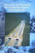
Assessing and Managing the Ecological Impacts of Paved Roads
by National Research Council of the National AcademiesAll phases of road development—from construction and use by vehicles to maintenance—affect physical and chemical soil conditions, water flow, and air and water quality, as well as plants and animals. Roads and traffic can alter wildlife habitat, cause vehicle-related mortality, impede animal migration, and disperse nonnative pest species of plants and animals. Integrating environmental considerations into all phases of transportation is an important, evolving process. The increasing awareness of environmental issues has made road development more complex and controversial. Over the past two decades, the Federal Highway Administration and state transportation agencies have increasingly recognized the importance of the effects of transportation on the natural environment. This report provides guidance on ways to reconcile the different goals of road development and environmental conservation. It identifies the ecological effects of roads that can be evaluated in the planning, design, construction, and maintenance of roads and offers several recommendations to help better understand and manage ecological impacts of paved roads.
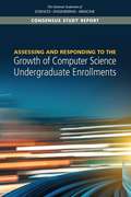
Assessing and Responding to the Growth of Computer Science Undergraduate Enrollments
by Engineering Medicine National Academies of SciencesThe field of computer science (CS) is currently experiencing a surge in undergraduate degree production and course enrollments, which is straining program resources at many institutions and causing concern among faculty and administrators about how best to respond to the rapidly growing demand. There is also significant interest about what this growth will mean for the future of CS programs, the role of computer science in academic institutions, the field as a whole, and U.S. society more broadly. Assessing and Responding to the Growth of Computer Science Undergraduate Enrollments seeks to provide a better understanding of the current trends in computing enrollments in the context of past trends. It examines drivers of the current enrollment surge, relationships between the surge and current and potential gains in diversity in the field, and the potential impacts of responses to the increased demand for computing in higher education, and it considers the likely effects of those responses on students, faculty, and institutions. This report provides recommendations for what institutions of higher education, government agencies, and the private sector can do to respond to the surge and plan for a strong and sustainable future for the field of CS in general, the health of the institutions of higher education, and the prosperity of the nation.
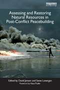
Assessing and Restoring Natural Resources In Post-Conflict Peacebuilding: Assessing And Restoring Natural Resources In Post-conflict Peacebuilding (Post-Conflict Peacebuilding and Natural Resource Management)
by David Jensen Steve LonerganWhen a country emerges from violent conflict, the management of the environment and natural resources has important implications for short-term peacebuilding and long-term stability, particularly if natural resources were a factor in the conflict, play a major role in the national economy, or broadly support livelihoods. Only recently, however, have the assessment, harnessing, and restoration of the natural resource base become essential components of postconflict peacebuilding. This book, by thirty-five authors, examines the experiences of more than twenty countries and territories in assessing post-conflict environmental damage and natural resource degradation and their implications for human health, livelihoods, and security. The book also illustrates how an understanding of both the risks and opportunities associated with natural resources can help decision makers manage natural resources in ways that create jobs, sustain livelihoods, and contribute to economic recovery and reconciliation, without creating new grievances or significant environmental degradation. Finally, the book offers lessons from the remediation of environmental hot spots, restoration of damaged ecosystems, and reconstruction of the environmental services and infrastructure necessary for a sustainable peace. Assessing and Restoring Natural Resources in Post-Conflict Peacebuilding is part of a global initiative to identify and analyze lessons in post-conflict peacebuilding and natural resource management. The project has generated six books of case studies and analyses, with contributions by practitioners, policy makers, and researchers. Other books address highvalue resources, land, water, livelihoods, and governance.
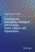
Assessing and Stimulating a Dialogical Self in Groups, Teams, Cultures, and Organizations
by Hubert HermansThis book presents 9 theory-based and practice-oriented methods for assessing and stimulating a multi-voiced dialogical self in the context of groups, teams, cultures, and organizations. All of these methods are based on Dialogical Self Theory. The book deals with the practical implications of this theory as applied in the areas of coaching, training, and counselling. A number of chapters focus on the process of positioning and dialogue on the level of the self, while other chapters combine self-processes with group work, and still others find their applications in leadership development and team-work in organizations. For each of the nine methods, the chapters present theory, method, case-study and discussions and make clear what kind of problems can be tackled using the method discussed. Specifically, the book discusses the following methods: A Negotiational Self Method for assessing and solving inner conflicts; a Self-Confrontation Method used to assess and stimulate personal meaning construction in career counselling; a Method of Expressive Writing in the context of career development; a Composition Method for studying the content and organization of personal positions via verbal and non-verbal procedures; a Dialogical Leadership Method that investigates and stimulates dialogical relationships between personal positions in the self of leaders in organizations; a Personal Position Repertoire Method that combines the assessment of personal positions with focus group discussions; a Team Confrontation Method for investigating collective and deviant positions or voices in organizational teams; a Method for Revising Organizational Stories with a focus on their emotional significance: and a Technique for Assessing and Stimulating Innovative Dialogue between Cultural Positions in global nomads.
Assessing and Teaching Reading Composition and Pre-Writing, K-3, Vol. 1
by K. Michael Hibbard Elizabeth WagnerThe performance tasks in this book are linked directly to instructional strategies and include holistic rubrics, analytic rubrics, and assessment lists. They can be photocopied and distributed to your students.